Method of predicting stroke evolution utilising mri

a magnetic resonance imaging and evolution technology, applied in image enhancement, instruments, antibody medical ingredients, etc., can solve the problems of time-consuming methods, difficult to predict the final size of the stroke, and deprived cerebral nervous tissue of oxygen,

Inactive Publication Date: 2004-06-03
THE UNIV OF QUEENSLAND
View PDF4 Cites 48 Cited by
  • Summary
  • Abstract
  • Description
  • Claims
  • Application Information

AI Technical Summary

Problems solved by technology

Typically, a person suffers an ischemic infarction or stroke when a blood vessel is blocked, causing cerebral nervous tissue to be deprived of oxygen.
The prediction of the final size of the stroke, i.e. the final volume of dead tissue is very difficult.
However, these methods are time consuming to implement and require highly skilled practitioners.
Further, there is a limited time window of opportunity for the administration of thrombolytic or neuroprotective therapy.

Method used


Image

  • Method of predicting stroke evolution utilising mri
  • Method of predicting stroke evolution utilising mri
  • Method of predicting stroke evolution utilising mri

Examples


Embodiment Construction

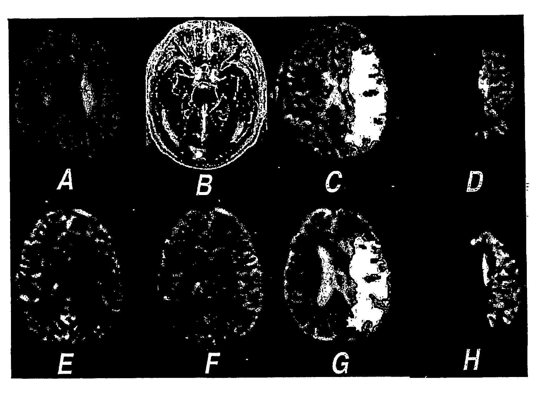
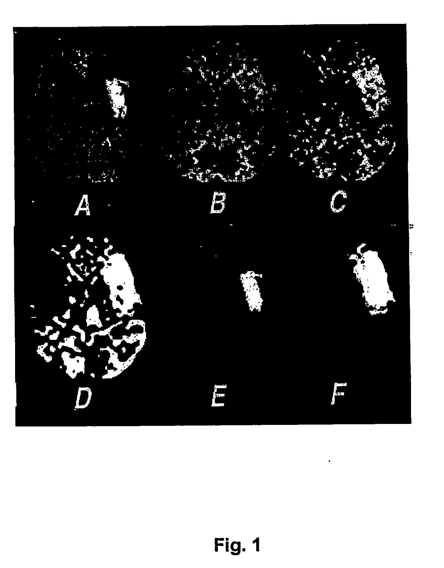
[0022] The method of predicting stroke evolution according to the preferred embodiment involves the computerised processing of brain scan images obtained shortly after the onset of stroke symptoms.

[0023] First, input magnetic resonance diffusion and perfusion images are acquired in the acute phase of stroke. Appropriate diffusion images can be acquired with standard diffusion- weighted MRI sequences.sup.1 or diffusion tensor imaging (DTI) methods..sup.2 The methodology of the preferred embodiment of this invention has been developed to process isotropically weighted diffusion images (DWI) generated from diffusion tensor images by the method of Sorensen et al..sup.3 However, the method would be applicable to process standard diffusion-weighted images where the lesion appears hyperintense.sup.4 or images of the apparent diffusion coefficient of water (ADC)..sup.5,6

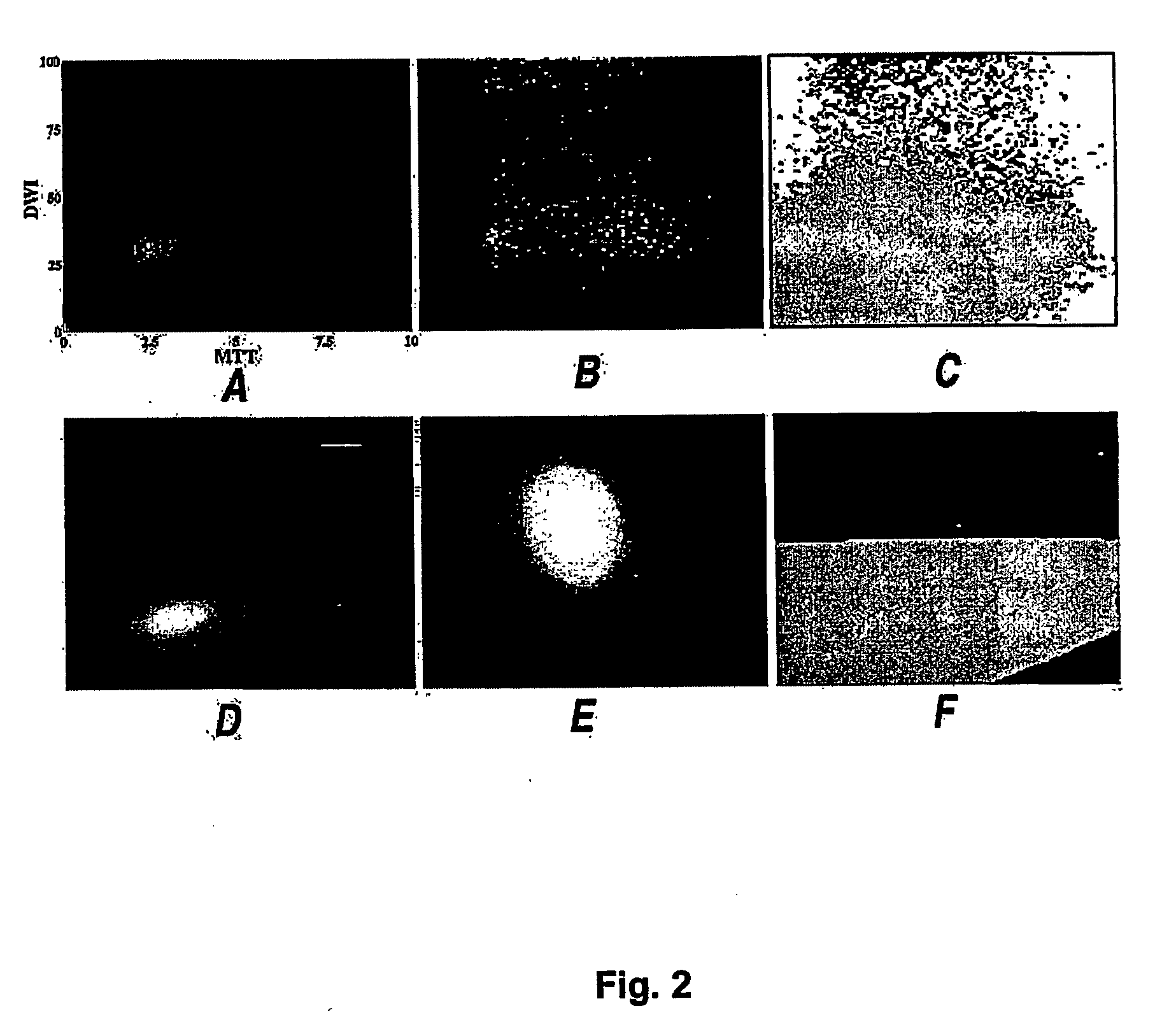
[0024] Perfusion images are defined as maps of cerebral blood flow (CBF), cerebral blood volume (CBV) and mean transit tim...


PUM

PropertyMeasurementUnit
thicknessaaaaaaaaaa
thicknessaaaaaaaaaa
thicknessaaaaaaaaaa

Abstract

A method predicting stroke evolution uses magnetic resonance diffusion and perfusion images obtained shortly after the onset of stroke symptoms to automatically estimate the eventual volume of dead cerebral tissue resulting from the stroke. The diffusion and perfusion images are processed to extract region(s) of interest presenting tissue at risk of infarction. A midplane algorithm is also used to calculate ratio and diffusion and perfusion measures for modelling infarct evolution. A parametric normal classifier algorithm is used to predict infarct growth using the calculated measures.

Description

[0001] THIS INVENTION relates to a method for predicting infarct evolution using magnetic resonance imaging (MRI) and image processing. In particular, the invention is directed to an automated method for estimating the volume of dead nervous tissue resulting from a stroke, using imaging information obtained shortly after the onset of stroke symptoms.[0002] Typically, a person suffers an ischemic infarction or stroke when a blood vessel is blocked, causing cerebral nervous tissue to be deprived of oxygen. In the initial few hours after a stroke, there is usually a significantly reduced blood supply to a region of nervous tissue due to a blocked or nearly-blocked blood vessel which would otherwise supply oxygen to that tissue. The nervous tissue deprived of adequate blood supply does not necessarily die immediately. It can often die over the next 18 hours or so. The prediction of the final size of the stroke, i.e. the final volume of dead tissue is very difficult.[0003] If the stroke ...

Claims


Application Information

Patent Timeline
Patent Type & Authority Applications(United States)
IPC IPC(8): C12N15/09A61B5/026A61B5/029A61B5/055A61K31/7088A61K35/76A61K39/12A61K39/39A61K48/00A61P31/20A61P35/00G06T7/00
CPCA61B5/0263A61B5/029A61B5/055G06T2207/30016A61B5/7267G06T7/0012A61B5/7264A61P31/20A61P35/00G16H50/20
Inventor ROSE, STEPHEN EDWARDGRIFFIN, MARK PHILIPJANKE, ANDREW LINDSAYCHALK, JONATHAN BRANDONMCLACHLAN, GEOFFREY JOHNPEEL, DAVIDWANG, DEMING
Owner THE UNIV OF QUEENSLAND
Why Eureka
Try Eureka