
Method and system for the analysis of high density cells samples

a high density cell and sample technology, applied in the field of cell array formation, can solve the problems of high variability of current state of the art methods for dispensing drops of cells onto microscope slides, non-uniform dispersal of cells across the surface of substrates, and considerable overlap of cells

Active Publication Date: 2005-01-06
BIODOT
View PDF62 Cites 67 Cited by
  • Summary
  • Abstract
  • Description
  • Claims
  • Application Information

AI Technical Summary

Benefits of technology

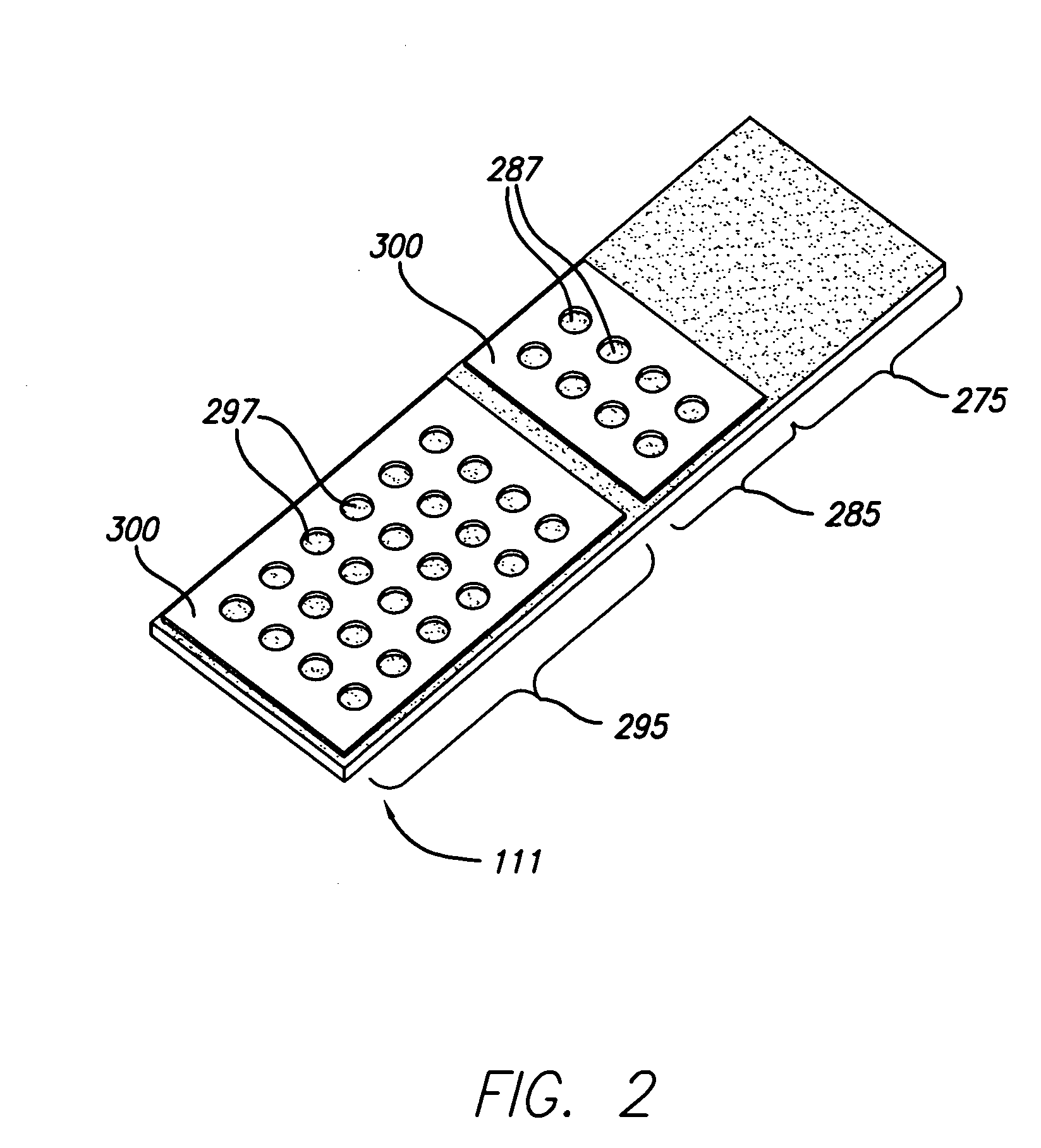
[0010] The cell arrays of the present invention overcome many of the drawbacks associated with current methods for conducting diagnostic assays. For example, the cell arrays formed according to the present invention may comprise as many as about 1000 discrete cell samples suitable for diagnostic analysis on a single substrate. Additionally, the cell arrays of the present invention are particularly suited for high-throughput diagnostic analyses, such as, for example, immunohistochemistry (IHC), fluorescent in situ hybridization (FISH), and cytogenetic analyses. Yet another advantage of the cell arrays of the present invention is the ability to conduct the desired diagnostic analysis on a single substrate that includes positive or negative control samples on the same substrate as the test samples.

Problems solved by technology

Unfortunately, the current state of the art methods for dispensing drops of cells onto microscope slides suffer from high variability due to the individual techniques employed by various operators.
However, these methods are considered more of an art than a science and frequently result in considerable overlap of cells and non-uniform dispersal of the cells across the surface of the substrate.
Overlap and non-uniformity may lead to inefficient or insufficient interaction between a test reagent and a target, which may ultimately lead to poor detection and a misdiagnosis.
The variability arising from the application of cell samples to the surface of the substrate may lead to increased variability in the results of the clinical assays and ultimately to inaccurate diagnoses.

Method used


Image

  • Method and system for the analysis of high density cells samples
  • Method and system for the analysis of high density cells samples

Examples


example 1

Cell Array Formation

[0083] In order to conduct clinical diagnostic tests on several patient samples simultaneously, it is often desirable to map (replicate or transform) one or more microplate arrays into a high density cell array of discrete cell samples arranged substantially in a monolayer on a single substrate. The substrate containing the cell array can then be subjected to any number of clinical diagnostic tests that rely on detection of a reaction between a test reagent and a predetermined analyte within the cells of the array, particularly diagnostic assays that benefit from an even and uniform distribution of cells.

[0084] In one example, samples from three cell lines, CaSki, HeLa, and T24, were formed in a cell array arranged substantially in a monolayer on a single substrate. The cell array was formed with 4 discrete cell samples from each of the 3 cells lines, i.e., the cell array contained 12 discrete cell samples. Prior to forming the cell array, the cell lines had an...

example 2

Dispensing Through a Cover Fluid

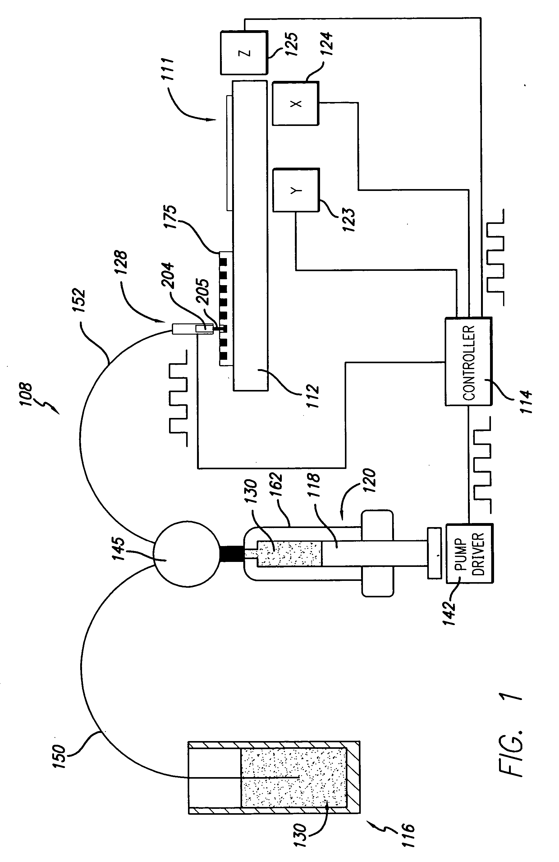
[0090] Some embodiments described herein relate generally to dispensing droplets and in particular to methods for mixing, aspirating, transporting, metering and dispensing droplets of cell suspension or test reagent below the surface of a cover fluid using without contacting the dispenser with the surface of the cover fluid in order to for a cell assay or to conduct a diagnostic assay. Advantageously, evaporation of valuable test regents and cell samples is substantially prevented or reduced by using a cover fluid. Another advantage, in the case of miscible reagents, is that the droplet velocities provide good mixing. Yet another advantage is that, in the non-contact dispensing scheme, the nozzle or tip is not immersed in the cover fluid, thereby facilitating cleaning.

[0091] Mineral oil may be used as a cover fluid over the cell array. Mineral oil is frequently used as an evaporation barrier. Typically, the cell array is formed on the substrate, and...

example 3

Florescent In Situ Hybridization Analysis

[0092] In another embodiment of the invention, cell arrays are formed by replicating one or more microplate arrays onto or into a high density array on a substrate, such as, for example, a glass slide. A cell suspension containing cytogenetic preparations of 104 different cell cultures was placed into a 384 well plate. The density of the cell suspension was approximately 1×10⁸ cells / mL. Approximately 500-1000 cells were mixed, aspirated, transported, metered, and dispensed through the dispensing apparatus onto the glass slide. The apparatus dispensed a drop volume of 100 nL. The cells comprising the array formed according to the methods described herein were counted and confirmed to be arranged substantially in monolayer on the substrate.

[0093] Additionally, cell arrays formed according to the methods described herein were subjected to FISH analysis using several commercially available DNA probes, including CEP3, CEP7, CEP17, 9p21, X and Y ...


PUM

PropertyMeasurementUnit
Diameteraaaaaaaaaa
Lengthaaaaaaaaaa
Volumeaaaaaaaaaa

Abstract

Methods for forming cell arrays of multiple cell samples arranged substantially in a monolayer on a single substrate particularly suited for diagnostic analysis are disclosed. The cell arrays are formed with a high-speed dispensing apparatus capable of dispensing small volumes in precise, complex patterns. Also disclosed are substrates upon which cell arrays may be formed, and methods for conducting diagnostic analyses on the formed cell arrays.

Description

CROSS-REFERENCE TO RELATED APPLICATION [0001] This application claims priority to U.S. Patent Application Ser. No. 60 / 477,807, filed Jun. 12, 2003, the teachings of which are incorporated herein in their entirety.BACKGROUND OF THE INVENTION [0002] 1. Field of the Invention [0003] The present invention relates generally to a method for forming cell arrays on a substrate to facilitate high-throughput diagnostic analysis of a plurality of cell samples and, in particular, to cell arrays in which the discrete cell samples are arranged substantially in a monolayer with positive and negative controls, as well as test samples, arranged on the same substrate. More particularly, the present invention relates to cell arrays comprising multiple test and control samples arranged on a single substrate upon which immunohistochemistry, fluorescent in situ hybridization, and cytogenetic analyses may be conducted. [0004] 2. Description of the Related Art [0005] Clinical analysis of cell samples obtai...

Claims


Application Information

Patent Timeline
IPC IPC(8): B01J19/00B05D3/00C12M1/34C12M3/00C12N5/00C12N5/02C12N5/06C12N5/16C12Q1/68G01NG01N33/48G01N33/53G01N33/567G01N33/569G01N35/10
CPCB01J19/0046Y10S436/809B01J2219/00378B01J2219/00389B01J2219/00527B01J2219/00547B01J2219/00576B01J2219/00585B01J2219/00596B01J2219/00605B01J2219/0061B01J2219/00612B01J2219/00619B01J2219/00637B01J2219/00641B01J2219/00659B01J2219/00689B01J2219/00691B01J2219/00743G01N33/56966G01N35/1016B01J2219/00315B01J2219/00698B01J2219/00398C12Q1/6841G01N33/57415
Inventor MOORE, MATHEWREYES, MIRIAMBAUNOCH, DAVIDTISONE, THOMASO'FARRELL, BRENDAN
Owner BIODOT
Learn More