System and method for the detection of brain iron using magnetic resonance imaging

Inactive Publication Date: 2005-01-27
GENERAL ELECTRIC CO
View PDF8 Cites 20 Cited by
  • Summary
  • Abstract
  • Description
  • Claims
  • Application Information

AI Technical Summary

Benefits of technology

[0007] In a first aspect, a method for detecting iron in the brain using magnetic resonance imaging (MRI) is provided. The method for detecting iron in the brain using magnetic resonance imaging (MRI) comprises generating a substantially high magnetic field strength within the MRI system, acquiring magnetic resonance (MR) images by a pulse sequence adapted to create a magnetic field map of the brain for use in enhancing brain iron deposits, and, characterizing regions of interest within using the magnetic field map to detect statistically relevant quantities of brain iron deposits to indicate a given disease.
[0008] In a second aspect,

Problems solved by technology

However, this imaging phenomenon has not been widely used for diagnostic purposes because of the difficulty in making diagnostic inferences due to the limited sensitivity of standard MR scanners and the complex and irregular shapes of the affected brain regions.
Although these deposits are often too small to be imaged as individual

Method used


Image

  • System and method for the detection of brain iron using magnetic resonance imaging
  • System and method for the detection of brain iron using magnetic resonance imaging
  • System and method for the detection of brain iron using magnetic resonance imaging

Examples


Embodiment Construction

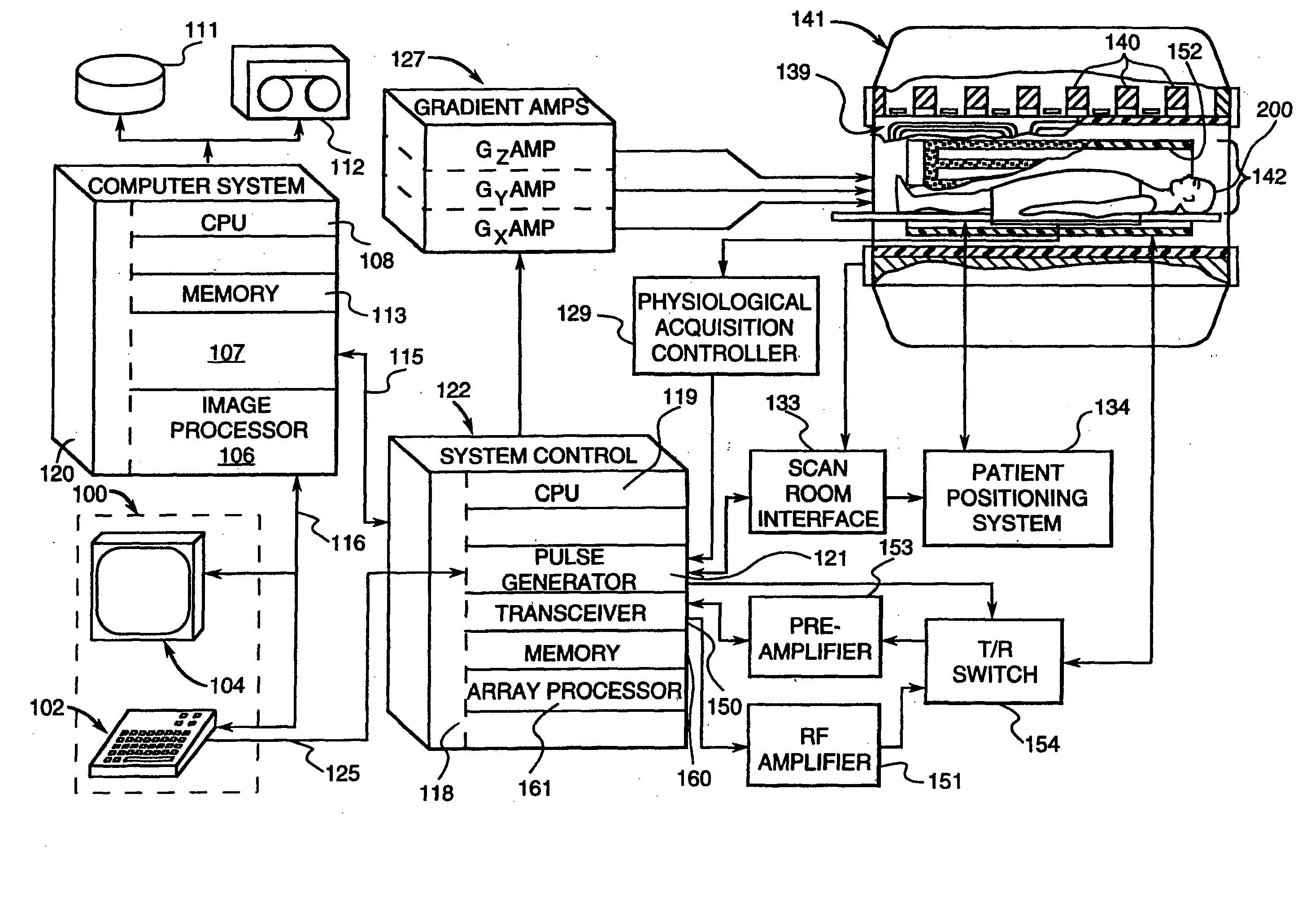
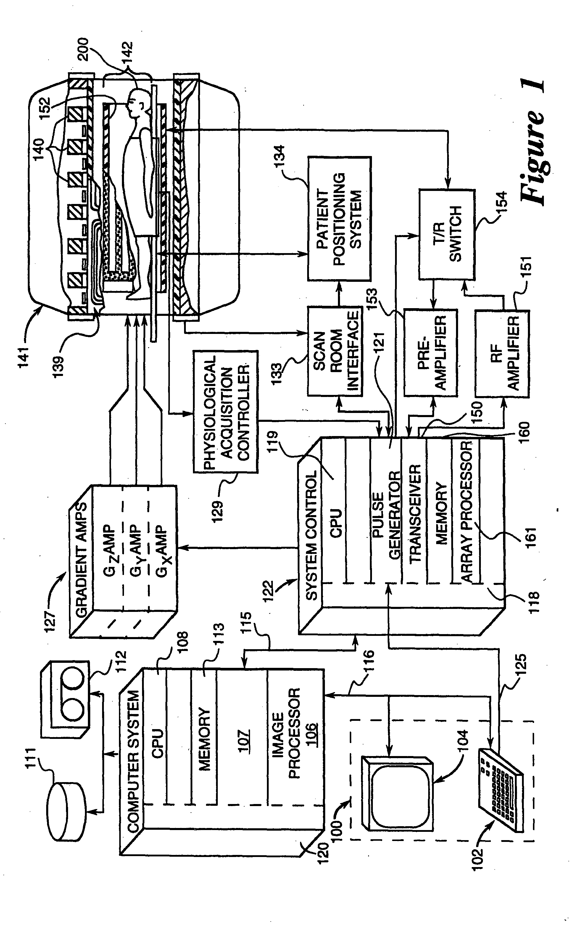
[0012] MRI scanners, which are used in various fields such as medical diagnostics, typically use a computer to create images based on the operation of a magnet, a gradient coil assembly, and a radio frequency coil(s). The magnet creates a uniform main magnetic field that makes nuclei, such as hydrogen atomic nuclei, responsive to radio frequency excitation. The gradient coil assembly imposes a series of pulsed, spatial-gradient magnetic fields upon the main magnetic field to give each point in the imaging volume a spatial identity corresponding to its unique set of magnetic fields during the imaging pulse sequence. The radio frequency coil(s) creates an excitation frequency pulse that temporarily creates an oscillating transverse magnetization that is detected by the radio frequency coil and used by the computer to create the image.

[0013] Generally, very high field strength is characterized as greater than 1.5 Tesla (1.5 T). In recent years, there has been an increase in usage of M...


PUM


Abstract

A method and system for detecting iron using magnetic resonance imaging (MRI) is provided. The method comprises generating a substantially high magnetic field strength within the MRI system, acquiring magnetic resonance (MR) images by a pulse sequence adapted to create a magnetic field map of the brain for use in enhancing brain iron deposits, and, characterizing regions of interest using the magnetic field map to detect statistically relevant quantities of brain iron deposits to indicate a given disease.

Description

BACKGROUND OF THE INVENTION [0001] The invention relates to magnetic resonance imaging (MRI) and image processing methods. More particularly, the invention relates to detection of brain iron using MRI and image processing techniques. [0002] It has been known for some time that specific regions of the brain contain deposits of iron in a storage pool consisting of iron atoms in a mineral matrix associated with and largely surrounded by associated proteins. The total complex of mineralized iron and proteins is referred to as ferritin or in other cases as hemosiderin. It has also been recognized that these deposits are to some extent capable of being visualized on MR images because of the tendency of the magnetized iron atoms to alter the local magnetic field and to thereby to reduce the MR signal from protons in water molecules and other compounds in their vicinity of the iron deposits. This effect is referred to as iron-dependent shortening of the local T2 relaxation time. It is known...

Claims


Application Information

Patent Timeline
IPC IPC(8): G01R33/54A61B5/055G01R33/465G01R33/483
CPCG01R33/483A61B5/055A61B5/4082
Inventor CLINE, HARVEY ELLISWATKINS, RONALD DEANSCHENCK, JOHN FREDERICK
Owner GENERAL ELECTRIC CO
Why Eureka
Try Eureka