Multifactorial assay for cancer detection

a multi-factorial assay and cancer detection technology, applied in the field of multi-factorial assay for cancer detection, can solve the problems of lack of specificity and sensitiveness of ca-125 for detecting early stage disease, and the death of ovarian cancer, and achieve the effect of rapid and early detection of ovarian cancer

Inactive Publication Date: 2005-03-31
UNIVERSITY OF PITTSBURGH
View PDF7 Cites 68 Cited by
  • Summary
  • Abstract
  • Description
  • Claims
  • Application Information

AI Technical Summary

Benefits of technology

[0010] A method for rapid, early detection of ovarian cancer is provided. The method provides the opportunity to simultaneously test a broad panel of angiogenic factors and repeat such testing at multiple time points with use of only, for example and without limitation, 50 μl of serum or plasma per time point.

Problems solved by technology

Epithelial ovarian cancer is so deadly in part because of a lack of effective early detection methods.
However, CA-125 is neither sensitive nor specific for detecting early stage disease.
Screening using transvaginal ultrasound, Doppler and morphological indices has shown some encouraging results but, used alone, it currently lacks the specificity required of a screening test for the general population (Karayiannakis, A. J., et al., Clinical significance of preoperative serum vascular endothelial growth factor levels in patients with colorectal cancer and the effect of tumor surgery.
However, it, too, is of questionable effectiveness in the general population.
Additionally, this approach is very expensive and could only be applied to high-risk population.
However, each single factor was only weakly associated with early stage disease.
All previous testing of serum markers of cancer patients was performed using ELISA, which is very expensive and requires a separate kit for each individual cytokine.

Method used


Image

  • Multifactorial assay for cancer detection
  • Multifactorial assay for cancer detection
  • Multifactorial assay for cancer detection

Examples


example 1

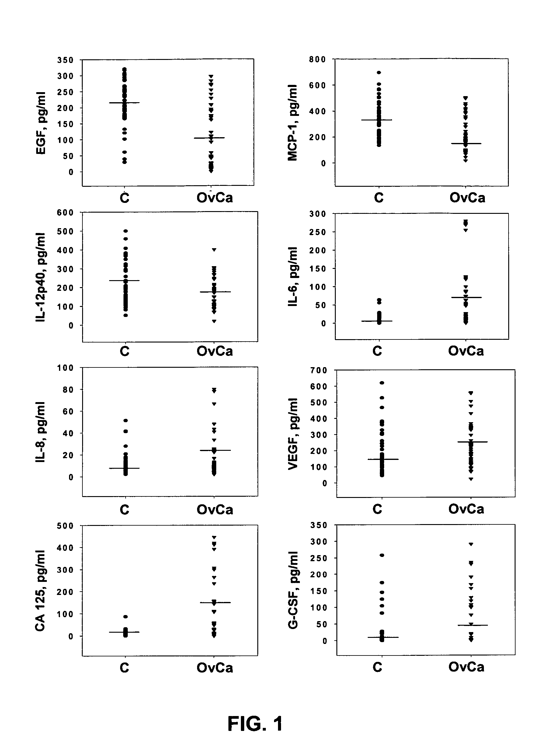
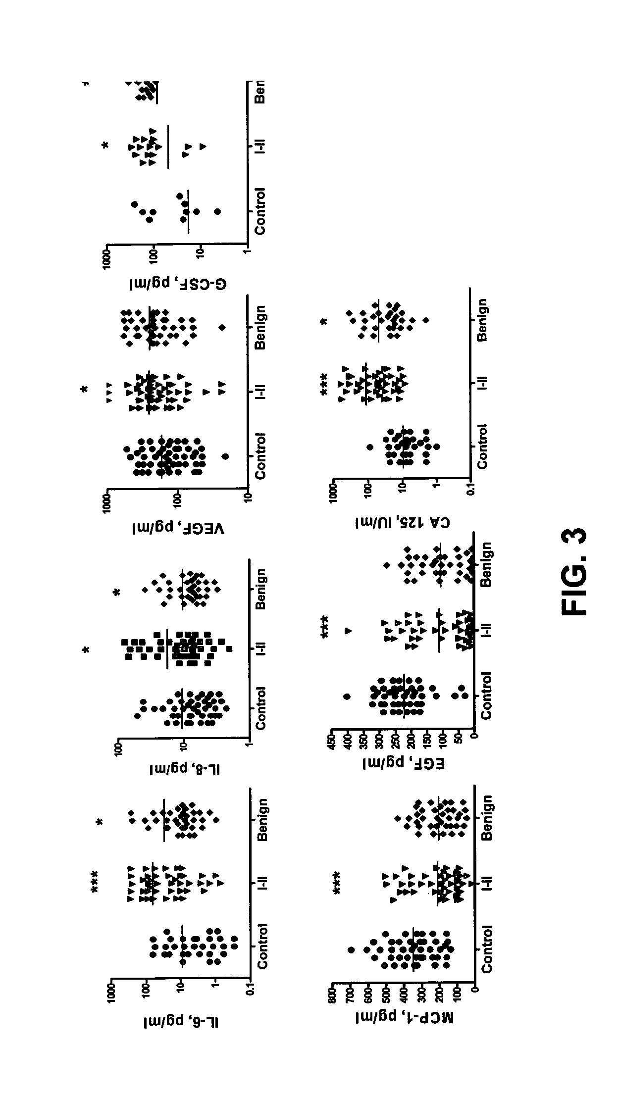
[0040] Patient Population. Serum samples from 55 patients diagnosed with early (I-II) stages ovarian cancer, 55 patients with benign pelvic masses, and 55 healthy age-matched controls were tested. Serum samples from patients with early stages (I-II) ovarian cancer and women with benign pelvic disease, were provided by the Gynecologic Oncology Group (GOG) (Cleveland, Ohio). Consent and blood specimens from all participants were obtained under IRB Protocol. Charts were reviewed by clinical oncologist to verify gynecologic diagnoses and ovarian cancer staging. Pathology slides for ovarian cancer cases were reviewed by a pathologist to verify histology and grade. All major types of epithelial ovarian cancer and benign pelvic conditions were represented. Table A summarizes patient data. Control serum samples from healthy, age-matched women were received from the Allegheny County Case-Control Network under the IRB Protocol.

TABLE APatient characteristicsPatient GroupAgeHistologic TypesCo...

example 2

Purification of Circulating Antibodies

[0066] Antigen-specific (monospecific) circulating antibodies, or populations of two or more such circulating antibodies can be purified, without limitation, according to the following protocol, thereby facilitating the assays for determining serum concentrations of specific circulating antibodies. The Ig purified in this manner can be used as a control for accurately quantitating individual circulating antibodies.

[0067] Purified antigens of interest, for example, IL-6, IL-8, EGF, EGFR, VEGF, Her2 / neu, PDGF, PDGFR, survivin, Fas, FasL, CA-125, CA 15-3, CA 19-9, CA 72-4, CEA, MUC-1, PSA; AFP, bHCG (human chorionic gonadotropin), transglutaminase, c-myc, N-Ras, K-Ras, p53; cyclin B, cyclin D, Akt1 (v-akt murine thymoma viral oncogene homolog 1), and others can be covalently coupled to carboxylate-modified polystyrene beads (Cat. No. CLB4, Sigma Chemical Co.) using, without limitation, the above-described protocols for coupling proteins to Luminex...

example 3

Serum Cytokine Analysis

[0069] Patient populations. Patient populations are described in Example 1. In this study, fewer samples from each group were utilized (Table I).

TABLE IPatient characteristicsPatient GroupAgeHistologic TypesControlRange 36-76N = 45Median 46Early StageRange 34-88Papillary serous carcinoma (n = 13)Ovarian CancerMedian 46Carcinoma, endometroid (n = 10)N = 44Carcinoma, mucinous (n = 7)Carcinoma, poorly differeniated (n = 6)Adenocarcinoma, serous (n = 5)Carcinoma, clear cell (n = 3)Benign TumorsRange 28-87Adenofibroma, serous (n = 1)N = 37Median 44.5Brenner tumor (n = 1)Crystadenofibroma, serous (n = 2)Cyst, paratubal (n = 2)Cyst, serous (n = 1)Cyst, simple (n = 3)Cystadenofibroma, serous (n = 3)Cystadenoma, mucinous (n = 8)Cystadenoma, serous (n = 9)Endometriosis (n = 1)Fibrosis (n = 1)Ovary benign (n = 3)Mucinous benign (n = 2)

[0070] Multiplex LabMap tassays for EGF, IL-6, IL-8, G-CSF, VEGF, CA-125 and MCP-1 were performed substantially as described in Example...


PUM

PropertyMeasurementUnit
volumeaaaaaaaaaa
pHaaaaaaaaaa
pHaaaaaaaaaa

Abstract

Provided are methods for the rapid detection of ovarian cancer. The methods employ a multiplex immunoassay to detect levels of two or more of the markers EGF, G-CSF, IL-6, IL-8, CA-125, VEGF, MCP-1, anti-IL6, anti-IL8, anti CA-125, anti-c-myc, anti-p53, anti-CEA, anti-CA 15-3, anti-MUC-1, anti-survivin, anti-bHCG, anti-osteopontin, anti-PDGF, anti-Her2 / neu, anti-Akt1, anti-cytokeratin 19, cytokeratin 19, EGFR, CEA, kallikrein-8, M-CSF, FasL, ErbB2 and Her2 / neu in a sample of the patient's blood, where the presence of abnormal levels of two or more of the markers indicates the presence of ovarian cancer in the patient. An array also is provided to quantitate levels of these markers in a patient's blood. Also provided is a method of predicting onset of clinical ovarian cancer comprising determining the change in concentration over time of two or more of anti-Her2 / neu, anti-MUC-1, anti-c-myc, anti-p53, anti-CA-125, anti-CEA, anti-CA 72-4, anti-PDGFRα, IFNγ, IL-6, IL-10, TNFα, MIP-1α, MIP-1β, EGFR and Her2 / neu in a patient's blood.

Description

CROSS REFERENCE TO RELATED APPLICATIONS [0001] This application claims priority under 35 U.S.C. §119(e) to U.S. Provisional Patent Application No. 60 / 495,547, filed Aug. 15, 2003, which is incorporated herein by reference in its entirety.BACKGROUND [0002] 1. Field of the Invention [0003] Methods and reagents for a multifactorial assay for the rapid, early detection of cancer. [0004] 2. Description of the Related Art [0005] Ovarian cancer represents the third most frequent cancer of the female genital tract. The majority of early-stage cancers are asymptomatic, and over three-quarters of the diagnoses are made at a time when the disease has already established regional or distant metastases. Despite aggressive cytoreductive surgery and platinum-based chemotherapy, the 5-year survival for patients with clinically advanced ovarian cancer is only 15 to 20 percent, although the cure rate for stage I disease is usually greater than 90 percent (Holschneider, C. H. and J. S. Berek, Ovarian ...

Claims


Application Information

Patent Timeline
Patent Type & Authority Applications(United States)
IPC IPC(8): A61BA61K38/00C12Q1/68G01N33/53G01N33/574G01N33/58
CPCB82Y5/00B82Y10/00G01N33/588G01N33/57449B82Y15/00
Inventor LOKSHIN, ANNA E.GORELIK, ELIESER
Owner UNIVERSITY OF PITTSBURGH
Why Eureka
Try Eureka