

Molecular markers for the diagnosis of Alzheimer's disease
a technology of alzheimer's disease and molecular markers, applied in the field of molecular markers, can solve the problems of insufficient understanding, data currently controversial, and not yet known, and achieve the effect of improving the diagnostic accuracy and accuracy of clinical tests
- Summary
- Abstract
- Description
- Claims
- Application Information
AI Technical Summary
Benefits of technology
Problems solved by technology
Method used
Image

Examples
example 1
Methods
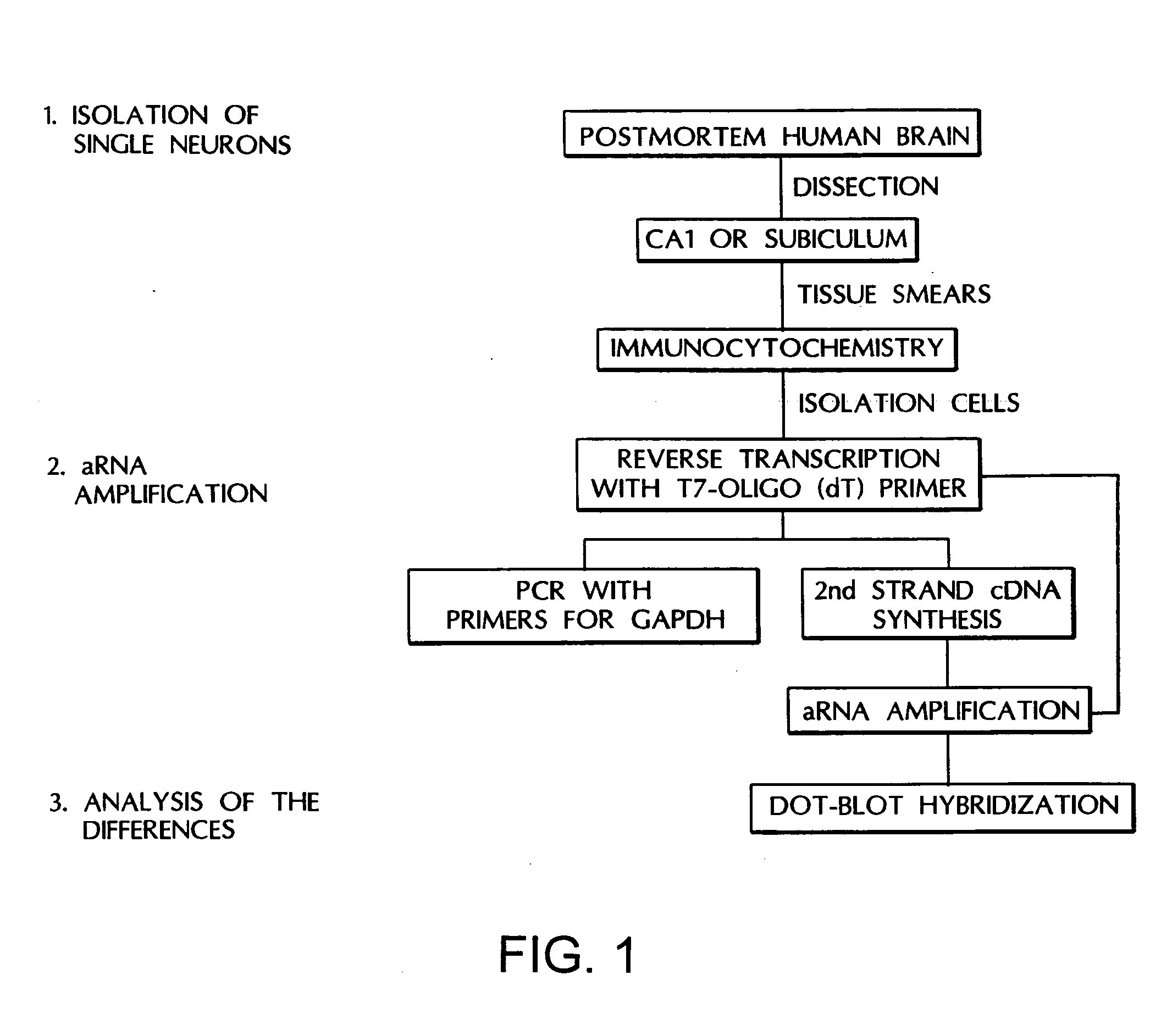
[0104] Collection of Tissue: The single cell approach (FIG. 1) was based on the methods of Eberwine, J. et al., “Analysis of Gene Expression in Single Live Neurons,”Proc. Natl. Acad. Sci. U.S.A., 89:3010-3014 (1992), which is hereby incorporated by reference) with modifications in tissue processing. Human brain tissue for this study was obtained from the Rochester Alzheimer's Disease Center. Criteria inclusion as a normal control case have been previously reported (Cheetham, J. E. et al., “Gap-43 Message Levels in Anterior Cerebellum in Alzheimer's Disease,”Mol. Brain Res., 36:145-151 (1996), which is hereby incorporated by reference). Human brain tissue was collected directly at autopsy with a maximum post mortem delay of 16 hours. The samples were transferred into ice cold phosphate-buffered saline (PBS) and maintained on ice until processed for cell spreads.
[0105] Preparation of cell spreads and storage: The CA1 or subiculum regions of post mortem human hippocampus were ...
example 2
Single Cell Analysis of Tissue from Alzheimer's Disease Patients
[0113] Single cell analyses of tissue from Alzheimer's disease patients have been undertaken since neurons appear to be in different stages of the disease even within one microenvironment of AD tissue. The single cell method has the advantage of two molecular techniques, in situ hybridization and aRNA analysis which enable the ability to determine changes in message levels within individual neurons. In situ hybridization provides the ability to analyze a large number of neurons within numerous sections of tissue, and to determine message levels by grain counting. While a powerful tool, one significant drawback of in situ hybridization is the length of time to obtain data for each individual message chosen. To address this issue, the aRNA technique has been developed, to provide the ability to simultaneously test a large number of messages within individual neurons. It is anticipated that the more rapid, broader analysi...
example 3
Dissecting the NFT-Free Neuronal Population
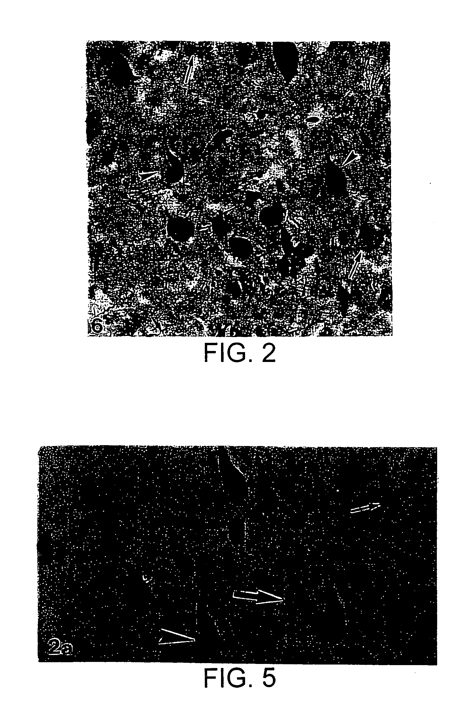
[0118] A double immunocytochemistry method combined with in situ hybridization was developed to enable the ability to located neurons with phosphorylated tau epitopes which did not contain NFT. The sections resulting from this technique exhibit the first antigen identified with brown reaction product, the message identified by emulsion grains and the second antigen identified with blue reaction product.
[0119] In the first study (FIGS. 5A and B), double immunocytochemistry to identify NFT-free neurons with diffuse phosphorylation of the serine 396 & 404 epitope of tau (recognized by PHF-1) was combined with in situ hybridization for synaptophysin message. The antibodies were chosen since NFT are reliably identified by Mab 69, and PHF-1 is believed to be one of the most sensitive markers of an earlier stage of the AD disease. FIG. 5A demonstrates PHF-1 “only”. neurons in blue (large arrow), neurons containing NFT in brown (arrowhead), and N...
PUM
| Property | Measurement | Unit |
|---|---|---|
| pH | aaaaa | aaaaa |
| pH | aaaaa | aaaaa |
| volume | aaaaa | aaaaa |
Abstract
Description
Claims
Application Information