Cell fixation and use in phospho-proteome screening

Inactive Publication Date: 2005-04-21
LAB OF AMERICA HLDG
View PDF6 Cites 72 Cited by
  • Summary
  • Abstract
  • Description
  • Claims
  • Application Information

AI Technical Summary

Benefits of technology

[0019] Thus, drugs can be now screened for their effect on signal transduction cascades and DNA content in a variety of cell types simultaneously. The same techniques can be used to monitor a particular patient's response to a treatment regime, and can be used to monitor the response o

Problems solved by technology

(7) Murray M, et al., ORTHO Permeafix fixation is not suitable for the flow cyto

Method used


Image

  • Cell fixation and use in phospho-proteome screening
  • Cell fixation and use in phospho-proteome screening
  • Cell fixation and use in phospho-proteome screening

Examples


Example

EXAMPLE 1

Fixative Method

[0035] For analysis of whole blood by flow cytometry, including evaluation based on light scatter, surface epitopes, internal epitopes, and nucleic acid content, the following protocol was employed:

[0036] 1. Sterilely place 100 μl of whole blood or 50 μl of whole marrow in each of the 2 tubes for each antibody combination desired.

[0037] 2. Sterilely add 2 ml of standard culture media to each tube and vortex briefly to mix.

[0038] 3. Add 4 times saturating amount (as determined by titration) of desired anti-surface-epitope antibodies to each tube.

[0039] 4. Add any growth factors or inhibitors required by study at this point. In this case, activation of the MAP Kinase cascade was studied, requiring stimulation of the cells with PMA.

[0040] 5. Add 50 μl of stock PMA solution to the “PMA” tubes and vortex briefly to mix. Start timer immediately and place tubes in 37° C., 5% CO2 incubator for the exact pulse time required (either determined by previous studie...

Example

EXAMPLE 2

Heat Fixation

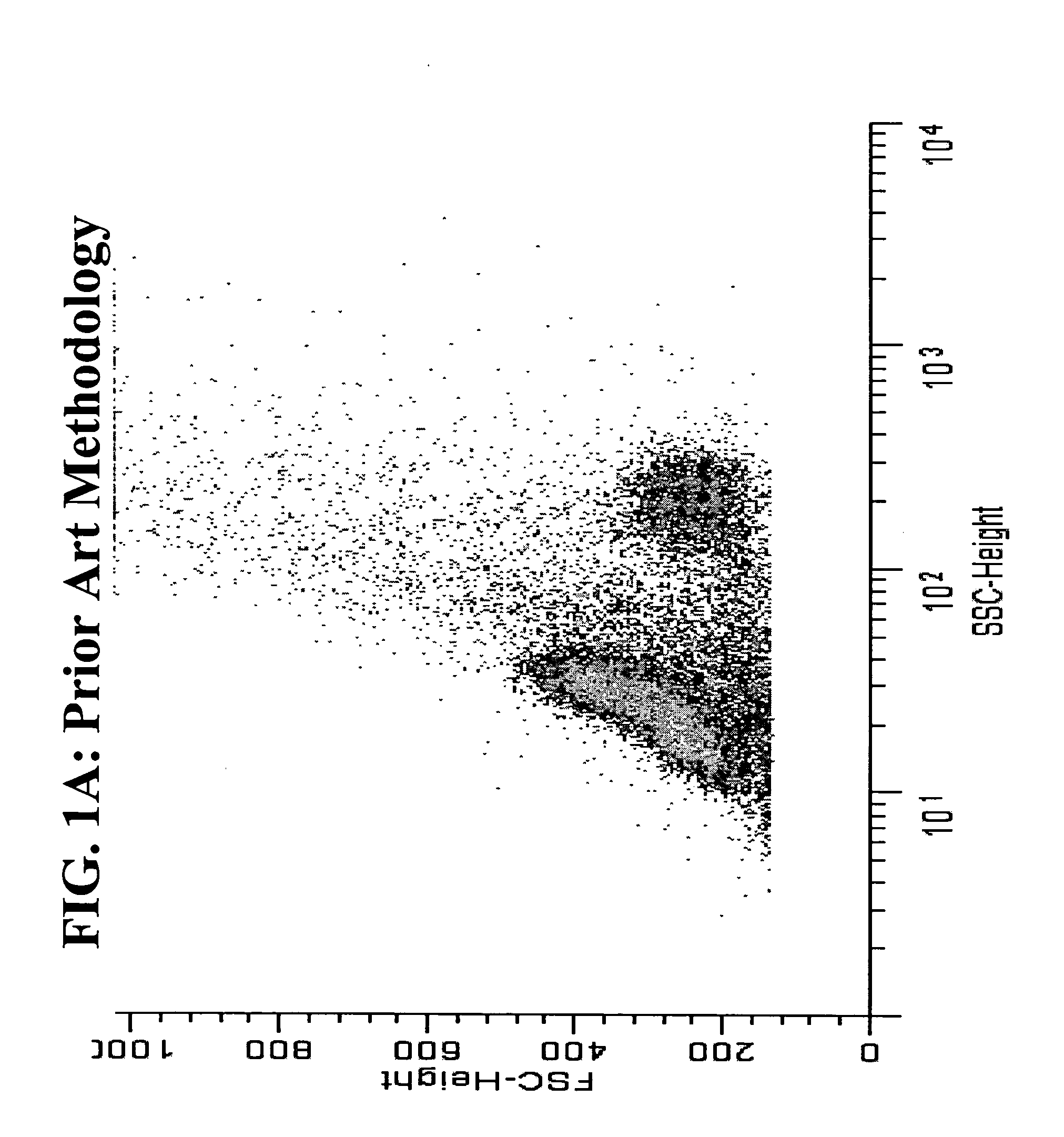
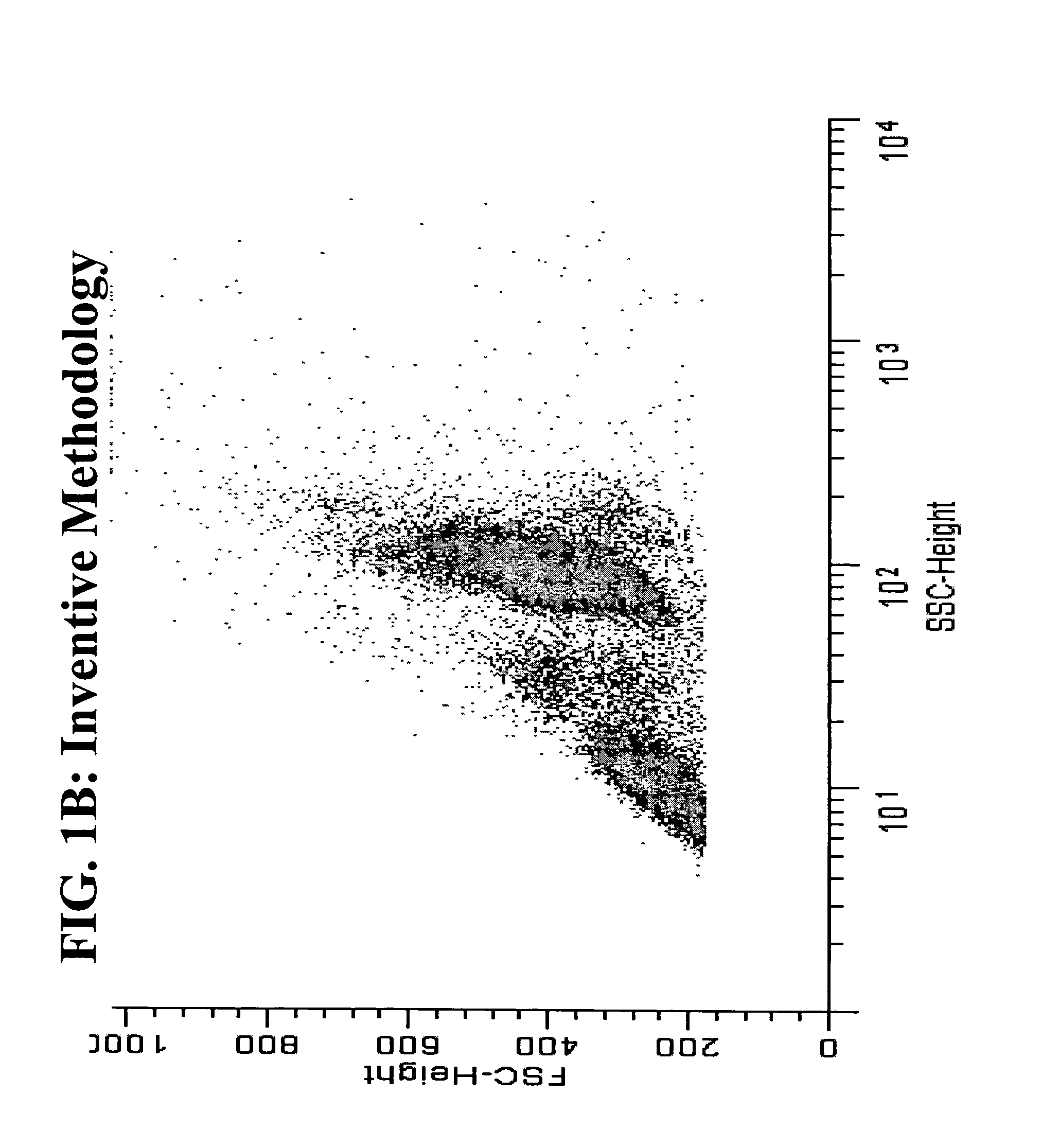
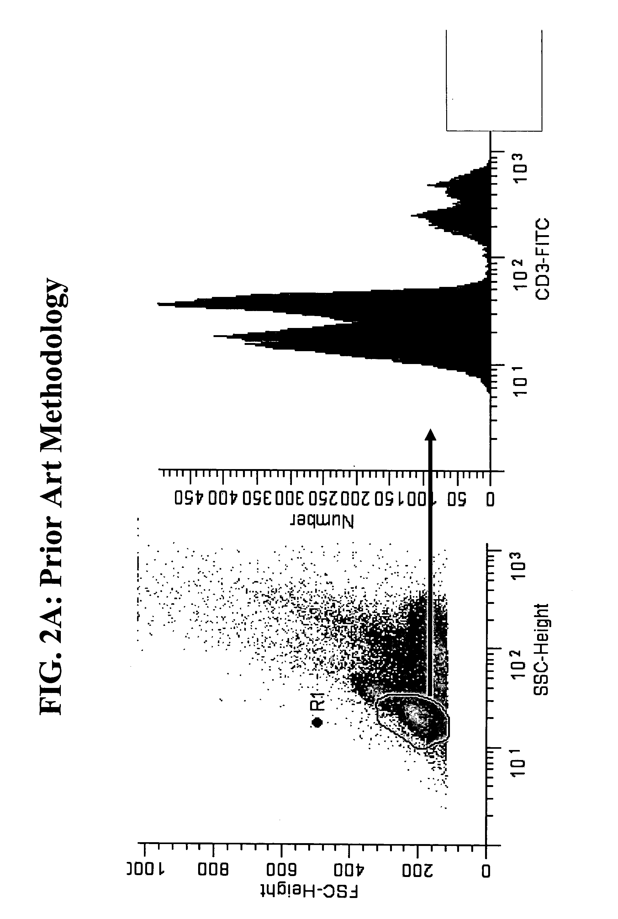
[0052] The use of OPF or PermiFlow™ as a fixative, permeation and lysis reagent was introduced in the original publications describing the product (4a, 4b, 5). However, to our knowledge this reagent has not been previously used for the detection of phospho-epitopes. Chow (1) and Perez (2) have studied phospho-epitopes by flow cytometry, but with the older paraformaldehyde / methanol based fixatives. The use of paraformaldehyde and methanol fixation is known to be detrimental to many surface epitopes and is emphasized in both the Chow (1) and Perez (2) articles.

[0053] The use of heat in antigen retrieval systems in immuno-histochemistry is well documented, but is typically not used in flow cytometry because researchers desire to slow down or stop metabolic activity and preserve intact cell morphology and status during fixation. Therefore, fixation is typically performed on ice or at room temperature. We have compared room temperature fixation to 43° C. fixation...


PUM

PropertyMeasurementUnit
Temperatureaaaaaaaaaa
Fractionaaaaaaaaaa

Abstract

A new method of cell fixation using a mild denaturing heat and the fixative PermiFlow™ or similar fixative that preserves cell morphology, light scatter profiles, nucleic acid content, cell surface epitopes and unmasks internal epitopes previously not available. The method can be combined with analysis of each of these parameters by flow cytometry and thus has application in drug screening and patient or tumor monitoring protocols.

Description

PRIOR RELATED APPLICATIONS [0001] This application claims the benefit of U.S. Provisional Application No. 60 / 512,834, filed Oct. 20, 2003.FEDERALLY SPONSORED RESEARCH STATEMENT [0002] Not applicable. REFERENCE TO MICROFICHE APPENDIX [0003] Not applicable. FIELD OF THE INVENTION [0004] The invention relates to a new method of fixing cells. The resulting fixed cells have retained immuno-detectable surface antigens, intracellular antigens, cell morphology, light scattering properties, and nucleic acid profiles. Thus, the cells can be used in a variety of applications previously not possible because prior art fixation methods degraded one or more of these properties. Applications of the new fixation method include drug screening methods. For example, it is possible to screen whole blood for drugs that affect the signal transduction pathways, for example MAP KINASE pathways. Other applications include diagnostic uses such as typing tumors or monitoring tumor responsiveness to a treatment...

Claims


Application Information

Patent Timeline
IPC IPC(8): G01N1/30
CPCG01N1/30Y10T436/25375Y10T436/108331Y10T436/2525Y10T436/25125
Inventor SHULTS, KEITH E.FLYE, ANGEL L.
Owner LAB OF AMERICA HLDG
Why Eureka
Try Eureka