
Ultrasonographic system and ultrasonography

a diagnostic system and ultrasonic technology, applied in ultrasonic/sonic/infrasonic image/data processing, instruments, tomography, etc., can solve the problems of hardly diagnosing structural changes in cardiac muscle, hardly being helpful in tissue characterization of cardiac muscle, and available diagnostic equipment cannot follow variations in the region, etc., to achieve accurate diagnosis of heart diseases

Inactive Publication Date: 2005-06-09
JAPAN SCI & TECH CORP
View PDF8 Cites 37 Cited by
  • Summary
  • Abstract
  • Description
  • Claims
  • Application Information

AI Technical Summary

Benefits of technology

[0036] It is an object of the present invention to provide an ultrasonic diagnostic system that is capable of acquiring detailed information on local tissue characteristics of cardiac muscle by utilizing ultrasonic backscatter IB with a view to more accurate diagnosis of heart diseases.
[0037] It is another object of the invention to provide an ultrasonic diagnostic method that permits acquisition of detailed information on local tissue characteristics of cardiac muscle by utilizing ultrasonic backscatter IB with a view to more accurate diagnosis of heart diseases.

Problems solved by technology

While known methods include diagnosis using ultrasonic integrated backscatter besides invasive biopsy, available diagnostic apparatuses cannot follow variations in the region of interest, because they fix the region of interest in the heart wall.
Accordingly these apparatuses can mainly sense signals of around 1 Hz due to density variations of myocardial fibers, but can hardly diagnose structural changes in cardiac muscle.
Ultrasonic diagnostic methods for heart diseases extensively used today are mostly based on morphological aspects of the heart, such as evaluation of its wall thickness or the cardiac output, but can hardly be helpful in the tissue characterization of cardiac muscle.
Since this is an invasive technique, it imposes heavy physical and mental burdens on the subject, and therefore cannot be repetitively applied.
However, it is difficult to quantitatively assess it on the image.
However, as the heart wall moves translationally along with pulsation, and the wall thickness varies with the myocardial extension and contraction, the position and size of the ROI should be varied from one point of time to another, and therefore it is difficult by this method to measure the IB from the same region of the heart wall all the time.
Similarly, when the vibration of an aorta is determined as a displacement velocity by the conventional Doppler method, the conditions for accuracy are satisfied theoretically, but the pulsation of the blood vessel has so significant an influence that it is difficult to extract a minute vibration superimposed over this relatively large amplitude.
Earlier studies on ultrasonic backscatter IB took note only of the difference between the maximum and the minimum of IB in one cardiac cycle, but paid no sufficient attention to the variation of the IB value at each point of time in one cardiac cycle.

Method used


Image

  • Ultrasonographic system and ultrasonography
  • Ultrasonographic system and ultrasonography
  • Ultrasonographic system and ultrasonography

Examples


Embodiment Construction

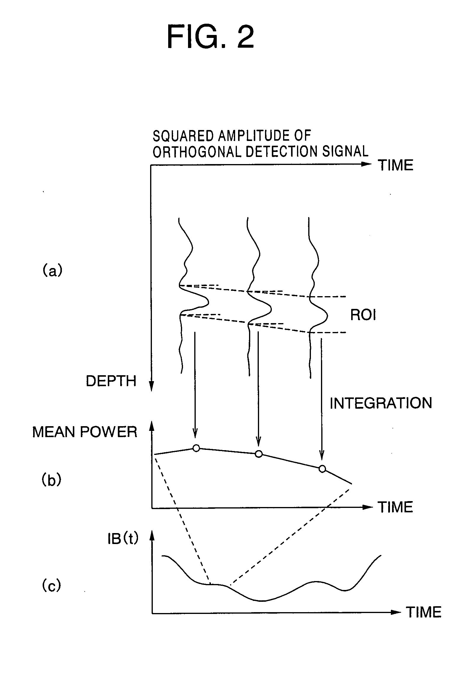
[0076] We earlier measured ultrasonic integrated backscatter IB from the heart wall of a healthy person at a repeated transmission frequency of a few kHz, and discovered, in addition to the already known CV synchronized with the heart beat, a component varying at a frequency of tens to hundreds of Hz superimposed over the CV. The present invention is made on the basis of this finding, and makes it possible to obtain the average power of IB from the region of interest by measuring the ultrasonic integrated backscatter IB at a high repeated transmission frequency of a few kHz, and to supply that variation frequency or variable cycle for displaying.

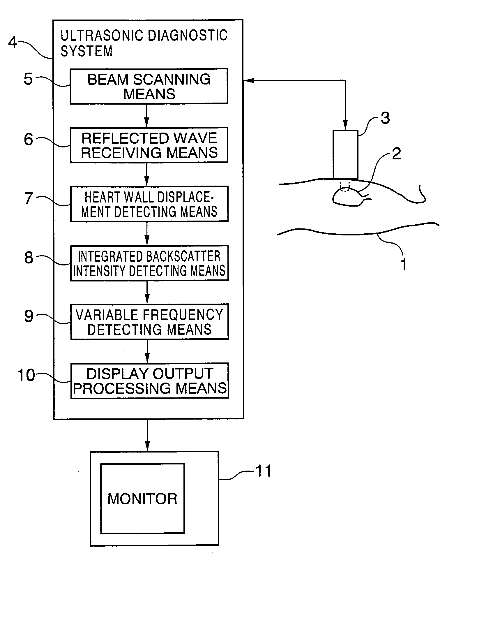
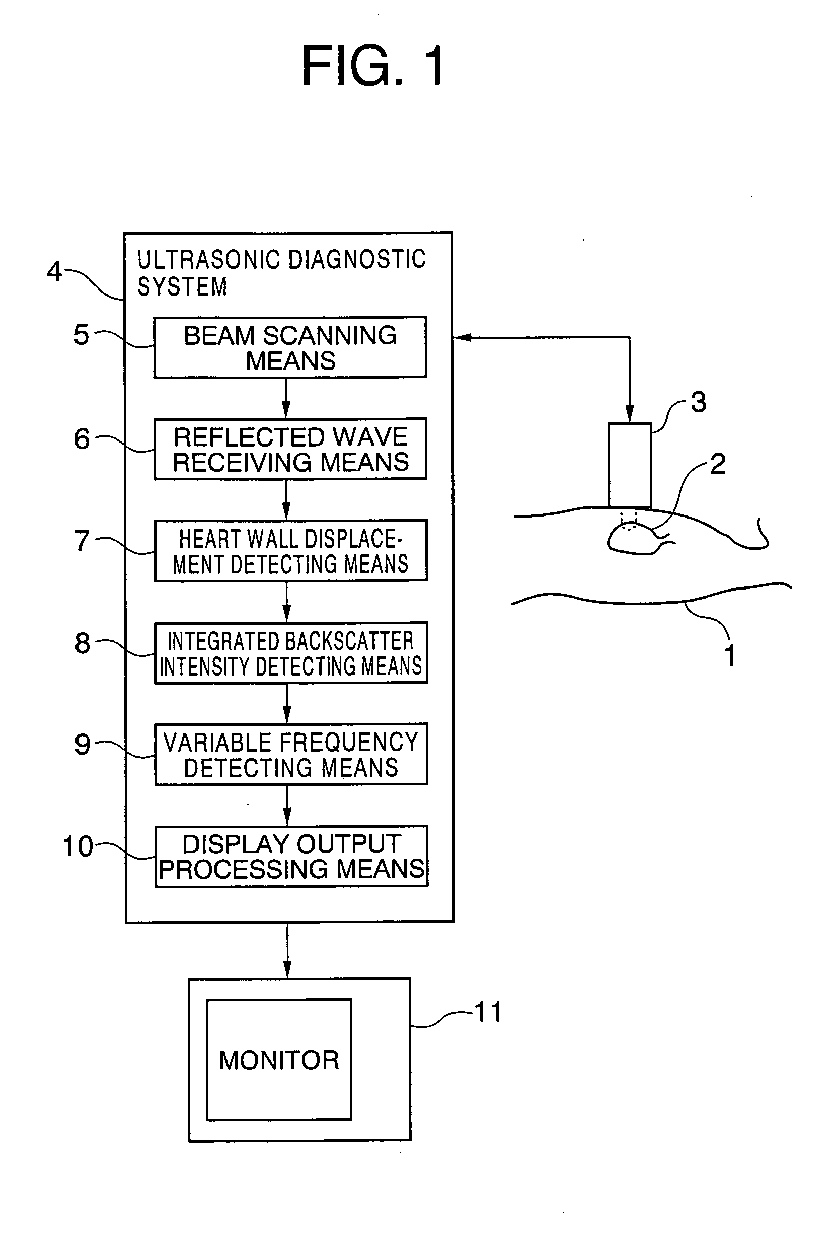
[0077]FIG. 1 schematically shows an ultrasonic diagnostic system according to the present invention. In FIG. 1, the object of diagnosis is the heart 2 of a subject 1. An ultrasonic pulse is transmitted from the body surface of the subject 1 by using an ultrasonic probe 3, and its reflected wave is received. An ultrasonic diagnostic system 4...


PUM


Abstract

An ultrasonic diagnostic system prepares a diagnostic data including image by transmitting ultrasonic pulses to a living tissue, and receiving and analyzing reflected wave of the ultrasonic pulses, and has an analytical processing unit to measure a backscattering intensity by using a scattering wave from a region of interest in the living tissue on a basis of the reflected wave which is received, and to detect a variation frequency of the measured backscattering intensity to obtain the diagnostic data to be available.

Description

BACKGROUND OF THE INVENTION [0001] 1. Field of the Invention [0002] This invention relates to an ultrasonic diagnostic system and an ultrasonic diagnostic method, and more particularly to an ultrasonic diagnostic system and an ultrasonic diagnostic method which enable a lesion on a living tissue, such as the heart, to be diagnosed by a noninvasive measurement using ultrasonic, by which local degeneration of myocardial structure in any region of the heart wall is identified, visually displayed and thereby enabled to be diagnosed. [0003] 2. Description of the Related Art [0004] In diagnosing heart diseases due to the malfunctioning of cardiac muscle, such as hypertrophic cardiomyopathy, dilated cardiomyopathy or ischemic cardiac diseases including myocardial infarction, the tissue characteristics of cardiac muscle should be identified. While known methods include diagnosis using ultrasonic integrated backscatter besides invasive biopsy, available diagnostic apparatuses cannot follow v...

Claims


Application Information

Patent Timeline
Patent Type & Authority Applications(United States)
IPC IPC(8): A61B8/14A61B8/08G06T1/00G06T7/40
CPCA61B8/0858
Inventor KANAI, HIROSHIKOIWA, YOSHIROTANAKA, MOTONAO
Owner JAPAN SCI & TECH CORP
Learn More