Device for end-to-side anastomosis

a technology of end-to-side anastomosis and end-to-side anastomosis, which is applied in the field of end-to-side anastomosis, can solve the problems of preventing the flow of original blood to be perfectly restored to the vessel, reducing the effectiveness of re-establishing flow to the vessel, and dangerous to the health of patients, so as to reduce the time required for the anastomosis operation. , the effect of learning in

Inactive Publication Date: 2005-07-07
NEWMAN MEDICAL
View PDF6 Cites 8 Cited by
  • Summary
  • Abstract
  • Description
  • Claims
  • Application Information

AI Technical Summary

Benefits of technology

[0026] A first advantage of the invention is that contact between the connecting parts and the blood vessel is limited to the vessel wall, leaving the inside surface free, and there is therefore no contact with the blood inside the vessel.
[0027] Another advantage is that there is no contact between the device and the cut edge of the wall of the anastomosed vessel, which permits the intima of the two vessel parts to be joined in optimum manner.
[0028] A yet further aim of the invention is to propose a simplified anastomosis apparatus whose use can be learned in a very short time and which permits the formation of anastomoses of constant quality.
[0029] A still further aim of the invention is to propose an anastomosis apparatus that reduces the time required for the anastomosis operation.

Problems solved by technology

This method, commonly known as suture anastomosis, has several disadvantages, the main one being that the suture produces a scar which, however small, prevents original flow from being perfectly restored to the vessel.
This is also a drawback in the case of lymphatic vessels which have a similar layered structure.
A second drawback is due to the fact that blood vessel suturing must be done by hand by a specialized surgeon because it is a complex operation that cannot be left to surgeons who are not highly skilled in this specific art.
This surgical technique (mechanical anastomosis by eversion), although simpler than suture anastomosis, is even less effective in re-establishing flow to the vessel.
Besides this, mechanical anastomosis by eversion has other serious drawbacks which may be dangerous to a patient's health.
One drawback is due to the fact that everting the vessel ends may cause the ends to break, especially in the case of vessels larger than 3 mm in diameter whose walls are hardened by arteriosclerosis.
Another complication, specific to blood vessels, is caused by the fact that the inside layers of the vessel ends, after being turned inside out and placed side by side, are no longer exposed to the blood flow and therefore tend to atrophy, which in turn leads to narrowing of the blood vessel and reduction of the blood flow through it.
The smaller the diameter of the vessel being operated on, the more serious this problem is.
This produces an extremely dangerous condition which exposes the anastomosis to risk of immediate occlusion caused by the formation of a blood clot and narrowing of the anastomotic lumen.
Indeed, it has been found that contact between metal and blood tends to cause stenosis, that is to say, abnormal narrowing of the blood vessel, which may lead to total occlusion of the vessel in a period of 6 months to 1 year.
This solution has, however, produced serious problems because in many cases, the chemically coated metal parts in contact with the blood favor the formation of thrombi and the onset of acute thromboses that come sharply to a crisis, exposing patients to the risk of heart failure and, in some cases, leading to death.
Another drawback of prior art anastomosis devices is the complexity of applying them, especially to the side portions of the vessel.
This means that the surgical operation involved tend to be very long, placing the patient at risk not only on account of the technical difficulty of the operation itself but also because of the likelihood of infection which increases with the increased length of time during which the body organs are exposed to the environment.

Method used


Image

  • Device for end-to-side anastomosis
  • Device for end-to-side anastomosis
  • Device for end-to-side anastomosis

Examples


Embodiment Construction

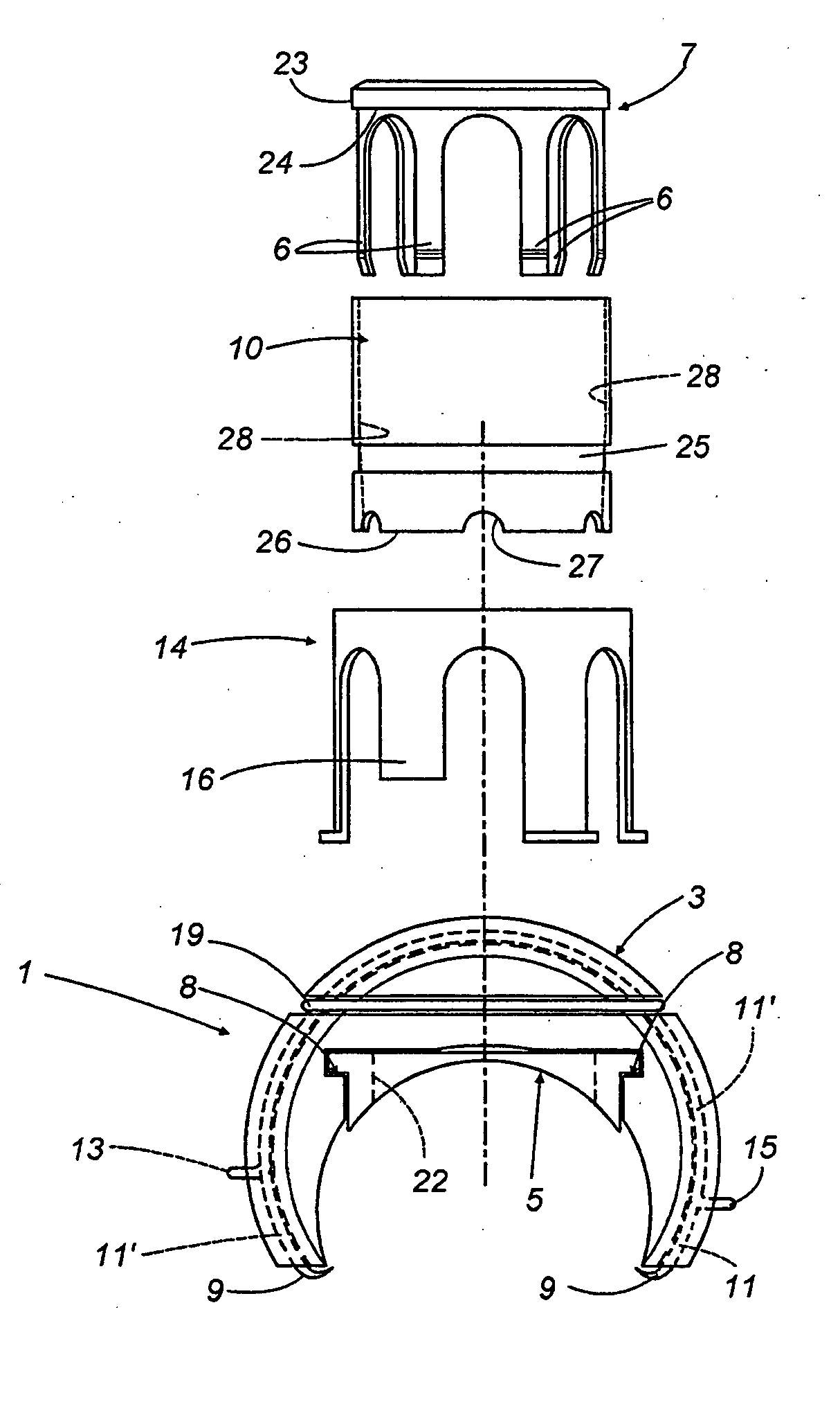
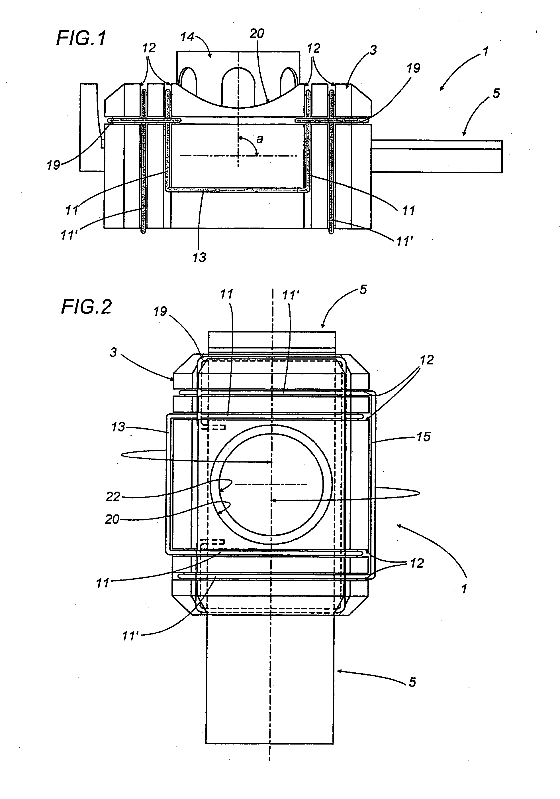
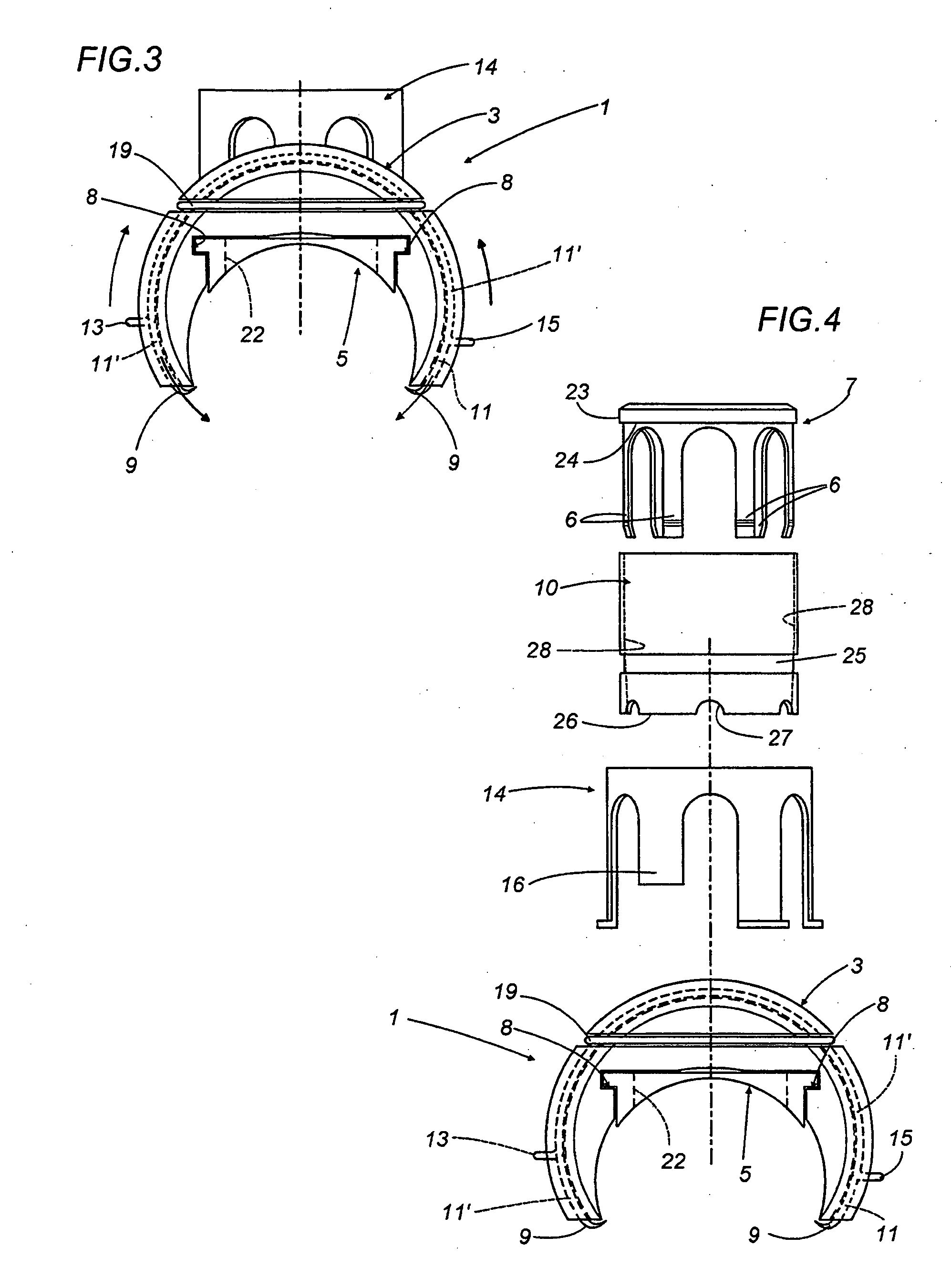
[0040] With reference the accompanying drawings, a device according to the invention comprises a body 1, clearly shown in FIGS. 1 to 3, for connecting a side wall portion 4 of an anastomosed vessel.

[0041] In the preferred embodiment described here, the body 1 comprises an approximately semicylindrical or “saddle-shaped” element 3 designed to be placed at least partly over the portion 4 and having a central hole 20 in which there is positioned and fixed a cylindrical element 14 whose axis “a” makes an angle, preferably, of 90°, 45° or 30° with the longitudinal axis of the saddle-shaped portion 3 or of the portion 4 of the anastomosed vessel.

[0042] The body 1 also comprises a gate valve 5 slidable lengthwise along internal grooves 8 made in the saddle 3 and having a hole 22 in it, this hole 22 being such that, during use, it is alternately aligned with and offset from the hole 20 in the saddle 3, in such a manner as to open and close the hole 20, respectively.

[0043] The inside of t...


PUM


Abstract

A device for forming an end-to-side anastomosis of blood vessels or ducts includes a first engagement structure (7) for engaging an end portion (2) of a blood vessel or of a prosthesis, a second engagement structure (35) for stably engaging a side portion (4) of the blood vessel or prosthesis, and a mechanism (10, 14) for reciprocally connecting the first and second engagement structures (7, 35). The second engagement structure (35) includes one or more piercing elements (9) adapted to only partially penetrate the side wall (4) of the blood vessel or prosthesis so as to avoid direct contact between the elements (9) and the blood inside the vessel or prosthesis.

Description

BACKGROUND OF THE INVENTION [0001] The present invention relates to a device and method for anastomosis of blood vessels. [0002] In surgery, the term “anastomosis” is used to mean a connection made surgically between two hollow structures. [0003] This invention relates in particular to end-to-side anastomosis, where an end portion of a blood vessel is connected to a side portion of a vessel wall. [0004] This specification expressly refers to vascular anastomosis applied to the human body, without thereby restricting the scope of the invention. [0005] The earliest method of anastomosing two parts of a blood or lymphatic vessel consists in suturing the free ends of the vessel with thread. [0006] This method, commonly known as suture anastomosis, has several disadvantages, the main one being that the suture produces a scar which, however small, prevents original flow from being perfectly restored to the vessel. [0007] In this regard, it should be stressed that blood vessels are made up...

Claims


Application Information

Patent Timeline
Patent Type & Authority Applications(United States)
IPC IPC(8): A61B17/11A61B17/32
CPCA61B17/11A61B2017/1135A61B2017/1107A61B17/32053
Inventor BORGHI, ENZOTOZZI, PIERGIORGIO
Owner NEWMAN MEDICAL
Why Eureka
Try Eureka