

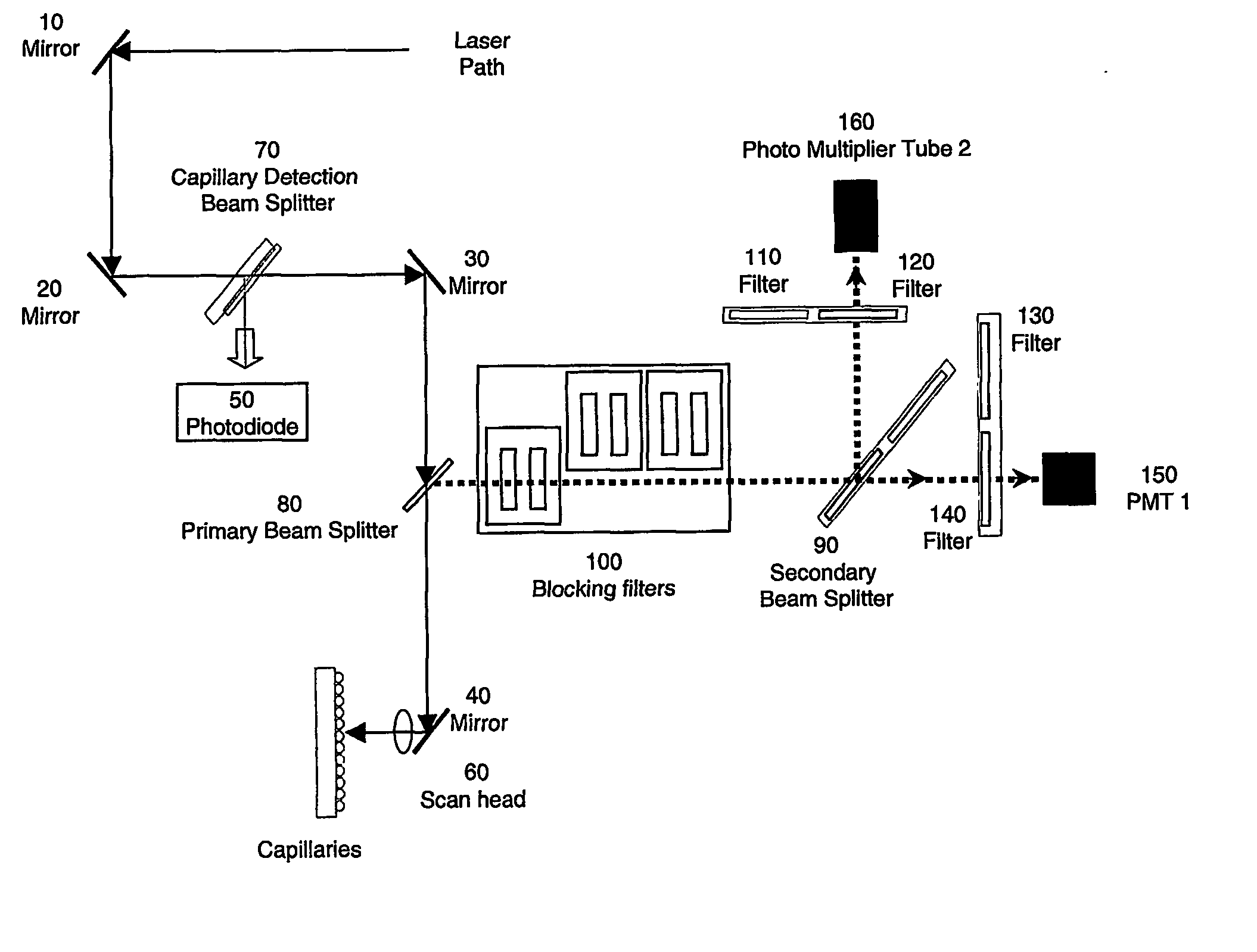
Multiplexed capillary electrophoresis systems
a capillary electrophoresis and multi-capillary technology, applied in the direction of fluorescence/phosphorescence, liquid/fluent solid measurement, peptides, etc., can solve the problems of gel-to-gel irreproducibility, band broadening, loss of resolution, etc., to achieve high throughput detection, high sensitivity, and high resolution
- Summary
- Abstract
- Description
- Claims
- Application Information
AI Technical Summary
Benefits of technology
Problems solved by technology
Method used
Image

Examples
example 1
Protein Separation by Multiplex CGE
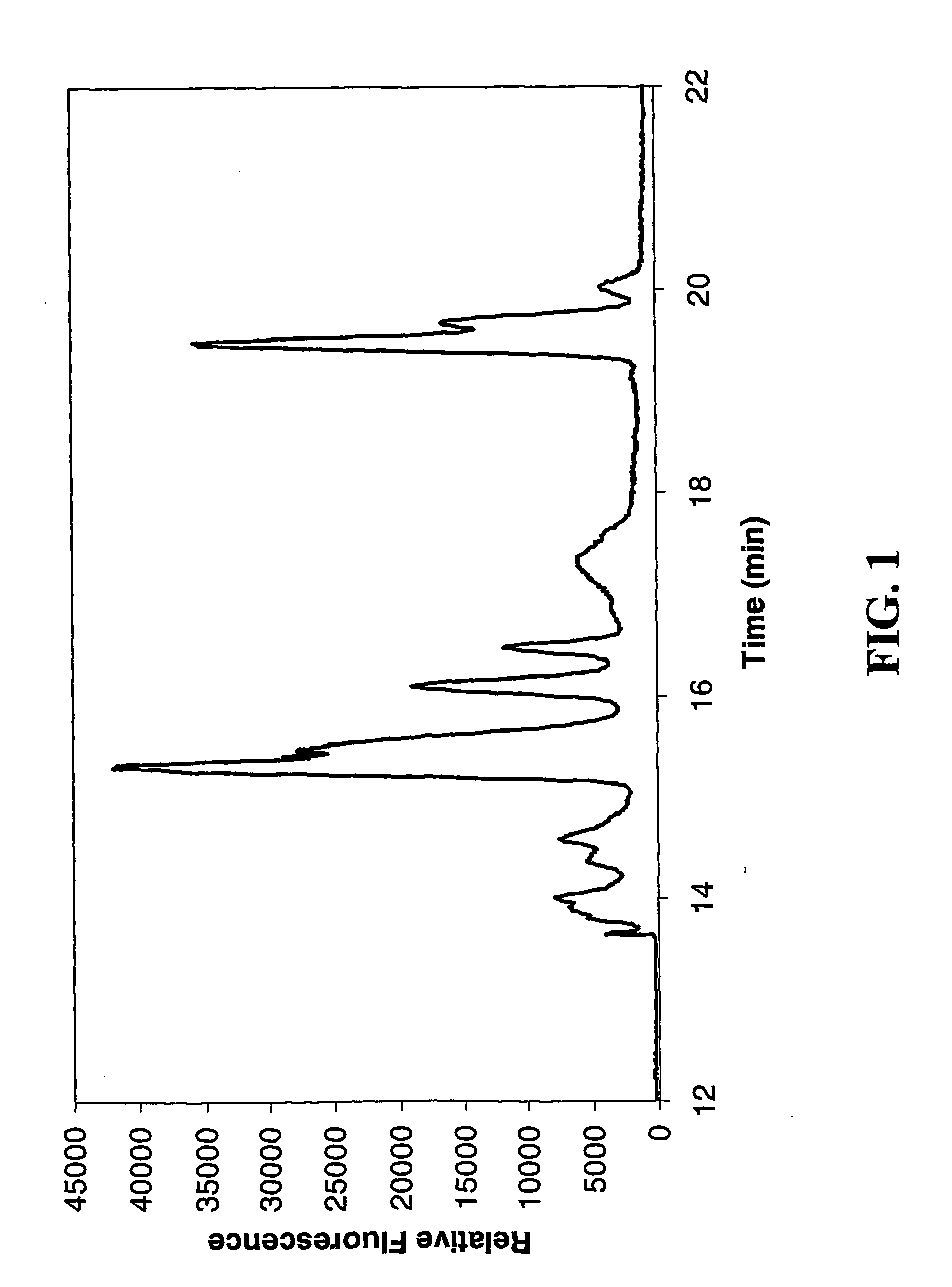
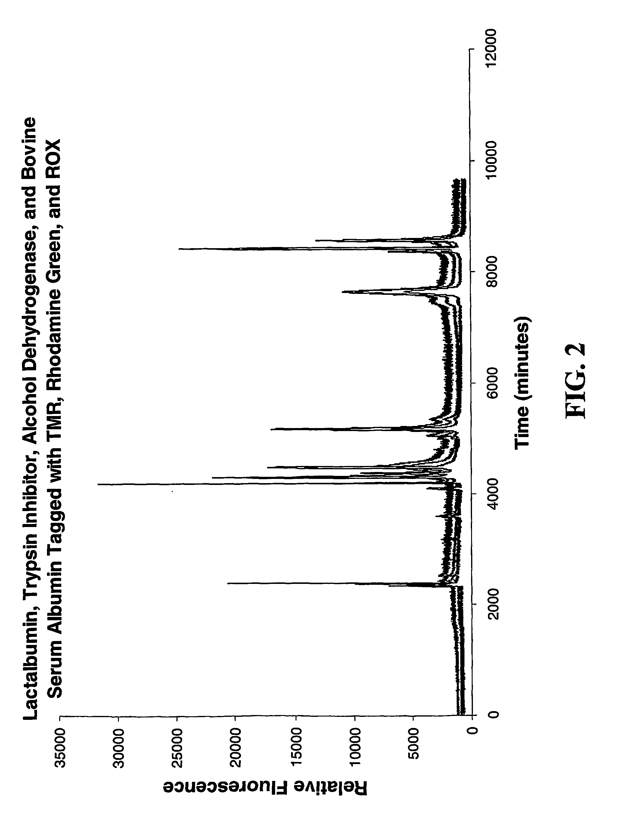
[0049] An unmodified MegaBACE 1000™ instrument was used for this separation. The 60 cm long capillaries were coated with linear polyacrylamide, then filled with a separation medium of 1% guaran sieving matrix, in 50 mM Tris, 50 mM HEPES, 4 mM SDS. Fluorescently labeled protein standards (Sigma, catalog number F3401) The labeled proteins were loaded onto the capillary columns by electrophoretic injection, with an injection time of 3 seconds at 10 kV. The protein standards were separated by electrophoresis over a period of 20 minutes at 12 kV. FIG. 1 shows a representative separation.
example 2
Two-Dimension Separation of Rat Liver Proteins by HPLC-CGE
[0050] In this separation, a MegaBACE 1000™ system was used to perform CGE as the second dimension separation, and an AKTA™ Explorer was used to perform HPLC as the first dimension. Protein samples were prepared from rat liver tissue which had been homogenized with polytrone in a buffer containing 8 M urea, 4%; (w / v) CHAPS, 20 mM TRIS, 10 mg / mL dithiothreitol (DTT), and 17.4 mg / mL phenylmethylsulfonyl floride (PMSF). The samples were incubated for one hour, and then centrifuged to remove the insoluble material.
[0051] Two buffers were prepared for the separation: buffer A: 10 mM phosphate buffer, and buffer B: 75% acetonitrile in 10 mM phosphate buffer, pH 6.5. The separation was performed on a Sephasil C4, 5 μm ST 4.6 / 100 mm column. The gradient used was as follows: first, 4 ml 100% A were introduced, then a 34 ml gradient to 100% B, and finally 12 ml 100% B. The effluent was collected into 180 fractions of 200 μl each in a...
example 3
Two-Dimensional Separation of Rat Liver Proteins by IEF-CGE
[0052] Protein samples were prepared from rat liver tissue as in the previous example. In this separation, isoelectric focusing (IEF) was performed on a drystrip (Amersham Biosciences, part number 17-6002-44, 24 cm Immobiline Drystrip, pH 3-10), in the conventional manner. The strip was then sectioned, ground, and the proteins in each section was extracted into 10 mM Tris 5 mM SDS buffer (pH 8.5). The sections were then analyzed in parallel by size sieving on a MegaBACE 1000™ system (2 kV, 40 second injection, 10 kV run voltage), separated in 15% Dextran matrix with a 10 mM Tris 5 mM SDS buffer (pH 8.5), on 60 cm long capillaries. Shown in FIG. 4 is the CGE separation profile generated from one IEF fraction.
PUM
| Property | Measurement | Unit |
|---|---|---|
| voltage | aaaaa | aaaaa |
| pH | aaaaa | aaaaa |
| volume | aaaaa | aaaaa |
Abstract
Description
Claims
Application Information