Method for mapping and eliminating T cell epitopes

a technology of t cell epitopes and mapping methods, applied in the field of immunology, can solve the problems of reducing immunogenicity, affecting the efficacy of therapy, and reducing immunogenicity, so as to reduce immunogenicity, reduce immunogenicity, and high immunogenicity

Inactive Publication Date: 2005-08-18
MERCK PATENT GMBH
View PDF1 Cites 23 Cited by
  • Summary
  • Abstract
  • Description
  • Claims
  • Application Information

AI Technical Summary

Problems solved by technology

There are many instances in which the efficacy of a therapeutic protein is limited by an undesirable immune reaction to the therapeutic protein.
Several mouse monoclonal antibodies have shown promise as therapeutic agents in a number of human disease settings, but in some cases have failed due to the induction of significant degrees of a human anti-murine antibody (HAMA) response in the patient [Schroff, R. W. et al.
In such situations where these human proteins are immunogenic, there is a presumed breakage of immunological tolerance that would otherwise have been operating in these subjects to these proteins.
In such cases, the therapeutic replacement protein may function immunologically as a foreign molecule from the outset, and where the individuals are able to mount an immune response to the therapeutic, the efficacy of the therapy is likely to be significantly compromised.
In addition, the computational approaches to T cell epitope prediction have in general not been capable of predicting epitopes with DP or DQ restriction.
However, such techniques are not adapted for the screening multiple potential epitopes to a wide diversity of MHC allotypes, nor can they confirm the ability of a binding peptide to function as a T cell epitope.
These reagents and procedures are used to identify the presence of T cell clones from peripheral blood samples from human or experimental animal subjects that are able to bind particular MHC-peptide complexes and are not adapted for the screening multiple potential epitopes to a wide diversity of MHC allotypes.
Such a technique requires careful application of cell isolation techniques and cell culture with multiple cytokine supplements to obtain the desired immune cell sub-sets (dendritic cells, CD4+ and or CD8+ T cells) and is not conducive to rapid through-put screening using multiple donor samples.
However, none of the procedures provide a unified method for the detection of biologically relevant epitopes in proteins of human origin, nor are the known techniques readily applicable to the detection of epitopes of significance to a wide population of MHC allotypes.

Method used


Image

  • Method for mapping and eliminating T cell epitopes
  • Method for mapping and eliminating T cell epitopes
  • Method for mapping and eliminating T cell epitopes

Examples


example 1

[0081] The interaction between MHC, peptide and T cell receptor (TCR) provides the structural basis for the antigen specificity of T cell recognition. T cell proliferation assays test the binding of peptides to MHC and the recognition of MHC / peptide complexes by the TCR. In vitro T cell proliferation assays of the present example, involve the stimulation of peripheral blood mononuclear cells (PBMCs), containing antigen presenting cells (APCs) and T cells. Stimulation is conducted in vitro using synthetic peptide antigens, and in some experiments whole protein antigen. Stimulated T cell proliferation preferably is measured using 3H-thymidine (3H-Thy) and the presence of incorporated 3H-Thy assessed using scintillation counting of washed fixed cells.

[0082] Buffy coats from human blood stored for less than 12 hours were obtained from the National Blood Service (Addenbrooks Hospital, Cambridge, UK). Ficoll-paque was obtained from Amersham Pharmacia Biotech (Amersham, UK). Serum free AI...

example 2

[0088] An epitope map for the human protein interferon α2 (IFNα) was derived using the method of EXAMPLE 1. In all respects the method was as per EXAMPLE 1 except that synthetic peptides were as given in Table 2 (below) and incubation with the PBMC preparations was at a concentration of 10 μM.

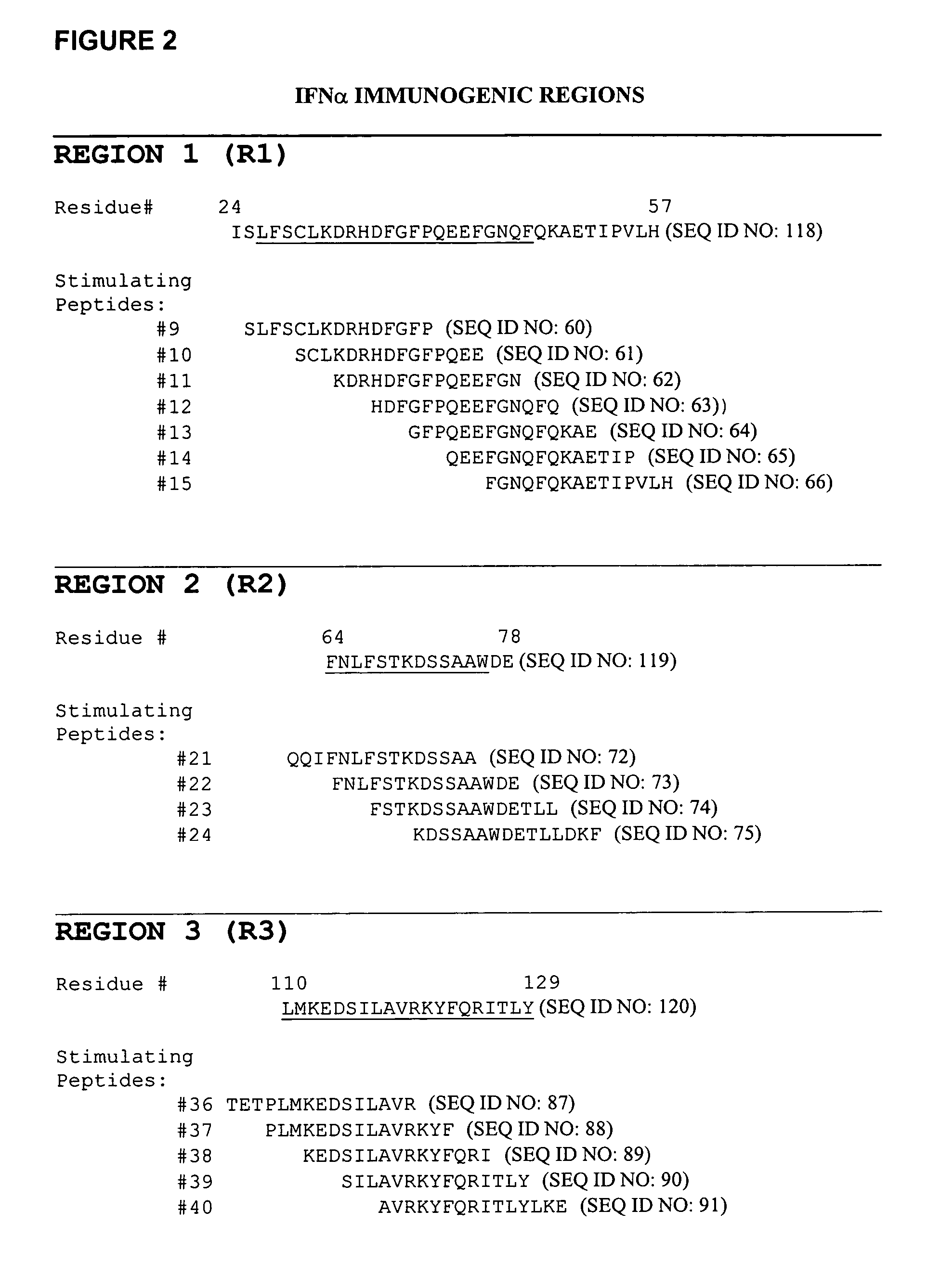
[0089] Mapping T cell epitopes in the IFNα sequence resulted in the initial, preliminary identification of three immunogenic regions R1, R2, R3. This was determined by T cell proliferation to seven, four and five overlapping peptides respectively as shown in FIG. 2. Region 3 is considered to contain a potential immunodominant T cell epitope as proliferation is scored in two thirds of donors that responded to IFNα peptides.

TABLE 2IFNα peptidesPeptideIDIFNα2b; 15merSEQ IDNumbersequenceNO:1CDLPQTHSLGSRRTL522PQTHSLGSRRTLMLL533HSLGSRRTLMLLAQM544GSRRTLMLLAQMRRI555RTLMLLAQMRRISLF566MLLAQMRRISLFSCL577AQMRRISLFSCLKDR588RRISLFSCLKDRHDF599SLFSCLKDRHDFGFP6010SCLKDRHDFGFPQEE6111KDRHDFGFPQEEFGN6212HDFGFPQ...

example 3

Protocol for Conducting a Time Course T Cell Activation Assay

[0090] A general protocol for conducting a time course T cell activation assay comprises the following steps: [0091] 1. Thaw 1 vial of PBMC per donor [0092] 2. Resuspend cells at 2-4×10⁶ cells / ml (in AIM V). [0093] 3. Transfer 1 ml to 3 wells of a 24 well plate (giving a final concentration of 2-4×10⁶ PBMC / well), since it is usual to test the antigen at two different concentrations and compare against a non-antigen treated control (e.g. 10-50 μg / ml protein or 1-5 μM peptide). [0094] 4. Make stock solutions of antigens typically 100 μg / ml for proteins and 2-10 μM for peptides. Add 1 ml of antigen to each well to give a final concentration 10-50 μg / ml protein or 1-5 μM peptide. [0095] 5. Incubate for 5 days. [0096] 6. Gently resuspend the cells in the 2 ml cultures by pipetting and from each condition remove 100 μl cells and place into a well of 96 well plate (round bottom), repeat this three time of reach culture conditio...


PUM

PropertyMeasurementUnit
concentrationaaaaaaaaaa
volumeaaaaaaaaaa
concentrationaaaaaaaaaa

Abstract

The invention provides methods for the identification of immunogenic regions within the amino acid residue sequence of a polypeptide, such as a therapeutic protein or a fragment thereof. The method comprises the steps of: (i) culturing, in vitro, an aliquot of peripheral blood monocyte cells (PBMC) isolated from a donor in the presence of a peptide for a period of up to about 7 days, the amino acid residue sequence of the peptide being identical to at least a portion of the amino acid residue sequence of the polypeptide of interest, the peptide being selected from a library of peptides, the amino acid residue sequences of the individual peptides of the library collectively encompassing the entire amino acid residue sequence of the polypeptide of interest; culturing the T cell aliquot from step (i) for an additional period of up to about 3 days in the presence of a T cell proliferation-stimulating cytokine to expand the number of T cells therein; (iii) culturing the T cell aliquot from step (ii) for a period of about 4 days in the presence of autologous irradiated PBMC from the same donor and in the presence of an additional amount of the peptide sufficient to re-prime the T cells within the PBMC with the peptide; (iv) determining the level of T cell proliferation of the re-primed T cells relative to an established baseline control level of proliferation; and (v) repeating steps (i) through (iv) with each peptide of the library of peptides to thereby identify at least one immunogenic region within the amino acid residue sequence of the polypeptide of interest.

Description

CROSS-REFERENCES TO RELATED APPLICATIONS [0001] This application is a continuation-in-part of International Application Serial No. PCT / EP03 / 06110, filed on Jun. 11, 2003, designating the United States, which is incorporated herein by reference.FIELD OF THE INVENTION [0002] The present invention relates to the field of immunology. The invention provides methods for the identification of determinants and epitopes on protein molecules that can evoke an immune response. In particular the invention is concerned with the identification of epitopes for T cells in therapeutic proteins. Additionally, the invention relates to a combined approach of using epitope mapping in concert with identification of MHC class II ligands comprising epitopes from said epitope mapping method and modification of the therapeutic protein to reduce the number epitopes present in the protein sequence. BACKGROUND OF THE INVENTION [0003] There are many instances in which the efficacy of a therapeutic protein is lim...

Claims


Application Information

Patent Timeline
Patent Type & Authority Applications(United States)
IPC IPC(8): C12N5/08G01N33/48G01N33/50G01N33/53G01N33/567G01N33/68G06F19/00
CPCG01N33/6878G01N33/5047
Inventor BAKER, MATTHEWCARR, FRANCISCARTER, GRAHAM
Owner MERCK PATENT GMBH
Why Eureka
Try Eureka