System and method for endoscopic optical constrast imaging using an endo-robot

Inactive Publication Date: 2005-09-01
SIEMENS MEDICAL SOLUTIONS USA INC
View PDF3 Cites 180 Cited by
  • Summary
  • Abstract
  • Description
  • Claims
  • Application Information

AI Technical Summary

Benefits of technology

[0015] In yet another exemplary embodiment of the present invention, a system for performing endoscopic optical imaging is provided. The system comprising: a capsule endoscope, comprising: a linear magnet for enabling the capsule endoscope to be moved inside a lumen of a patient; a camera for capturing images inside the lumen, an illuminator for illuminating the inside of the lumen; a controller for controlling the camera and the illuminator; a transceiver for performing one of transmitting the images captured by the camera and receiving commands from outside the patient; and a power supply for receiving an inductive charge from an externally applied magnetic field; and a magnetic control system, comprising: a first and second magnet tube for generating the magnetic field for contr

Problems solved by technology

Because the movement of the endoscope is brought about by a pushing action, the impact of the lens end against a wall of the gastrointestinal tract can be discomforting to a patient.
In addition, as the lens end enters a bend in the gastrointestinal tract, the wall can be damaged if too much force is applied.
These hindrances typically limit the endoscopic examination to non-convoluted regions of the gastrointestinal tract.
Thus, a medical practitioner who is analyzing data received from the capsule endoscope cannot control how

Method used


Image

  • System and method for endoscopic optical constrast imaging using an endo-robot
  • System and method for endoscopic optical constrast imaging using an endo-robot
  • System and method for endoscopic optical constrast imaging using an endo-robot

Examples


Example

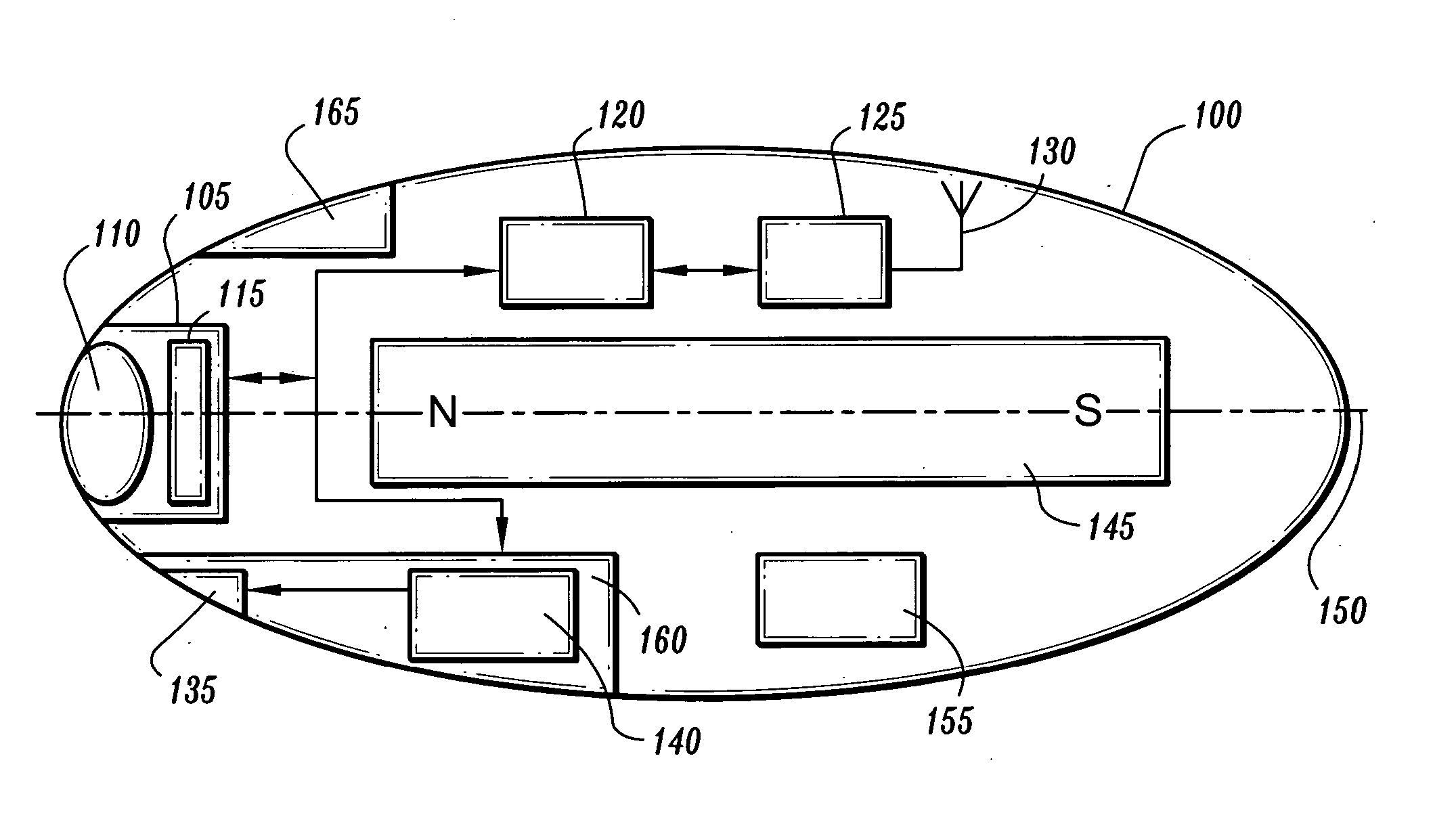
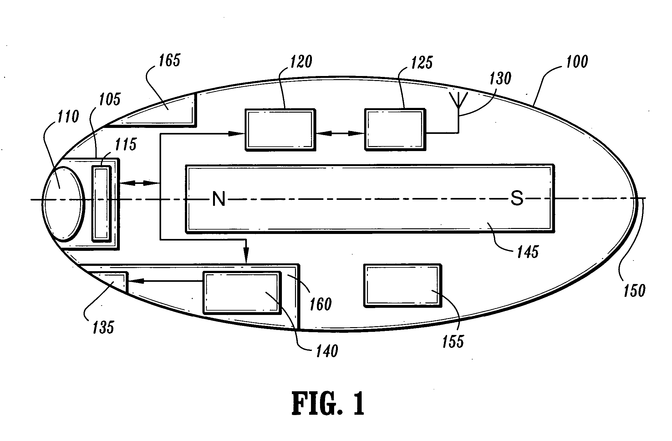
[0025]FIG. 1 is a block diagram of an endo-robot 100 according to an exemplary embodiment of the present invention. As shown in FIG. 1, the endo-robot 100 has an ellipsoidal housing in which a bar magnet 145 or a drivable approximately linear coil is co-linearly arranged about an axis 150. The endo-robot 100 includes a camera 105, for example, a video camera, having a lens 110 and an image sensor 115, for example, a charge coupled device (CCD) or complementary metal oxide semiconductor (CMOS) image sensor, for capturing images of the inside of a patient's body.

[0026] The endo-robot 100 also includes an illumination device 160, which includes an illuminator 135 and an illumination circuit 140. The illuminator 135 may be, for example, an infrared (IR) light emitting device, light emitting diode (LED), high-performance three-color LED or micro-fluorescent lamp for lighting an area surrounding the endo-robot 100 or for providing targeted phototherapy. Coupled to the camera 105 and the ...


PUM


Abstract

A system and method for endoscopic optical imaging using an endo-robot is provided. The method for performing endoscopic optical imaging using a capsule endoscope comprises: navigating the capsule endoscope through a lumen of a patient that has been introduced with an optical contrast agent; illuminating a portion of the lumen that is not penetrable by an external light source with light emitted from the capsule endoscope to enhance an image intensity of the portion of the lumen; and powering the capsule endoscope with an externally applied magnetic field.

Description

CROSS-REFERENCE TO RELATED APPLICATION [0001] This application claims the benefit of U.S. Provisional Application No. 60 / 548,540, filed Feb. 27, 2004, the disclosure of which is herein incorporated by reference.BACKGROUND OF THE INVENTION [0002] 1. Technical Field [0003] The present invention relates to an endoscopic examination, and more particularly, to endoscopic examination using an endo-robot. [0004] 2. Discussion of the Related Art [0005] An endoscopy is the examination and inspection of the interior of body organs, joints or cavities through an endoscope and has become a common practice in the medical diagnosis of many internal body diseases. The endoscope is a tubular device using fiber optics and a powerful lens system to provide lighting and visualization of the interior body organs, joints or cavities. During an endoscopy, the lens end or head of the endoscope is inserted into, for example, a gastrointestinal tract, and is moved along by an external pushing action. Becaus...

Claims


Application Information

Patent Timeline
IPC IPC(8): A61B1/04A61B5/06A61N5/06
CPCA61B1/00016B82Y10/00A61B1/00158A61B1/041A61B1/0638A61B1/0676A61B1/0684A61B5/06A61B5/4839A61B2560/0219A61N5/0601A61N5/062A61N2005/0605A61N2005/0609A61N2005/061A61N2005/0661B82Y5/00A61B1/00029A61B5/062
Inventor WILLIAMS, JAMES P.ABRAHAM-FUCHS, KLAUS
Owner SIEMENS MEDICAL SOLUTIONS USA INC
Why Eureka
Try Eureka