Method and apparatus for anatomically tailored k-space sampling and recessed elliptical view ordering for bolus-enhanced 3D MR angiography

a 3d mr angiography and k-space sampling technology, applied in the field of mr angiography, can solve the problems of reducing affecting the accuracy of mr angiography, and unable to image multiple stations using a single administration of contrast agents, so as to increase the dose sharing between stations and minimize scan time

Inactive Publication Date: 2005-09-15
CORNELL RES FOUNDATION INC
View PDF7 Cites 20 Cited by
  • Summary
  • Abstract
  • Description
  • Claims
  • Application Information

AI Technical Summary

Benefits of technology

[0009] The present invention uses a variable k-space sampling method that minimizes scan time by tailoring the acquisition according to the vascular anatomy of the station, hence increasing dose sharing between stations. In particular, in a three station scan of the lower extremities (abdomen, thigh, and calf), at least the central station is sampled such that the number of samples taken is less than would be taken at the Nyquist rate. The spacing is more sparse than that required by the Nyquist sampling theorem, to prevent the well-known imaging artifact of wrap-around (aliasing). The MR images of the region of interest are duplicated. Subtraction of a pre-contrast mask removes non-enhancing tissue (ref. Wang), while the limited extent of vessels allows duplicated vessels to be removed using volume of interest imaging.

Problems solved by technology

However, there is little sharing of contrast dose between stations, and venous enhancement can still occur.
However, sampling at the Nyquist rate and at the spacing interval required by the Nyquist theorem increases the acquisition time at a particular station, making it difficult to image multiple stations using a single administration of a contrast agent.
The rapid transit time of a contrast agent through the peripheral vascular anatomy also poses timing problems in other regions, such as the carotid artery.
Although 2D and 3D time-of-flight MRA have revolutionized the evaluation of the carotid arteries, these MRA techniques have important limitations related to motion and flow artifacts, long acquisition times, and the difficulty in imaging the entire vessel down to the aortic arch.
However, a major challenge in contrast enhanced imaging of the carotid arteries is due to rapid arterial-venous transit.
However, if the k-space center is acquired during the rising edge of arterial enhancement, reduced arterial signal intensity and arterial ringing artifacts may result.
Delaying the acquisition of the k-space center (while using the conventional centric-order view) too far into the scan produces a significant venous artifact due to rapid contrast transit.

Method used


Image

  • Method and apparatus for anatomically tailored k-space sampling and recessed elliptical view ordering for bolus-enhanced 3D MR angiography
  • Method and apparatus for anatomically tailored k-space sampling and recessed elliptical view ordering for bolus-enhanced 3D MR angiography
  • Method and apparatus for anatomically tailored k-space sampling and recessed elliptical view ordering for bolus-enhanced 3D MR angiography

Examples


second embodiment

[0034] The described process of sample size reduction and image processing may be implemented by software. In the invention, the number of samples taken can correspond to the number of samples taken according to the Nyquist rate, but spaced further apart than in Nyquist sampling. This would extend the spatial frequency, thereby improving image resolution over standard Nyquist sampling.

Patient Studies of the Peripheral Extremities

[0035] In order to compare the present invention to a known protocol, 30 consecutive patients were recruited for evaluation of clinically suspected peripheral vascular disease at two identical MR imaging systems from October to December, 1999. The conventional protocol was used to image 16 patients at one facility (3 female, 13 male; ages 43 to 89 with a mean of 70). The method of the present invention was used for the remaining 14 (2 female, 12 male; ages 41 to 76 with a mean of 62) at the other facility. The choice of location and protocol was based on p...

embodiment 1

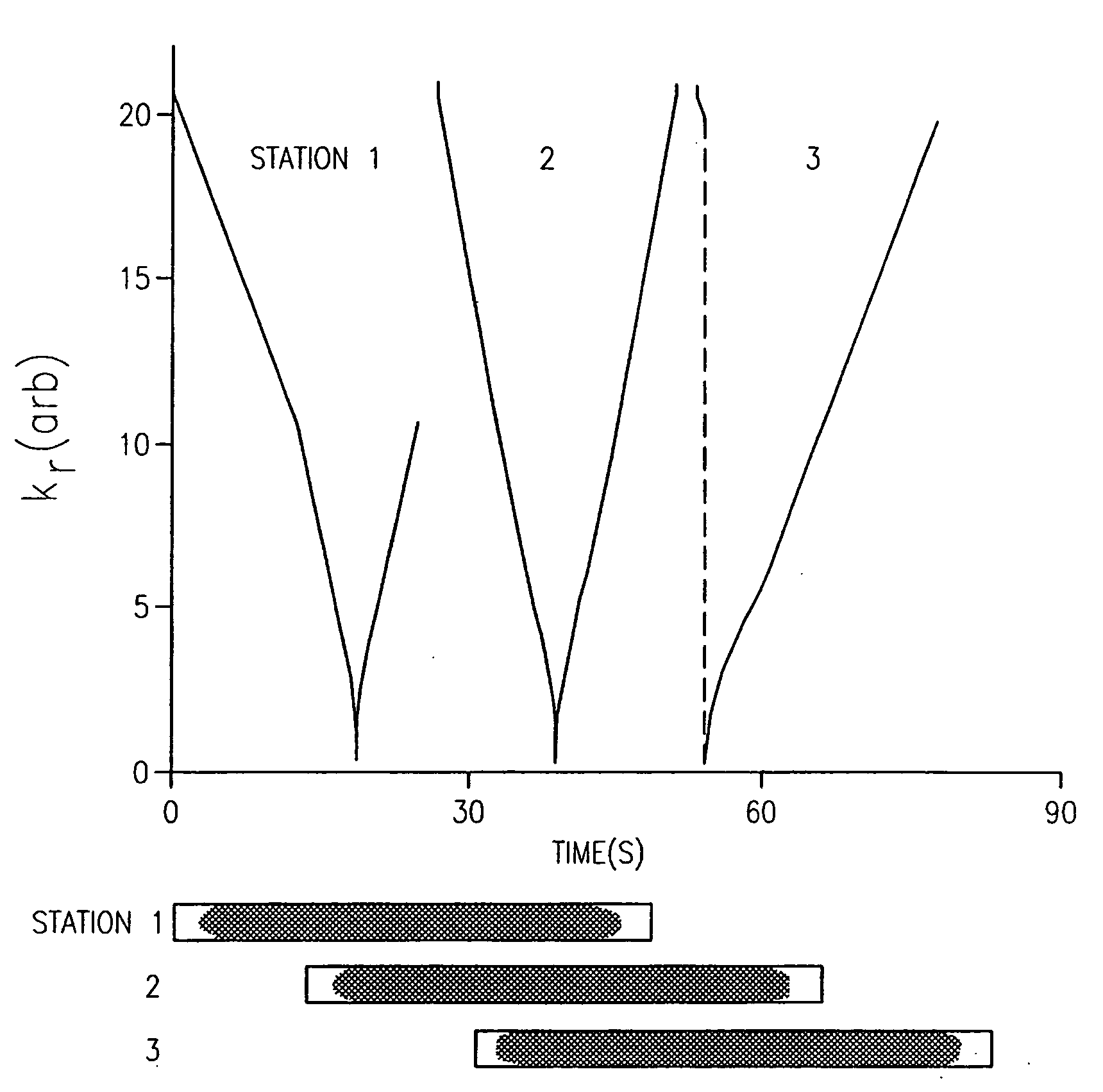
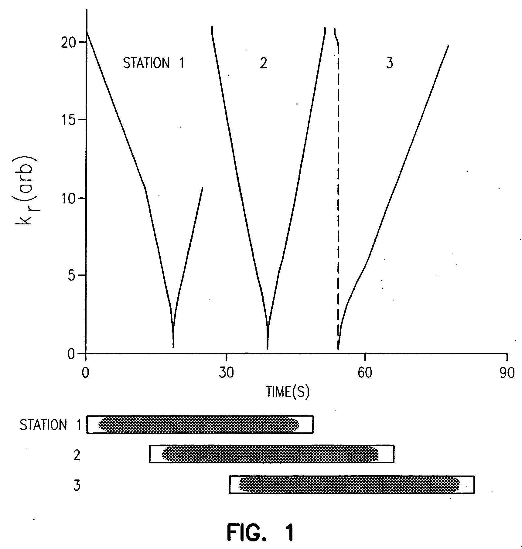
[0070] Then the recessed centric k-space sampling algorithm that matches the k-space sampling to the contrast enhancement curve is to allocate k-space point from the array K[ ] (as defined in the above embodiment 1) using the following index assignment:

[0071] Index=D[n] for the nth point to be acquired, with n=0, 1, . . . , N−1. (N is the total number of k-space points, as defined in embodiment 1).

[0072] Summarizing this scheme, the lowest spatial frequency data is acquired at the estimated maximum contrast enhancement, the next lowest spatial frequency data at the next highest contrast and so on.

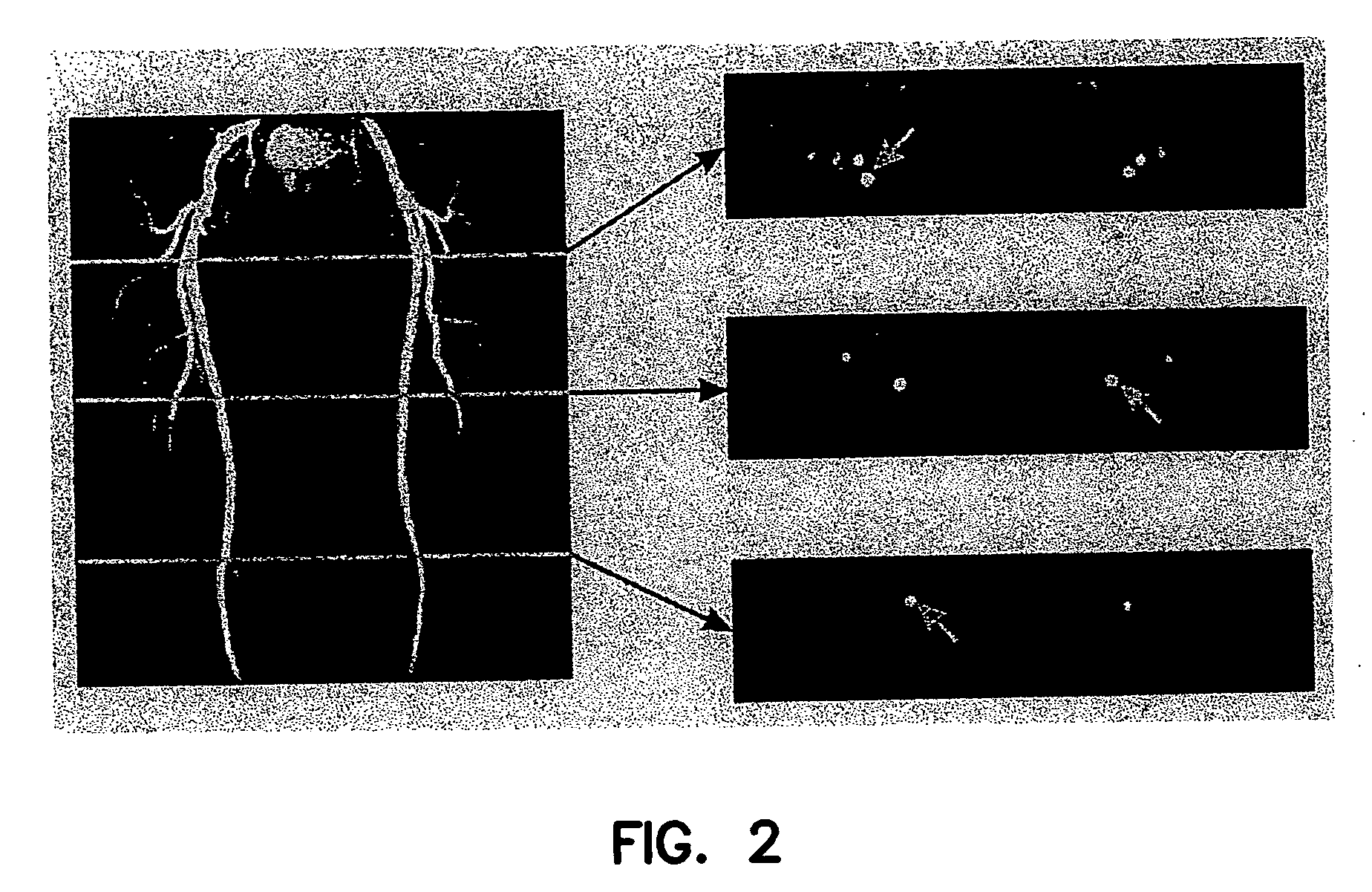
[0073] The 3D volume to be imaged is a coronal section prescribed graphically from an axial scout scan. The top portion of FIG. 7 shows the analysis of a dynamic 2D study. The arterial and venous signals were measured as a function of time after contrast injection. The arterial only phase is observed to last 8-10 seconds. Experimental data such as this may be used to accurately simulate t...


PUM


Abstract

Current bolus chase magnetic resonance angiography is limited by the imaging time for each station. Tailoring the density of k-space sampling along the anterior-posterior direction of the coronal station allows a substantial decrease in scan time that leads to greater contrast bolus sharing among stations and consequently a significant improvement in image quality. Fast arterial-venous transit in the carotid arteries requires accurate, reliable timing of the acquisition to the bolus transit to maximize arterial signal and minimize venous artifacts. The rising edge of the bolus is not utilized in conventional elliptical-centric view ordering because the critical k-space center must be acquired with full arterial enhancement. The invention provides a recessed elliptical-centric view ordering scheme is introduced in which the k-space center is acquired a few seconds following scan initiation. The recessed view ordering is shown to be more robust to timing errors in a patient studies.

Description

[0001] The invention was made with government support under R01HL60879 by the National Institutes of Health. The government has certain rights in the invention.FIELD OF INVENTION [0002] The present invention relates to MR angiography using a reduced sample set to decrease the time needed to acquire image data for arterial features. As a result of the time reduction, the MR data acquisition can be performed in conjunction with the arrival and flow of a contrast agent through the arteries, thereby reducing the amount of contrast agent introduced in a patient. The invention further relates to method for acquiring MR to better delineate arterial features from a background and venous artifacts. In particular, modification of k-space view ordering more accurately times data acquisition to contrast arrival and peak contrast enhancement in a region of interest. DESCRIPTION OF THE RELATED ART [0003] Generally, contrast-enhanced MRA offers many advantages in imaging, including reduced flow ar...

Claims


Application Information

Patent Timeline
Patent Type & Authority Applications(United States)
IPC IPC(8): A61B5/055G01R33/28
CPCG01R33/5601A61B5/055
Inventor WATTS, RICHARDWANG, YIPRINCE, MARTIN R.
Owner CORNELL RES FOUNDATION INC
Why Eureka
Try Eureka