Microvascular blood volume magnetic resonance imaging

a magnetic resonance imaging and microvascular technology, applied in the field of diagnostic imaging arts, can solve the problems of not providing a direct measure of blood volume, requiring contrast agents, and existing techniques generally not distinguishing between blood in large blood vessels

Active Publication Date: 2005-09-29
KENNEDY KRIEGER INST
View PDF30 Cites 23 Cited by
  • Summary
  • Abstract
  • Description
  • Claims
  • Application Information

AI Technical Summary

Benefits of technology

[0009] One advantage resides in measuring the parenchymal vascular space occupancy, which is more sensitive to physiological perturbations than is the total vascular volume which includes the large blood vessels.
[0010] Another advantage resides in providing me...

Problems solved by technology

The requirement of an administered contrast agent is a substantial disadvantage of these techniques.
Hence, BOLD does not provide a direct measure of the blood volume.
A disadvantage of both the invasive techniques and the BOLD techniques as applied to blood volume measurement is that these existing techniques generally do not differenti...

Method used


Image

  • Microvascular blood volume magnetic resonance imaging
  • Microvascular blood volume magnetic resonance imaging
  • Microvascular blood volume magnetic resonance imaging

Examples


Embodiment Construction

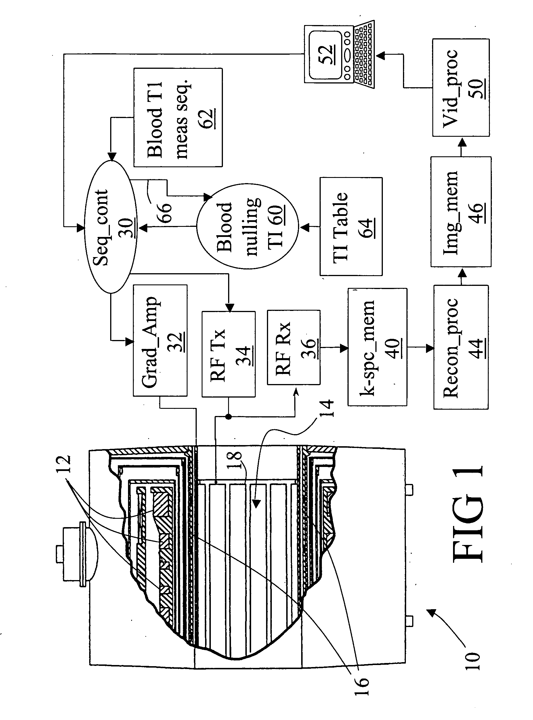
[0019] With reference to FIG. 1, a magnetic resonance imaging scanner 10 includes main magnet coils 12, which are preferably superconducting coils, although resistive main magnet coils or a permanent magnet can also be employed. The main magnet coils 12 are energized to generate a substantially uniform main magnetic field in an examination region 14. Magnetic field gradient coils 16 produce gradients in selected spatial directions to spatially encode magnetic resonances that are generated by energizing a radio frequency coil 18. In FIG. 1, a whole-body radio frequency coil 18 is shown; however, local coils such as head coils, phased radio frequency coil arrays, SENSE coils, and the like can be used instead of or in conjunction with the whole-body radio frequency coil 18 to excite magnetic resonances and / or to detect magnetic resonance echoes.

[0020] A magnetic resonance sequence controller 30 coordinates and controls a radio frequency transmitter 34 that is coupled to the whole-body...


PUM


Abstract

A magnetic resonance imaging system includes a magnetic resonance imaging scanner (10) that performs an inversion recovery magnetic resonance excitation sequence (70) having a blood-nulling inversion time (60) determined based on a blood T1 value appropriate for a selected magnetic field and blood hematocrit, whereby magnetic resonance of blood is substantially nulled. The inversion recovery excitation sequence (70) includes an inversion radio frequency pulse (74) applied with a small or zero slice-selective magnetic field gradient pulse to avoid inflow effects, and an excitation radio frequency pulse (80). The inversion pulse (74) and excitation pulse (80) are separated by the inversion time (60). The magnetic resonance imaging scanner (10) subsequently performs a readout magnetic resonance sequence (72) or spectroscopy sequence to acquire a magnetic resonance signal from tissue other than the nulled blood. A reconstruction processor (44) generates a reconstructed image from the acquired magnetic resonance signal.

Description

BACKGROUND OF THE INVENTION [0001] The following relates to the diagnostic imaging arts. It finds particular application in non-invasive measurement by magnetic resonance imaging of cerebral blood volumes, and will be described with particular reference thereto. However, it also finds application in measurement by magnetic resonance imaging of blood volumes in other tissues. [0002] In-situ measurement of blood volume is useful in various clinical, diagnostic, and research applications. Local cerebral blood volume changes, for example, correlate with local neuronal activity in the brain. Cerebral blood volume measurements during physiological stimulation thus provides a tool for functional studies of brain activity. Cerebral blood volume measurements can also provide information about impaired and / or damaged tissue in stroke victims, as well as about lesions in many disorders, including, but not limited to cancer, vascular disorders, and the like. Blood volume imaging of other organs...

Claims


Application Information

Patent Timeline
IPC IPC(8): G01R33/48A61B5/05A61B5/055G01R33/483G01R33/50G01R33/563
CPCG01R33/563A61B5/055G01R33/483G01R33/50
Inventor VAN ZIJL, PETER C.M.LU, HANZHANGGOLAY, XAVIER
Owner KENNEDY KRIEGER INST
Why Eureka
Try Eureka