Treating infectious diseases using ice inhibitors

Inactive Publication Date: 2005-10-20
VERTEX PHARMA INC
View PDF3 Cites 5 Cited by
  • Summary
  • Abstract
  • Description
  • Claims
  • Application Information

AI Technical Summary

Benefits of technology

[0013] Similarly bacterial load was reduced in the cornea at 7 days p.i. in mice treated with an ICE inhibitor vs. placebo without ciprofloxacin. An ICE inhibitor also reduced clinical scores after corneal infection induced by a clinical isolate-1025 or a ciprofloxacin resistant P. a

Problems solved by technology

Disease progresses rapidly to cause ulceration of the cornea and can potentially lead to permanent loss of vision from corneal scaring if not treated aggressively (Laibson, 1972).
The recent increased incidence of refractory bacterial keratitis resulting from antibiotic (especially fluoroquinolone) resistant P. aeruginosa strains (Chaudhry et al., 1999; Garg et al., 1999; Kunimoto et al., 1999; Landman et al., 2002), is of great concern and also may limit future therapeutic choices.
However, identification of the causative organism and response to antibacterial therapy (or antibiotic sensitivity) are the key restrictive factors that must be considered before initiating corticosteroid therapy.
B

Method used


Image

  • Treating infectious diseases using ice inhibitors
  • Treating infectious diseases using ice inhibitors
  • Treating infectious diseases using ice inhibitors

Examples


Example

Example 1

Animal Infection

[0094] Eight week old female B6 mice (The Jackson Laboratory, Bar Harbor, Me.) were used in these experiments. The left cornea of each anesthetized mouse was scarified with three parallel 1 mm incisions using a sterile 25⅝ gauge needle under a stereoscopic microscope. Scarified corneas were challenged topically with 1.0×10⁶ CFU / μl of P. aeruginosa (ATCC strain 19660 or clinical isolate-1025 or a ciprofloxacin resistant 19660 strain in a 5 μl dose as described before (Kwon and Hazlett, 1997). Eyes were examined macroscopically at 1 day post-infection (p.i.) and at times described below to ensure that all mice were similarly infected and to monitor the course of disease. All animals were treated humanely and in full compliance with the Association for Research in Vision and Ophthalmology resolution on usage and treatment of animals in research.

Example

Example 2

Bacterial Strains

[0095]P. aeruginosa strain 19660 was used as a standard laboratory strain and produces reproducible corneal pathology in the B6 mouse model (kernacki et al., 2000; Rudner et al., 2000). P. aeruginosa strain 1025 (KEI-1025) was isolated in 1999 from a human microbial keratitis case at the Kresge Eye Institute, Detroit, Mich. The laboratory-derived ciprofloxacin-resistant mutant was developed by serially passaging the wild-type P. aeruginosa strain 19660 on ciprofloxacin-containing Luria-Bertani (LB) broth to obtain ciprofloxacin resistance (Sanchez et al., 2002). The ciprofloxacin-resistant P. aeruginosa strain when compared with the parent strain exhibited a 100-fold increase in the minimum inhibitory concentration (MIC) of ciprofloxacin (0.25 mg / ml vs. 25 mg / ml) required for in vitro killing of the bacteria. The virulence of the ciprofloxacin-resistant (P. aeruginosa-19660) mutant was decreased compared to the parent strain during the in vitro generatio...

Example

Example 3

ICE Inhibitor Formulations

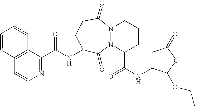
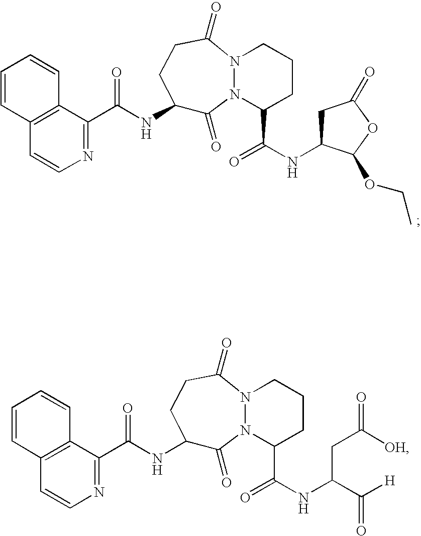
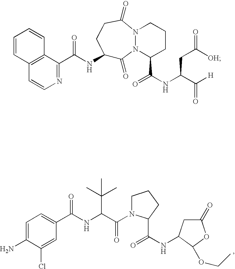
[0096] The ICE inhibitor used in these experiments displays potent inhibition of ICE (Ki=0.8 nM) and selectivity >100-fold vs. other non-ICE caspases. Four coded blinded formulations of vehicle (PBS) with or without ICE inhibitor (300 μM) were studied, for subconjunctival and topical administration. All formulations were found to be non-toxic to the eye in otherwise untreated mice and had no direct (in vitro) ability to kill bacteria.


PUM

PropertyMeasurementUnit
Antimicrobial propertiesaaaaaaaaaa
Stereoisomeraaaaaaaaaa

Abstract

This invention relates to methods and compositions for treating infectious and other diseases, particularly of the eye, by administering an ICE inhibitor. This invention also relates to methods for treating injuries, allergies, chemical irritations, or burns of the eye by administering an ICE inhibitor.

Description

CROSS-REFERENCE TO RELATED APPLICATION(S) [0001] This application claims the benefit under 35 U.S.C. § 119 of U.S. Provisional Application 60 / 526,362, filed Dec. 1, 2003, the entire contents of that application being incorporated herein by reference.FIELD OF THE INVENTION [0002] This invention relates to methods and compositions for treating infections and other diseases and disorders with an ICE inhibitor. BACKGROUND OF THE INVENTION [0003]Pseudomonas aeruginosa (P. aeruginosa) keratitis is a sight threatening corneal disease that accounts for approximately ¾ of reported cases of contact lens-associated microbial infection (Liesegang, 1997). Disease progresses rapidly to cause ulceration of the cornea and can potentially lead to permanent loss of vision from corneal scaring if not treated aggressively (Laibson, 1972). Tissue damage during Pseudomonas keratitis can occur from multiple microbial (Engel et al., 1998; Kernacki et al., 1995) and host-associated factors (Steuhl et al., 1...

Claims


Application Information

Patent Timeline
IPC IPC(8): A61K31/00A61K31/4015A61K31/4025A61K31/551A61K31/5513A61K38/04A61K38/05A61K38/06A61K38/55A61K45/06
CPCA61K31/00A61K31/4015A61K31/4025A61K31/551A61K31/5513A61K9/0048A61K38/06A61K38/55A61K45/06A61K38/05A61K2300/00A61P27/02A61P27/14A61P31/00A61P31/04A61P31/10A61P31/12A61P33/00
Inventor RANDLE, JOHNKU, GEORGEHAZLETT, LINDA
Owner VERTEX PHARMA INC
Why Eureka
Try Eureka