
Agents which regulate, inhibit, or modulate the activity and/or expression of connective tissue growth factor (ctgf) as a unique means to both lower intraocular pressure and treat glaucomatous retinopathies/optic neuropathies

a technology of connective tissue growth factor and ctgf, which is applied in the field of ocular conditions, can solve the problems of reducing the normal ability of aqueous humor to leave the eye without closing the space, progressive visual loss and blindness, and degrading the extracellular matrix of the optic nerve head, so as to reduce the intraocular pressure, inhibit expression and/or signaling, and reduce the effect of intraocular pressur

Inactive Publication Date: 2005-10-20
NOVARTIS AG
View PDF11 Cites 15 Cited by
  • Summary
  • Abstract
  • Description
  • Claims
  • Application Information

AI Technical Summary

Benefits of technology

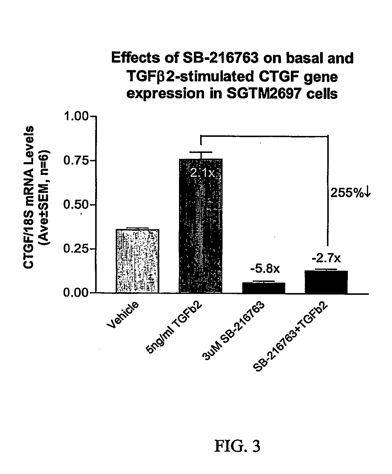
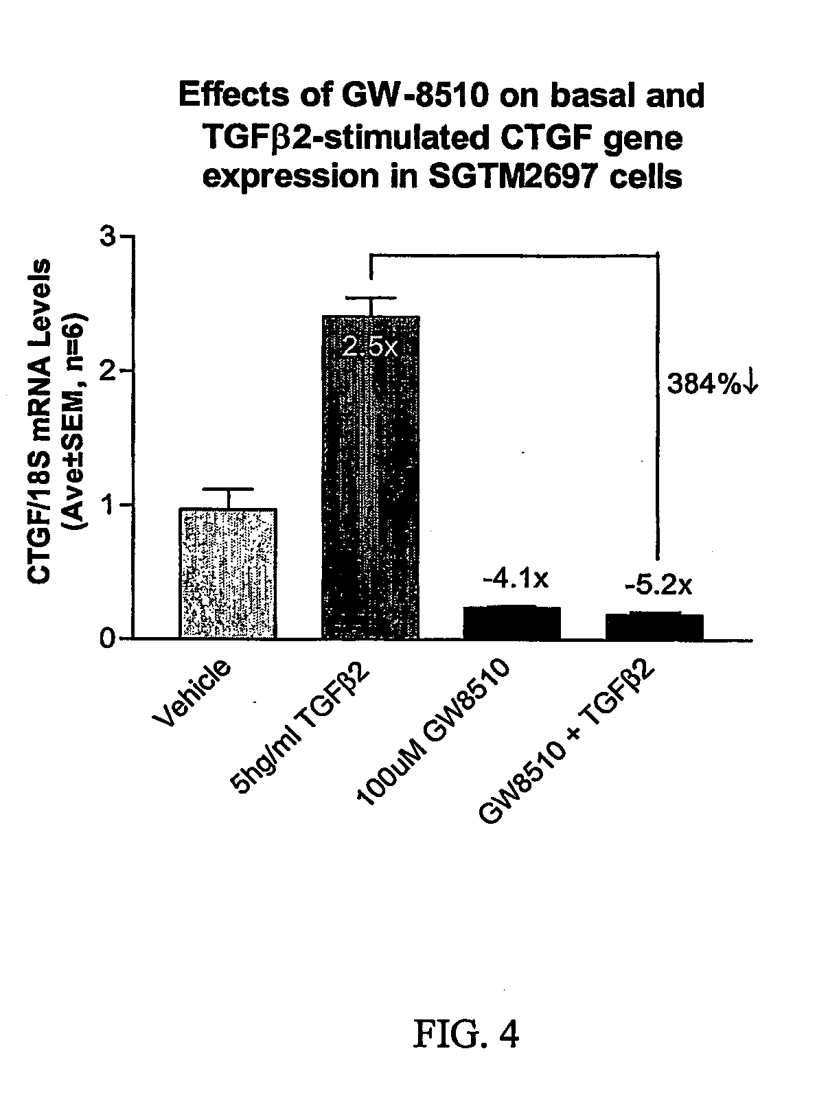
[0009] The present invention overcomes these and other drawbacks of the prior art by providing a method for lowering intraocular pressure and providing neuroprotection to a patient in need thereof by administering a therapeutically effective amount of a composition including at least one non-nucleotide or non-protein agent that inhibits expression and / or signaling of connective tissue growth factor (CTGF), and a pharmaceutically acceptable carrier. In another aspect, the invention provides a method for lowering intraocular pressure by administering to a patient a therapeutically effective amount of an agent that inhibits expression and / or signaling of CTGF. Preferably, the compositions for use in the method of the invention will lower intraocular pressure that is elevated due to an increased expression of CTGF or of a product of CTGF signaling.
[0011] The invention further provides a method for preventing the visual field loss associated with POAG by administering to a patient in need thereof a composition including a non-nucleotide or non-protein agent that modulates the expression and / or signaling of CTGF such that intraocular pressure is controlled and protection is provided to retinal ganglion cells or to the optic nerve head.

Problems solved by technology

The disease is characterized by the degeneration of the trabecular meshwork, leading to obstruction of the normal ability of aqueous humor to leave the eye without closure of the space (e.g., the “angle”) between the iris and cornea (Vaughan, D. et al., (1992)).
A characteristic of such obstruction in this disease is an increased intraocular pressure (“IOP”), resulting in progressive visual loss and blindness if not treated appropriately and in a timely fashion.
For example, a drug that effects the expression of enzymes that degrade the extracellular matrix of the optic nerve head would not likely prevent RGC death caused by excitotoxicity or neurotrophic factor deficit.
However, drugs that effect multiple metabolic pathways are more likely to produce undesirable side-effects.
These therapies lower IOP, but they do not directly address the pathogenic mechanisms, and the disease continues to progress.

Method used


Image

  • Agents which regulate, inhibit, or modulate the activity and/or expression of connective tissue growth factor (ctgf) as a unique means to both lower intraocular pressure and treat glaucomatous retinopathies/optic neuropathies
  • Agents which regulate, inhibit, or modulate the activity and/or expression of connective tissue growth factor (ctgf) as a unique means to both lower intraocular pressure and treat glaucomatous retinopathies/optic neuropathies
  • Agents which regulate, inhibit, or modulate the activity and/or expression of connective tissue growth factor (ctgf) as a unique means to both lower intraocular pressure and treat glaucomatous retinopathies/optic neuropathies

Examples


example 1

Expression of CTGF in Glaucomatous TM vs Non-glaucomatous TM

Microarray Screen

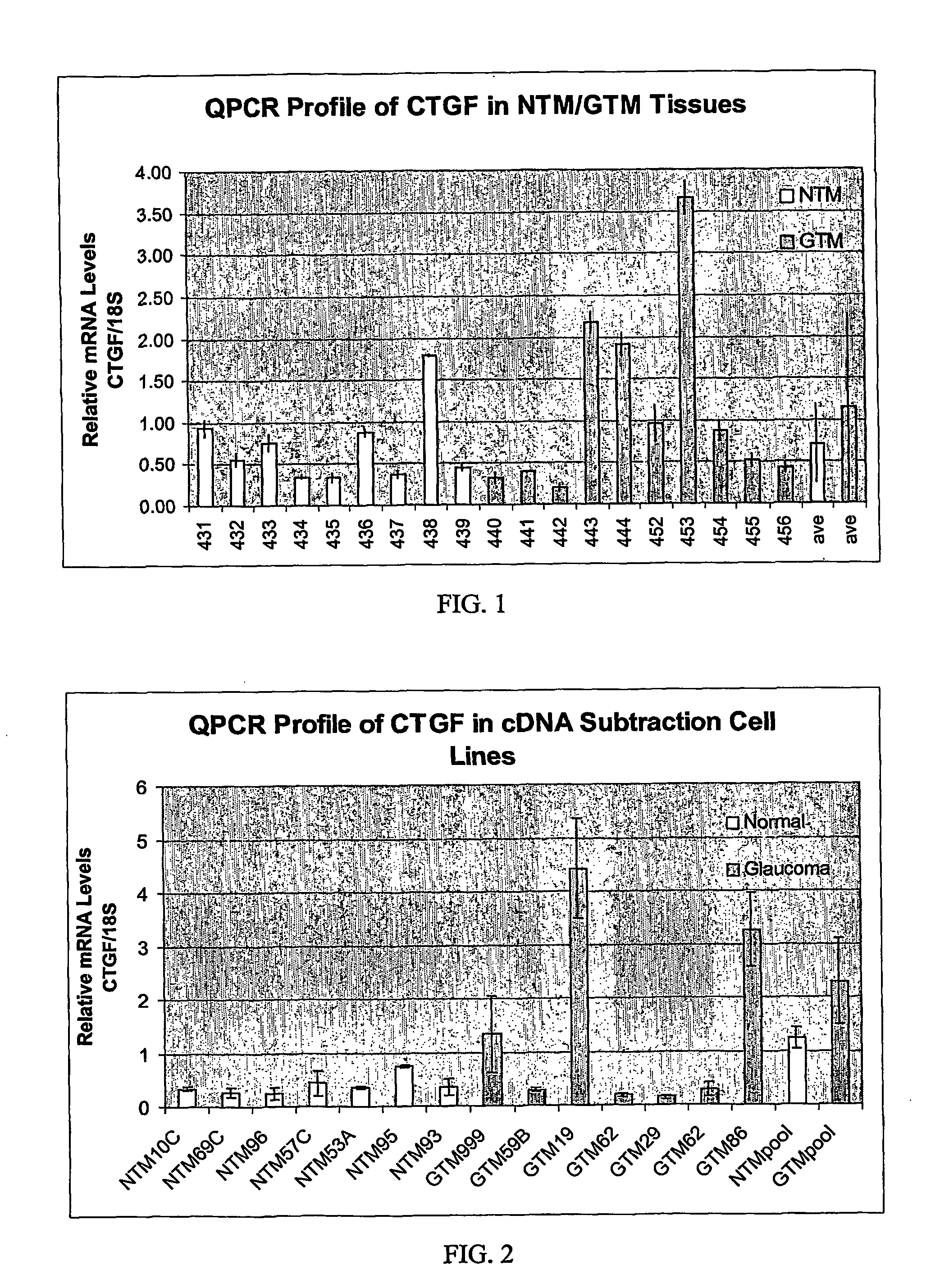
[0041] The mRNA transcript for CTGF was identified as being elevated in glaucomatous TM cells vs. normal TM cells by a GeneFilter® (Research Genetics) screen.

[0042] Five hundred ηg of total RNA (TRIzol Reagent; Invitrogen) was isolated from pooled (6 each) normal or glaucomatous TM cell lines as described (Shepard et al., IOVS 2001, 42:3173) and reverse-transcribed separately in the presence of 300 units of Superscript II RNase Hreverse transcriptase (Invitrogen), 100 μCi [(α-33P]dCTP (10 mCi / ml, 3,000 Ci / mmol; Amersham), 0.33 mM dATP, dGTP, dCTP (Promega), 3.3 mM DTT, 1× First Strand Buffer (Invitrogen), and 2 μg oligo-dT at 37° C. for 90 min. The radiolabeled cDNA was subsequently purified with Chroma-Spin 100 Columns (Clontech) according to the manufacturer's instructions. A single Genefilter® (GF211; Research Genetics) spotted with ≈4,000 known genes was used for hybridization with radiolabeled pro...

example 2

RNA Isolation and First Strand cDNA Preparation

[0046] Total RNA was isolated from TM cells using Trizol® reagent according to the manufacturer's instructions (Life Technologies). First strand cDNA was generated from 1 μg of total RNA using random hexamers and TaqMan® Reverse Transcription reagents according to the manufacturer's instructions (PE Biosystems, Foster City, Calif.). The 100 μl reaction was subsequently diluted 20-fold to achieve an effective cDNA concentration of 0.5 ng / ul.

Quantitative PCR

[0047] Differential expression of CTGF was verified by quantitative real-time RT-PCR (QPCR) using an ABI Prism® 7700 Sequence Detection System (Applied Biosystems). essentially as described (Shepard et al., IOVS 2001, 42:3173). Primers for CTGF amplification were designed using Primer Express software (Applied Biosystems) and anneal to adjacent exons of Genbank accession # NM—001901.1 (CAGCTCTGACATTCTGATTCGAA, nts 1667-1689 and TGCCACAAGCTGTCCAGTCT, nts 1723-1742) and generate a 76-...


PUM

PropertyMeasurementUnit
Fractionaaaaaaaaaa
Fractionaaaaaaaaaa
Pressureaaaaaaaaaa

Abstract

The present invention provides a method for lowering intraocular pressure and providing neuroprotection to a patient in need thereof by administering a therapeutically effective amount of at least one non-nucleotide or non-protein agent that inhibits expression and / or signaling of connective tissue growth factor (CTGF).

Description

BACKGROUND OF THE INTENTION [0001] 1. Field of the Invention [0002] The present invention relates to the field of ocular conditions involving neurodegeneration and / or elevated intraocular pressure. More specifically, the invention provides compositions that lower intraocular pressure and provide ocular neuroprotection. [0003] 2. Description of the Related Art [0004] There are a number of ocular conditions that are caused by, or aggravated by, damage to the optic nerve head, degeneration of ocular tissues, and / or elevated intraocular pressure. For example, “glaucomas” are a group of debilitating eye diseases that are a leading cause of irreversible blindness in the United States and other developed nations. Primary Open Angle Glaucoma (“POAG”) is the most common form of glaucoma. The disease is characterized by the degeneration of the trabecular meshwork, leading to obstruction of the normal ability of aqueous humor to leave the eye without closure of the space (e.g., the “angle”) be...

Claims


Application Information

Patent Timeline
IPC IPC(8): A61F9/007A61K31/00A61K31/202A61K31/404A61K31/426A61K31/427A61K31/4439A61K31/454A61K31/519A61K31/52A61K45/00A61P27/02A61P27/06C12N9/99
CPCA61K31/202A61K31/426A61K31/52A61K31/4439A61K31/519A61K31/427A61P9/12A61P27/02A61P27/06A61P43/00A61K38/18A61K38/43A61K48/00C12N9/99
Inventor FLEENOR, DEBRA L.SHEPARD, ALLANJACOBSON, NASREENPANG, IOK-HOUCLARK, ABBOT F.
Owner NOVARTIS AG
Learn More