Antitumor agents with the use of hsv

a technology of dendritic cell activator and anti-tumor agent, which is applied in the field of anti-tumor agent, tumor immunity inducer, t cell activator, and dendritic cell activator, can solve the problems of inability to use hsvgd for a cancer vaccine, pathologic condition that is extremely difficult to treat, and questionable side effects of hsvgd. , to achieve the effect of enhancing the antigen-presenting ability of dend

Inactive Publication Date: 2005-11-10
INST OF GENE & BRAIN SCI
View PDF4 Cites 9 Cited by
  • Summary
  • Abstract
  • Description
  • Claims
  • Application Information

AI Technical Summary

Benefits of technology

[0068] T cells were obtained from the lymph node of 6-week-old female C57BL/6 mice by the immunomagnetic bead method. Specifically, magnetic bead-coupled monoclonal antibodies (10 μl/107 cells each; manufactured by Miltenyi Biotec) against CD90, CD45R, MHC ClassII, and CD11c were incubated with the cells at 4° C. for 15 min. The T cell fraction was isolated by magnetically separating bead-bound cells using a magnetic cell separation system (MACS; manufactured by Miltenyi Biotec). The fraction (CD90+, CD45R−, MHC ClassII−, and CD11c−) was finally isolated as T cells. T cells (6×10⁴ cells/well) were cultured for 5 days in the presence or absence of anti-CD3 antibody (0.01 ng/ml) and/or HSV-1 gD protein (0.05 ng/ml; manufactured by ViroStat Inc.). After labeling the cultured lymphocytes with 3H-thymidine for about 12 hours, the amount (CPM) of 3H-thymidine incorporated was measured by the top counter. The result is shown in FIG. 8. As observed from FIG. 8, it was revealed that T cells are not activated by either of the anti-CD3 antibody that stimulates the T cell receptor or HSV-1 gD that is a ligand for herpes-virus entry mediator (HVEM), but can be activated only when they are stimulated by both HSVgD and the anti-CD3 antibody. It has been found that LIGHT, a member of TNF cytokine family, activates T cells via HVEM as a costimulatory factor (Nat Med 6, 283-289, 2000). Being a ligand for HVEM, HSVgD has been revealed to work for activation of T cells by acting as a costimulatory factor to T cells.
[0069] Mouse T cells were separated using the same method as that used in Example 7. T cells were plated at 6×10⁴ cells/well and cultured for 5 days in the presence or absence (control) of HSV-1 gD (0.2 or 0.4 ng) and/or anti-HVEM monoclonal antibody (0.01 ng/ml; provided by Dr Patricia G. Spear, Northwestern University, U

Problems solved by technology

However, nothing has been known of using HSVgD for a cancer vaccine.
Metastasis of cancer is a pathologic condition that is extremely difficult to treat; there have been no effective treatment to date.
Although it has been reported that

Method used


Image

  • Antitumor agents with the use of hsv
  • Antitumor agents with the use of hsv
  • Antitumor agents with the use of hsv

Examples


example 1

Viruses, Cell Lines, and Proliferation

[0060] As viruses, the KOS strain of HSV-1 and the 169 strain of HSV-2 (provided by Dr. Yoshiko Seto) were used. African green monkey kidney Vero cells (purchased from ATCC) were used for proliferation of HSVs. Vero cells were cultured in Dulbecco's modified Eagle's medium (DMEM) supplemented with 10% heat-inactivated fetal calf serum (IFCS), and in DMEM supplemented with 1% IFCS when viruses were proliferating. After repeating freeze-thaw of the cells in the virus buffer (150 mM NaCl-20 mM Tris pH7.5) and ultrasonicating the extract, viruses were recovered from the supernatant.

example 2

Inactivation of HSV

[0061] Test HSVs were subjected to inactivation treatment by ultraviolet treatment and heat treatment. The infection efficiency of the HSVs used before inactivation was 2×10⁸ plaque-formation units / ml. As inactivation by ultraviolet rays, the viruses were irradiated at 4 J / m2 for 30 min using 254 nm ultraviolet light on ice, according to the partly modified method of David C. J. et al. (J. virology, 62, 4605-4612, 1988). Subsequently, the UV-treated HSVs were subjected to heat treatment. As inactivation by heat treatment, thermal denaturation treatment of the HSVs were performed at 56° C. for 30 min according to the partly modified method of Hitsumoto Y et al. (Microbiol. Immunol., 27, 757-765, 1983).

example 3

In Situ Cancer Vaccine in a Mouse Bilateral Subcutaneous Tumor Model Using Inactivated HSV

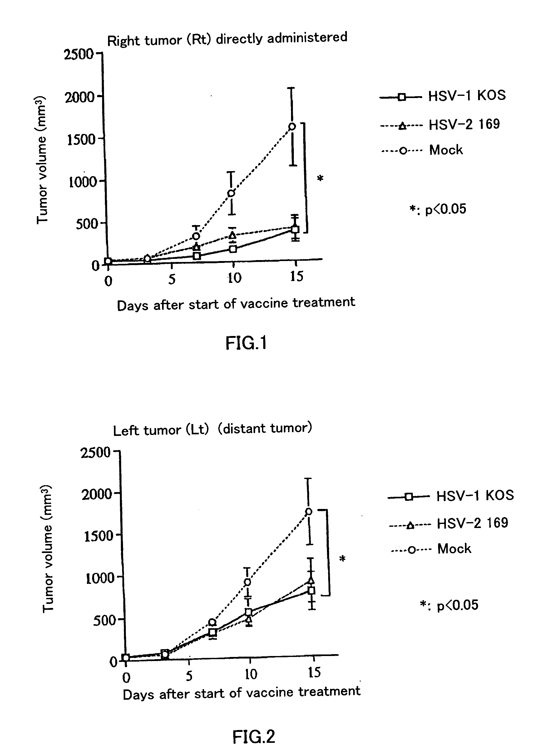
[0062] Six to eight-week old female BALB / c mice were anesthetized (with 0.25 ml of anesthetic liquid: 84% saline, 10% pentobarbital [50 mg / ml], 6% ethanol), and then 5×10⁵ CT26 cells / 100 μl was subcutaneously implanted bilaterally into mice. Treatment started when the diameter of the tumor reached about 5 mm. The extract of Vero cells used for virus preparation was UV-irradiated and heat-treated, and used as a control (hereinafter called “mock”). Either inactivated HSV or mock was administered only to the right tumor on days 0 and 3 after the start of treatment. Thereafter, the volumes of both of the tumors were periodically measured. Volume was calculated as major axis×(minor axis)2÷2. FIG. 1 shows the result of the periodical changes in tumor volume of the right tumor (Rt), into which CT 26 was directly administered (inoculated). FIG. 2 shows the result of the periodical changes in tumor vo...


PUM


Abstract

It is intended to provide highly safe antitumor agents which exhibit an antitumor effect on human remote tumors such as metastatic tumors too and by which an antitumor immune reaction enabling an immune therapy for cancer can be induced, tumor immunity inducers, T cell activators, dendritic cell activators, a method of treating cancer using the same, etc. Inactivated herpes simplex virus (inactivated HSV), herpes simplex virus glycoprotein D (HSVgD), etc. are employed as the active ingredients of antitumor agents, tumor immunity inducers, T cell activators or dendritic cell activators. As a specific example of the treatment for the above-described inactivation, citation may be made of a combination of UV-irradiation using ultraviolet light at 254 nm at 4 J/m2 for 30 minutes with heating at 56° C. for 30 minutes.

Description

TECHNICAL FIELD [0001] The present invention relates to antitumor agents, tumor immunity inducers, T cell activators, and dendritic cell activators that exhibit antitumor effects on human distant tumors such as metastatic tumors as well, and that contain an inactivated herpes simplex virus (inactivated HSV) or herpes simplex virus glycoprotein D (HSVgD) etc. as an effective ingredient. The present invention also relates to enhancement of antitumor effects, methods for treating distant tumors such as metastatic tumors, methods for inducing tumor immunity, methods for identifying tumor antigens, methods for activating T cells, and methods for activating dendritic cells based on the use of the above-mentioned agents, inducers, or activators. BACKGROUND ART [0002] Induction of tumor-specific immunity enables long-term prevention of recurrence of tumors. Such immunotherapy, however, basically depends on the presence or absence of tumor specific antigens and on whether or not a cytotoxic ...

Claims


Application Information

Patent Timeline
IPC IPC(8): A61K35/76A61K35/763A61K38/00A61K38/16A61K39/245A61P35/00A61P35/04A61P37/02A61P43/00C07K14/035C12N5/07C12N5/0783C12N5/0784C12N5/09C12N7/04
CPCA61K39/245A61K2039/5252A61K2039/57C07K14/005C12N7/00A61K41/0019C12N2710/16632C12N2710/16634C12N2710/16661A61K38/00A61K35/763C12N2710/16622A61K2039/5156A61K2039/585A61K39/12A61K41/17A61P35/00A61P35/04A61P37/02A61P43/00
Inventor TODA, MASAHIROKAWAKAMI, YUTAKAIIZUKA, YUKIHIKOUEDA, YOKOIWAHORI, YOSHIHIRO
Owner INST OF GENE & BRAIN SCI
Why Eureka
Try Eureka