Selection of bacterial inner-membrane anchor polypeptides

a technology of inner membrane and polypeptide, which is applied in the field of protein engineering, can solve the problems of difficult to produce clones after several rounds of panning, difficult to screen large libraries consisting of tens of millions or billions of clones, and complex screening of phage-displayed libraries

Inactive Publication Date: 2005-11-24
BOARD OF RGT THE UNIV OF TEXAS SYST
View PDF20 Cites 30 Cited by
  • Summary
  • Abstract
  • Description
  • Claims
  • Application Information

AI Technical Summary

Problems solved by technology

However, even with robotic automation and digital image systems for detecting binding in high density arrays, it is not feasible to screen large libraries consisting of tens of millions or billions of clones.
However, several spectacular successes notwithstanding, the screening of phage-displayed libraries can be complicated by a number of factors.
As a result, the clones isolated after several rounds of panning are frequently difficult to produce on a preparative scale in E. coli.
Second, although phage displayed proteins may bind a ligand, in some cases their un-fused soluble counterparts may not (Griep et al., 1999).
Third, the isolation of ligand-binding proteins and more

Method used


Image

  • Selection of bacterial inner-membrane anchor polypeptides
  • Selection of bacterial inner-membrane anchor polypeptides
  • Selection of bacterial inner-membrane anchor polypeptides

Examples


example 1

Demonstration of Anchored Periplasmic Expression to Target Small Molecules and Peptides

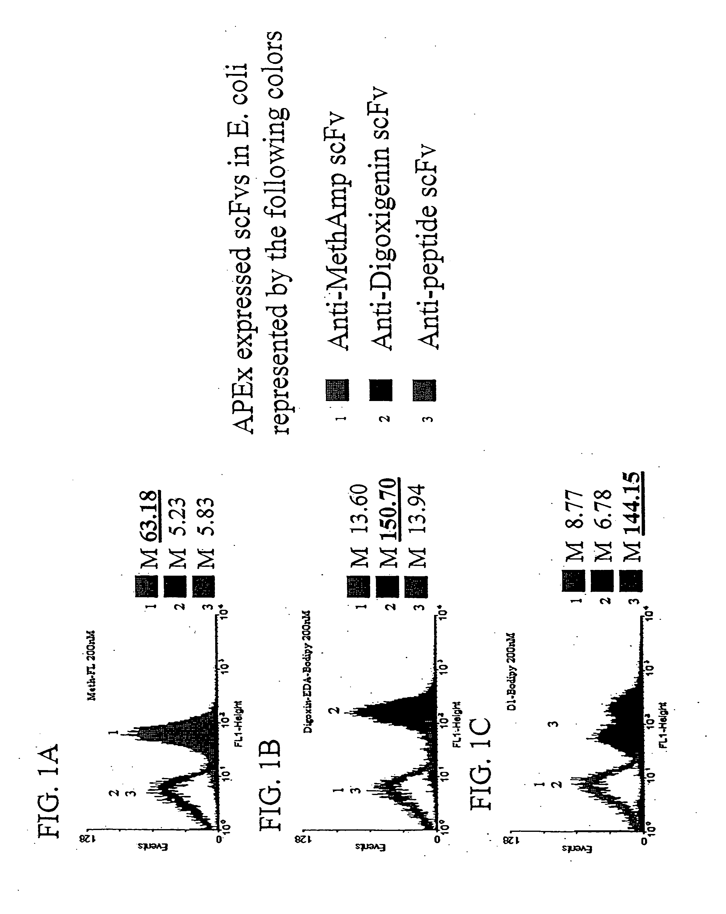
[0124] The ability of scFvs displayed by APEx to target small molecules and peptides is shown in FIGS. 1A-1B and in FIG. 1C, respectively. Three cultures of Escherichia coli containing fusions of the first six amino acids of NlpA (to serve as a inner membrane targeting sequence for APEx analysis) to either an anti-methamphetamine, anti-digoxin, or anti-peptide scfv were grown up and induced for protein expression as described below. Cells of each construct were then labeled in 5×PBS buffer with 200 nM concentrations of methamphetamine-FL (FIG. 1A), digoxigenin-bodipy (FIG. 1B), or 200 nM peptide(18mer)-BodipyFL (FIG. 1C). The data presented shows a histogram representation of 10,000 events from each of the labeled cell cultures. The results demonstrate the ability of scfvs displayed by APEx to bind to their specific antigen conjugated fluorophore, with minimal crossreactivity to non-specific liga...

example 2

Demonstration of Recognition of Ab Fragments by Anchored Periplasmic Expression

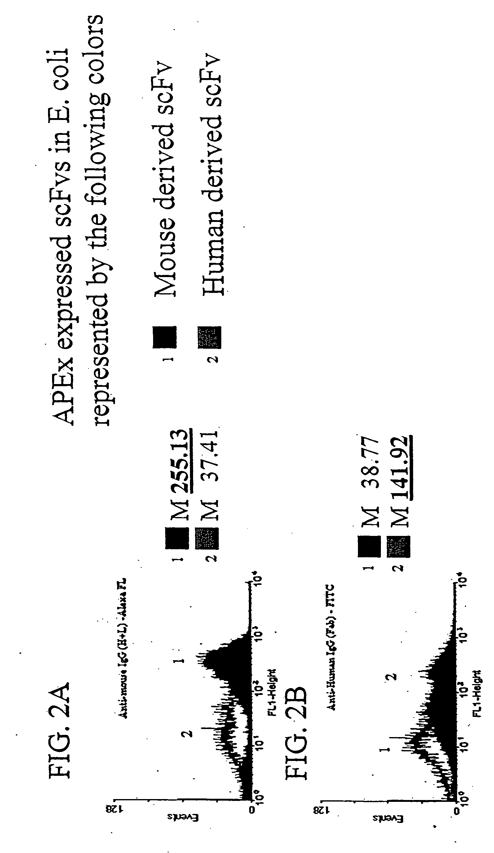
[0125] To demonstrate that the scFv is accessible to larger proteins, it was first demonstrated that polyclonal antibody serum against human Ab fragments or mouse Ab fragments would recognize scFvs derived from each displayed on the E. coli inner membrane by anchored periplasmic expression. Escherichia coli expressing a mouse derived scFv via anchored periplasmic expression (FIG. 2A) or expressing a human derived scFv via anchored periplasmic expression (FIG. 2B) were labeled as described below with either anti-mouse polyclonal IgG (H+ L)-Alexa-FL or anti-human polyclonal IgG (Fab)-FITC. Results (FIG. 2A, 2B) in the form of histogram representations of 10000 events of each demonstrated that the anti-human polyclonal (approximately 150 kDa in size) recognized the human derived scFv specifically while the anti-mouse polyclonal (150 kDa) recognized the mouse derived scFv.

example 3

Demonstration of the Ability of scFvs Displayed by Anchored Periplasmic Expression to Specifically Bind Large Antigen Conjugated Fluorophores

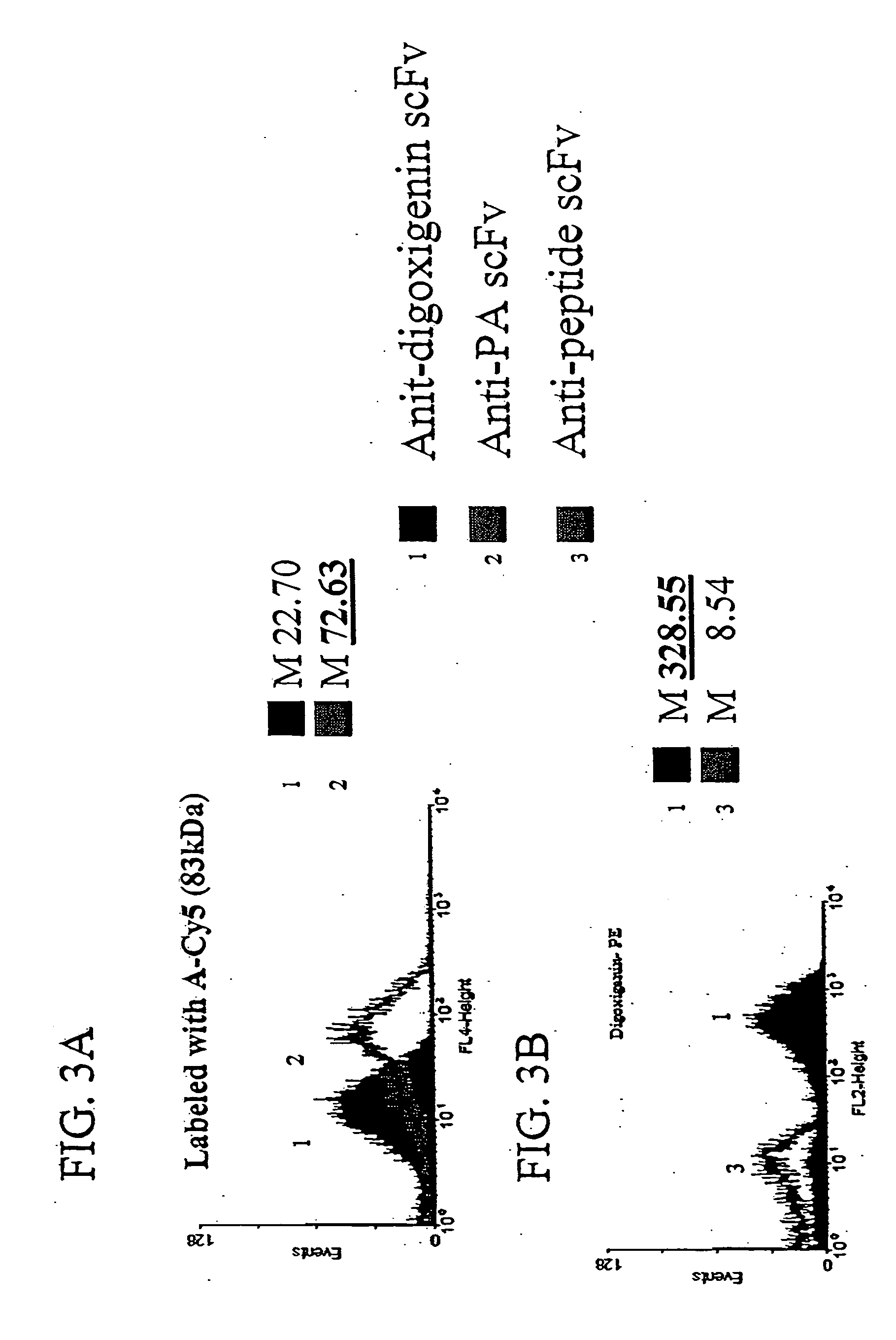
[0126] To demonstrate the ability of scFvs displayed via anchored periplasmic expression to specifically bind to large antigen conjugated fluorophores, E. coli were induced and labeled as described below expressing, via anchored periplasmic expression, an anti-protective antigen(PA) scFv (PA is one component of the anthrax toxin: a 83 kDa protein) or an anti-digoxigenin scFv. Histogram data of 10,000 events demonstrated specific binding to a PA-Cy5 antigen conjugated fluorophore as compared to the cells expressing the an anti-digoxigenin scFv (FIG. 3A). To further illustrate this point, digoxigenin was coupled to phycoerythrin(PE), a 240 kDa fluorescent protein. Cells were labeled with this conjugate as described below. It was found that E. coli (10,000 events) expressing the anti-digoxigenin scFv via anchored periplasmic expression were label...


PUM

PropertyMeasurementUnit
Volumeaaaaaaaaaa
Lengthaaaaaaaaaa
Affinityaaaaaaaaaa

Abstract

The invention overcomes the deficiencies of the prior art by providing a rapid approach for isolating polypeptides capable of anchoring heterologous polypeptides to a bacterial inner membrane. In the technique, libraries of candidate anchor polypeptides are expressed as fusions with a heterologous polypeptide that is capable of being detected when bound to the inner membrane. In bacteria expressing a functional anchor sequence, the heterologous polypeptide becomes bound to outer face of the inner membrane. Bacteria with the functional anchor sequence can be identified by removing the outer membrane to remove non-anchored heterologous polypeptide followed by detection of anchored heterologous polypeptide. Such bacteria may be detected in numerous ways, including use of direct fluorescence or secondary antibodies that are fluorescently labeled, allowing use of efficient techniques such as fluorescence activated cell sorting (FACS).

Description

[0001] The government may own rights in the present invention pursuant to the U.S. Army ARO MURI program; the Texas Consortium for Development of Biological Sensors; U.S. Department of Defense TransTexas BW Defense Initiative Grant No. DAA21-93C-0101 and in connection with contract number DADD17-01-D-0001 with the U.S. Army Research Laboratory. [0002] This application claims the priority of U.S. Provisional Patent App. No. 60 / 554,324, filed Mar. 18, 2004, and is a continuation-in-part of U.S. patent application Ser. No. 10 / 620,278, filed Jul. 15, 2003, which application claims the priority of U.S. Provisional Patent App. No. 60 / 396,058, filed Jul. 15, 2002.BACKGROUND OF THE INVENTION [0003] 1. Field of the Invention [0004] The present invention relates generally to the field of protein engineering. More particularly, it concerns methods for identification of bacterial inner membrane anchor sequences. [0005] 2. Description of Related Art [0006] The isolation of polypeptides that eith...

Claims


Application Information

Patent Timeline
IPC IPC(8): C12N1/21C12N15/10C12N15/74C40B40/02
CPCC07K2319/04C40B40/02C12N15/1086C12N15/1037
Inventor GEORGIOU, GEORGEJEONG, KI JUNHARVEY, BARRETTIVERSON, BRENT
Owner BOARD OF RGT THE UNIV OF TEXAS SYST
Why Eureka
Try Eureka