
Nanogene therapy for cell proliferation disorders

a cell proliferation and neoplasm technology, applied in the direction of genetic material ingredients, drug compositions, peptide/protein ingredients, etc., can solve the problems of aggressive cancers that are difficult to treat, disease remains difficult to effectively treat, and their long-term survival is poor, so as to reduce (partially or completely inhibit, slow down, slow down) uncontrolled cell growth and reduce cellular growth

Inactive Publication Date: 2005-12-01
UNIV OF SOUTH FLORIDA
View PDF54 Cites 43 Cited by
  • Summary
  • Abstract
  • Description
  • Claims
  • Application Information

AI Technical Summary

Benefits of technology

[0014] In another aspect, the present invention concerns a method of treating a cell proliferation disorder by administering a therapeutically effective amount of particles to a patient in need thereof. Accordingly, a method of reducing cellular growth by administering a therapeutically effective amount of particles of the invention is contemplated, in order to reduce (partially or completely inhibit, prevent, or slow) uncontrolled cell growth. In one embodiment, an effective amount of parti...

Problems solved by technology

Despite progress made in our understanding of the multiple risk factors associated with the development of lung cancer, and progress in developing novel approaches, this disease remains difficult to treat effectively.
Their long-term survival is poor and such aggressive cancers are difficult to treat because of drug-induced toxicity.
Exogenous recombinant IFNs have a shorter half-life in vivo, and systemic administration at moderate to high doses may cause substantial adverse effects (Gutterman, J. U. PNAS, 1994, 91:1198-1205; Antoniou, K. M. et al.
However, mucosal administration of IFN-γ pDNA has not been studied.
One problem associated with the pDNA approach is inefficient gene transfer in vivo, especially in slow and non-dividing cells such as epithelial cells (Mohapatra, S. S. Pediatr. Infect. Dis. J., 2003, 22(2 Suppl):S100-S103).

Method used


Image

  • Nanogene therapy for cell proliferation disorders
  • Nanogene therapy for cell proliferation disorders
  • Nanogene therapy for cell proliferation disorders

Examples


example 1

pIFN-γ Induces Apoptosis of HEp-2 Carcinoma Cells

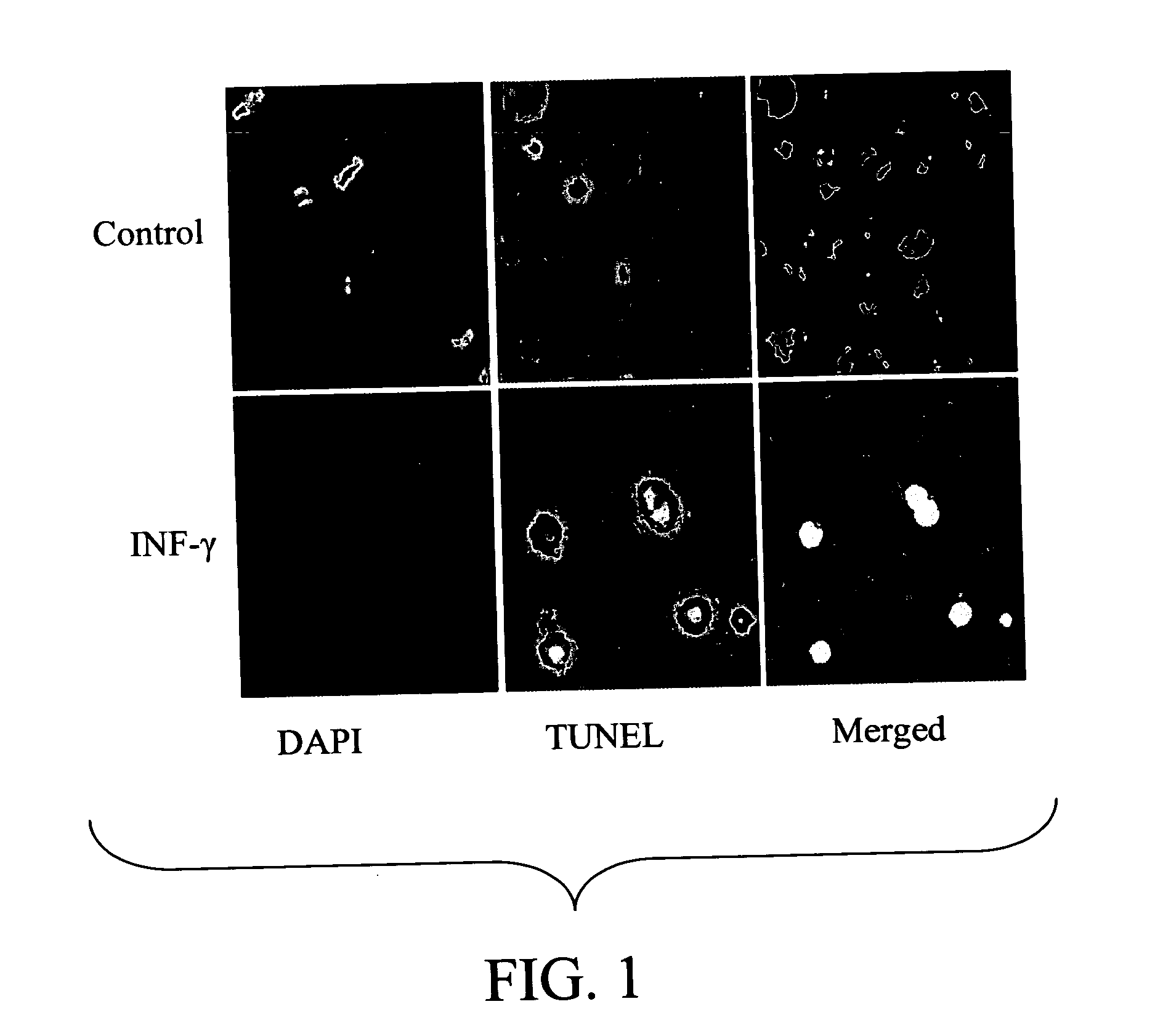
[0145] To determine the effect of overexpression of pIFN-γ on proliferation of A549 lung epithelial cells, cells were transfected with either pIFN-γ or empty vector, pVAX (control). Cell cycle analysis was performed using propidium iodide (PI) staining and flow cytometry 48 hours after transfection. No significant difference was observed between control and pIFN-γ-transfected cells in S1, Go-G1 and G2-M stages of the cell cycle (data not shown). However, an analysis of apoptosis using fluorescence microscopy cells transfected with pIFN-γ exhibited significantly higher apoptosis compared to cells transfected with either the control plasmid or a plasmid encoding pVAX (shown in FIG. 1).

[0146] Cells were seeded into 4-well slide dishes at 104 cells per well and allowed to grow to 75% confluency. Cells were treated for 20 hours with 1000 U / ml IFN-γ. After 24 hours, cells were fixed with 4% paraformaldehyde in PBS for 25 minutes at 4° C. ...

example 2

Microarray Analysis of Chitosan—pIFNgamma Treated Lungs

[0149] Using MU11KsubA and B chips (Affymetrix), which contain probes interrogating about 11,000 murine genes and ESTs (Unigene, Build-4), as well as EST clusters from TIGR (1.0 Beta), we have identified a total of 126 differentially expressed genes whose expression level is altered in the CIN treated mouse lung in the range of −10.6- to 152.4-fold. A noteworthy observation is the up-regulation of the expression of a number of IFN-inducible genes, immune response related genes, and genes involved in signal transduction, including STAT1 and STAT4.

TABLE 1GENE EXPRESSION ANALYSIS INBALB / c LUNG BY MICROARRAYMax. FoldCategory of GenesChangeGenesIFN-regulated12IFN-induced 15 KDa protein,IFN-activable protein 204,Eukaryotic initiation factor5, Mx protein, MIG, IP-10,Interferon regulatory factor(mirf7), interferon-activatableprotein, IFN-induced protein6-16 precursor, IFN-inducedguanylate-binding protein,HLA-associated protein i(phap...

example 3

Chitosan-Conjugated pIFN-γ Plasmid Prevents Metastasis of Lung Tumors in Nude Mice

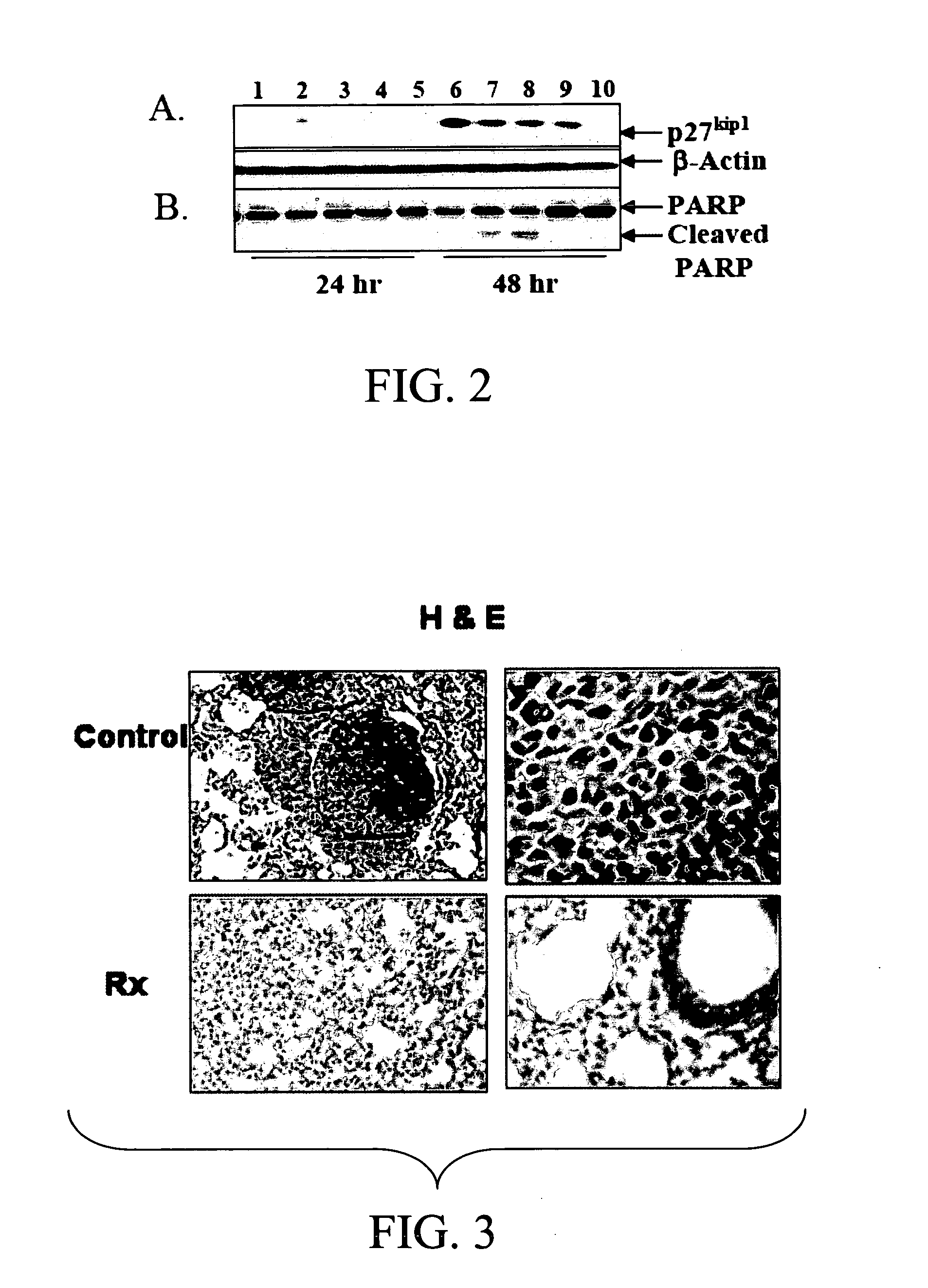
[0150] BALB / c nude mice were injected intravenously with 5×10⁶ A549 cells, then treated one day afterwards and at weekly intervals with pIFN-γ or control plasmid. After 4 weeks, mice were examined for lung histology. The control animals showed tumors, whereas no tumors were seen in the pIFN-γ-treated group (FIG. 3). These results indicate that pIFN-γ has the potential to decrease tumor metastasis.

[0151] The results indicated in FIG. 3 were obtained when BALB / c nude mice were injected with A549 cells (5×10⁶ cells / mouse) intravenously and one group treated with pIFN-gamma and another group with pVAX as control. The lungs of control mice showed numerous lung nodules in contrast to mice treated with pIFN-gamma, which showed very few tumors. The lungs were removed from mice treated with nanoparticles carrying empty plasmid pVAX (control) or with pIFN-gamma (Rx) and H & E stained. The lungs of control mice...


PUM

PropertyMeasurementUnit
Atomic weightaaaaaaaaaa

Abstract

The present invention concerns particles comprising a chitin component, such as chitosan or a derivative thereof, associated with a polynucleotide encoding an interferon (IFN) molecule, 2-5′ oligoadenylate synthetase (2-5 AS), or a combination thereof. Preferably, the chitin component comprises chitosan or a derivative thereof. The particles of the invention are useful for delivery and expression of the interferon-encoding and / or 2-5 AS-encoding polynucleotide within a host in vitro or in vivo. The invention further concerns pharmaceutical compositions comprising particles of the invention and a pharmaceutically acceptable carrier, and a method for producing particles of the present invention. The present invention further pertains to a method of inducing apoptosis in a cancer cell, such as a lung cancer cell, by contacting a target cancer cell in vitro or in vivo with an effective amount of particles of the invention. In one embodiment, a therapeutically effective amount of particles are administered to target cancer cells within a patient in vivo, for treatment of cancer, such as lung cancer. The particles and therapeutic methods of the invention provide anti-metastatic and anti-cancer therapeutics for cancer patients, particularly lung cancer patients.

Description

CROSS-REFERENCE TO RELATED APPLICATION [0001] This application claims benefit of U.S. Provisional Application Ser. No. 60 / 565,756, filed Apr. 27, 2004, which is hereby incorporated by reference herein in its entirety, including any figures, tables, nucleic acid sequences, amino acid sequences, and drawings.FIELD OF THE INVENTION [0002] This invention pertains to particles including a chitin component, a polynucleotide encoding an interferon molecule, such as IFN-gamma (IFN-γ), or an interferon-inducible molecule such as 2′-5′ oligoadenylate synthetase (2-5 AS) or interferon regulatory factor (IRF-1), or a combination of any of the foregoing, and the use of such particles for treatment of cell proliferation disorders, such as lung cancer. BACKGROUND OF THE INVENTION [0003] Lung cancer is one of the leading causes of death worldwide. Despite progress made in our understanding of the multiple risk factors associated with the development of lung cancer, and progress in developing novel ...

Claims


Application Information

Patent Timeline
IPC IPC(8): A61K9/00A61K9/127A61K9/16A61K9/50A61K9/51A61K38/17A61K38/21A61K38/53A61K48/00C07K14/57C12N15/88
CPCA61K9/0043A61K9/5161A61K38/1709A61K38/21A61K38/212C12N15/88A61K38/217A61K38/53A61K48/0025A61K48/0075C07K14/57A61K38/215A61P35/00
Inventor MOHAPATRA, SHYAM
Owner UNIV OF SOUTH FLORIDA
Learn More