Methods for rapid forensic analysis of mitochondrial DNA
a mitochondrial dna and forensic analysis technology, applied in the field of mitochondrial dna analysis, can solve the problems of inability to detect low levels of heteroplasmy in detection methods currently available to molecular biologists, difficult recording and comparing mtdna sequences, and potentially confusing,
- Summary
- Abstract
- Description
- Claims
- Application Information
AI Technical Summary
Problems solved by technology
Method used
Image
Examples
example 1
Nucleic Acid Isolation and Amplification
[0075] General Genomic DNA Sample Prep Protocol: Raw samples were filtered using Supor-200 0.2 μm membrane syringe filters (VWR International). Samples were transferred to 1.5 ml eppendorf tubes pre-filled with 0.45 g of 0.7 mm Zirconia beads followed by the addition of 350 μl of ATL buffer (Qiagen, Valencia, Calif.). The samples were subjected to bead beating for 10 minutes at a frequency of 19 l / s in a Retsch Vibration Mill (Retsch). After centrifugation, samples were transferred to an S-block plate (Qiagen, Valencia, Calif.) and DNA isolation was completed with a BioRobot 8000 nucleic acid isolation robot (Qiagen, Valencia, Calif.).
[0076] Isolation of Blood DNA—Blood DNA was isolated using an MDx Biorobot according to according to the manufacturer's recommended procedure (Isolation of blood DNA on Qiagen QIAamp® DNA Blood BioRobot® MDx Kit, Qiagen, Valencia, Calif.)
[0077] Isolation of Buccal Swab DNA—Since the manufacturer does not suppo...
example 2
Digestion of Amplicons with Restriction Enzymes
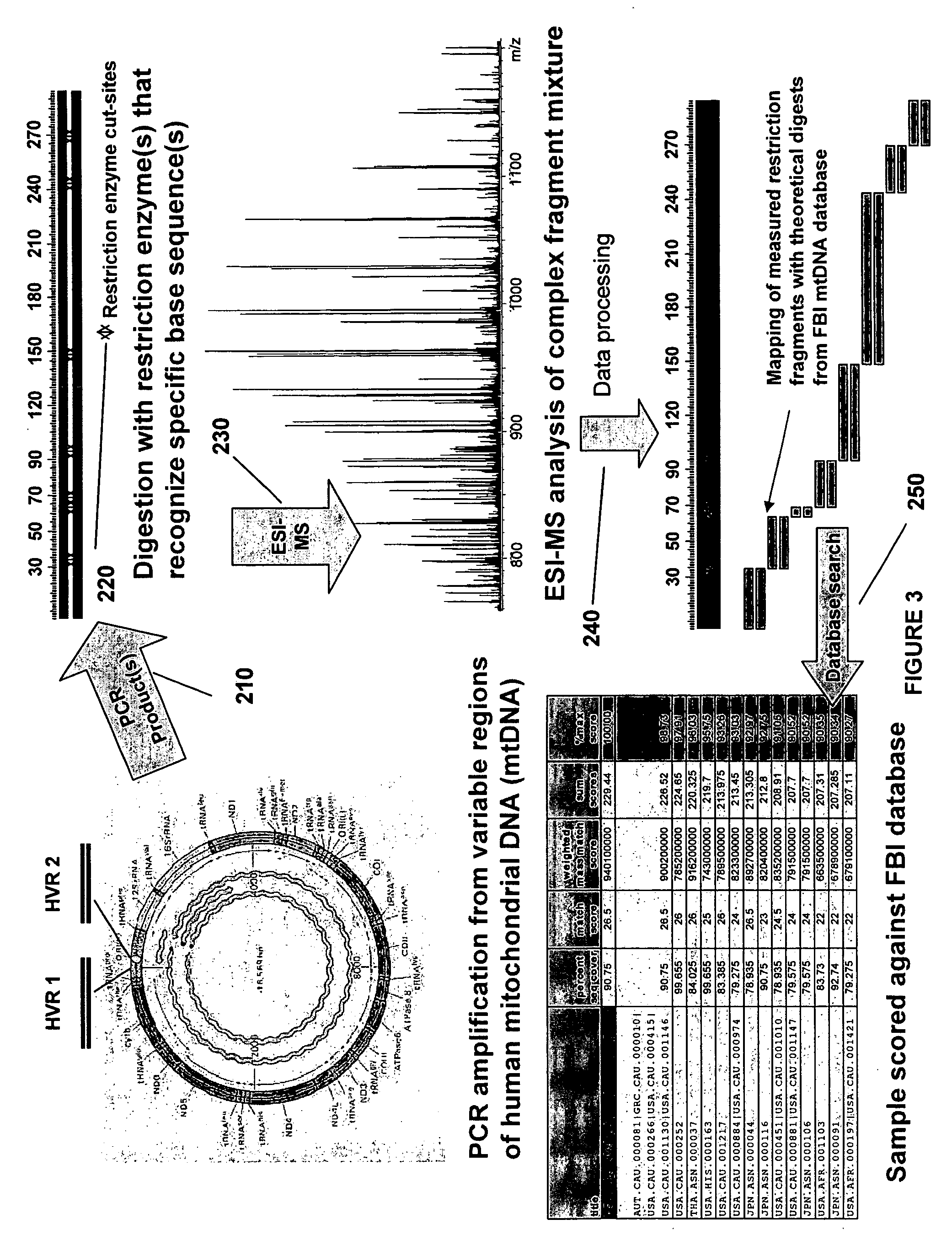
[0080] Reaction Conditions—The standard restriction digest reaction conditions outlined herein are applicable to all panels of restriction enzymes. The PCR reaction mixture is diluted into 2×NEB buffer 1+BSA and 1 μl of each enzyme per 50 μl of reaction mixture is added. The mixture is incubated at 37° C. for 1 hour followed by 72° C. for 15 minutes. Restriction digest enzyme panels for HV1, HV2 and twelve additional regions of mitochondrial DNA are indicated in Table 2.
TABLE 2mtDNA Regions, Coordinates and Restriction Enzyme Digest PanelsCOORDINATES RELATIVETO THE ANDERSONRESTRICTIONmtDNA REGIONSEQUENCE (SEQ ID NO: 72)ENZYME PANELHV1 (highly variable16050-16410RsaIcontrol region 1)HV2 (highly variable 29-429HaeIII HpaII MfeI SspIcontrol region 2)orHpaII, HpyCH4IV, PacI andEaeIREGION R1 (COX2,8162-8992DdeI MseI HaeIII MboIIntergenic spacer, tRNA-LYS, ATP6)REGION R2 (ND5)12438-13189DdeI HaeIII MboI MseIREGION R3 (ND6 tRNA-Glu,14629-15...
example 3
Nucleic Acid Purification
[0081] Procedure for Semi-Automated Purification of a PCR Mixture Using Commercially Available Zip Tips®—As Described by Jiang and Hofstadler (Y. Jiang and S. A. Hofstadler Anal. Biochem. 2003, 316, 50-57) an amplified nucleic acid mixture can be purified by commercially available pipette tips containing anion exchange resin. For pre-treatment of ZipTips® AX (Millipore Corp. Bedford, Mass.), the following steps were programmed to be performed by an Evolution™ P3 liquid handler (Perkin Elmer) with fluids being drawn from stock solutions in individual wells of a 96-well plate (Marshall Bioscience): loading of a rack of ZipTips®AX; washing of ZipTips®AX with 15 μl of 10% NH4OH / 50% methanol; washing of ZipTips® AX with 15 μl of water 8 times; washing of ZipTips® AX with 15 μl of 100 mM NH4OAc.
[0082] For purification of a PCR mixture, 20 μl of crude PCR product was transferred to individual wells of a MJ Research plate using a BioHit (Helsinki, Finland) multich...
PUM
| Property | Measurement | Unit |
|---|---|---|
| molecular weight | aaaaa | aaaaa |
| pH | aaaaa | aaaaa |
| total volume | aaaaa | aaaaa |
Abstract
Description
Claims
Application Information
 - R&D
- Intellectual Property
- Life Sciences
- Materials
- Tech Scout
- Unparalleled Data Quality
- Higher Quality Content
- 60% Fewer Hallucinations
Browse by: Latest US Patents, China's latest patents, Technical Efficacy Thesaurus, Application Domain, Technology Topic, Popular Technical Reports.
© 2025 PatSnap. All rights reserved.Legal|Privacy policy|Modern Slavery Act Transparency Statement|Sitemap|About US| Contact US: help@patsnap.com


