Internal control for in situ hybridization

a technology of in situ hybridization and control, which is applied in the direction of microorganism testing/measurement, fermentation, biochemistry apparatus and processes, etc., can solve the problems of false negative diagnosis, unreliable results, and negative ish results obtained for a particular target probe to be viewed as unreliabl

Inactive Publication Date: 2005-12-08
VENTANA MEDICAL SYST INC
View PDF4 Cites 14 Cited by
  • Summary
  • Abstract
  • Description
  • Claims
  • Application Information

AI Technical Summary

Benefits of technology

[0021] Specific preferred embodiments of the present invention will become evident from

Problems solved by technology

For example, improper preservation of cellular or tissue samples can result in target DNA degradation, leading to a false negative diagnostic result.
Unreliable results can also be obtained through the use of defective ISH detection reagents.
In general, any negative ISH result obtained for a particular target probe should be viewed as unreliable when an inadequate staining result is obtained with an Alu control probe.
The use of Alu probes as an ISH control, however, also presents several disadvantages.
Second, because Alu elements are short, intersperse

Method used


Image

  • Internal control for in situ hybridization
  • Internal control for in situ hybridization
  • Internal control for in situ hybridization

Examples


example 1

Preparation of Tissue and Cell Samples for Chromogenic In Situ Hybridization Analysis

[0041] Chromogenic in situ hybridization (CISH) analyses were performed using two human papilloma virus (HPV)-positive cell lines, CaSki (containing 200-600 copies of HPV 16) and HeLa (containing 10-50 copies of HPV 18), and one HPV-negative cell line (T24). Cell samples were fixed in 10% neutral buffered formalin, embedded in paraffin, and sectioned at 4-8 microns. Fixed cell samples were placed on Superfrost® Plus glass slides (VWR Scientific; West Chester, Pa.) prior to CISH analysis.

[0042] CISH analyses were also performed on cervical lesion cells of tissue biopsies and cervical smear samples prepared using commercially available liquid-based prep (LBP) systems from Cytyc Corp. (Boxborough, Mass.) and TriPath Imaging Inc. (Burlington, N.C.).

example 2

Preparation of Probes for Chromogenic In Situ Hybridization Analysis

[0043] HPV DNA probes for chromogenic in situ hybridization (CISH) analysis were prepared by cloning HPV DNA from genotypes 16, 18, 31, 33, 35, and 51 into plasmid vectors, as described in International Publication No. WO 00 / 24760.

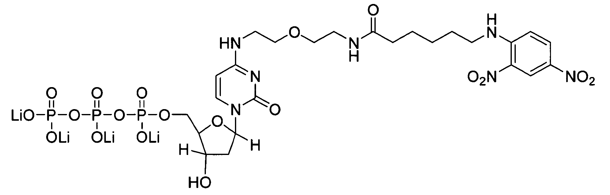
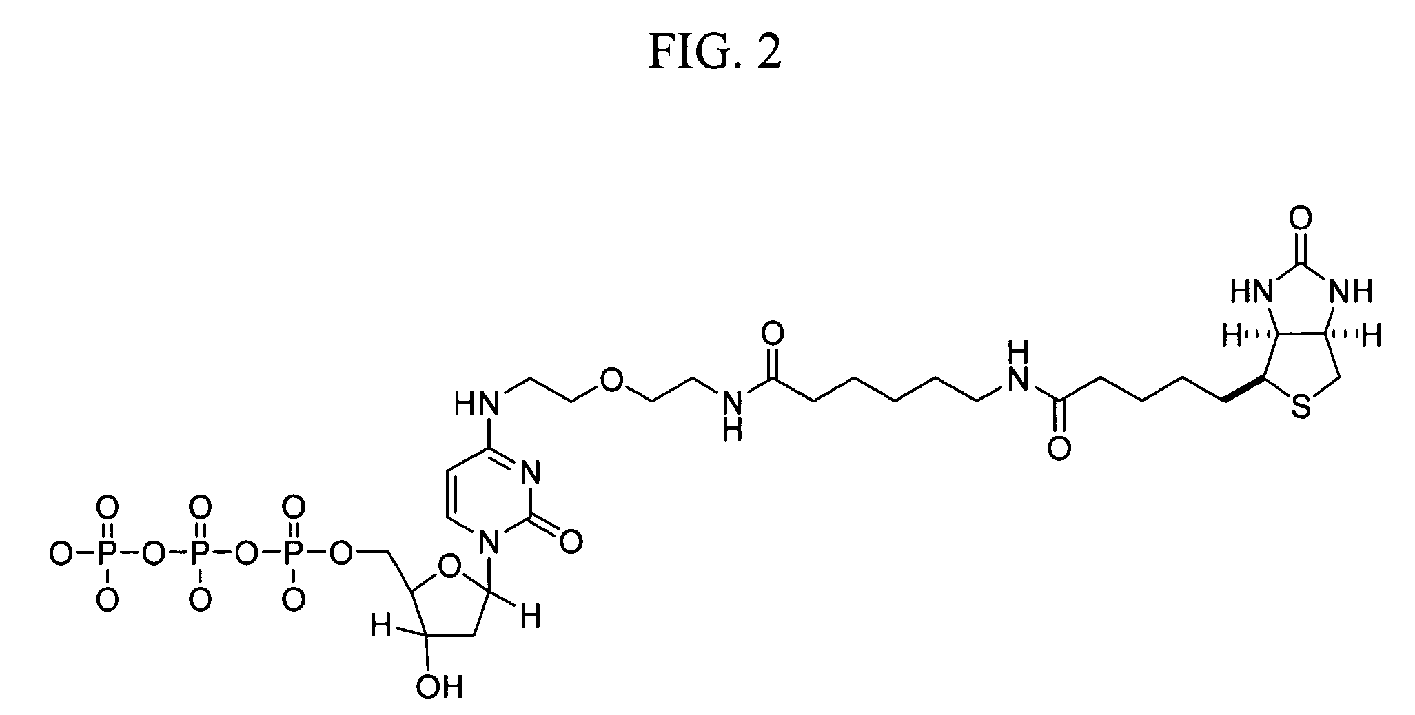
[0044] Mitochondrial DNA probes for CISH analysis were prepared by PCR amplification using the Expand Long Template PCR System (Roche Molecular Biochemicals; Indianapolis, Ind.) and primers shown in Table I. Amplification reactions containing 500 μM of each dNTP, 5 units of Taq Polymerase, 0.3 μM of each primer, 50 mM KCl, 2.75 mM Mg2Cl, 10 mM Tris-HCl, pH 8.5, and a DNA template from the human cell line, C33A, were performed at 94° C. for 2 minutes for one cycle and at 94° C. for 10 minutes, 55° C. for 30 minutes, and 68° C. for 15 minutes for 35 cycles. Amplification products were separated on a 0.6% agarose gel and analyzed using an α-imager. Products having the expected size were obt...

example 3

Analysis of Nuclear and Mitochondrial DNA Targets by Chromogenic In Situ Hybridization Using Identical Haptens and Detection Systems

[0047] CISH analysis of nuclear and mitochondrial DNA targets using identical haptens and detection systems was performed as follows. CaSki and cervical lesion cells of tissue biopsies were prepared as described in Example 1. Samples included formalin-fixed / paraffin-embedded tissues, formalin-fixed / paraffin-embedded tissue culture cell pellets, fixed tissue culture cells on Cytospin-prepared slides, and fixed cervical cells prepared with using the ThinPrep Pap Test specimen collection system (Cytyc Corp.). HPV and mitochondrial DNA probes were prepared and labeled with biotin-dCTP by nick translation as described in Example 2.

[0048] CISH was performed on a BenchMark® automated slide stainer (Ventana Medical Systems, Inc.). The degree of hybridization between the HPV and mitochondrial DNA probes and their respective targets was determined using one of ...


PUM

PropertyMeasurementUnit
Fractionaaaaaaaaaa

Abstract

The invention provides a method for monitoring the quality of in situ hybridization analysis of a nuclear DNA target in a tissue or cell sample using a mitochondrial DNA probe as an internal control. The invention also provides a reagent for in situ hybridization detection of a nuclear DNA target and a mitochondrial DNA target in a tissue or cell sample.

Description

BACKGROUND OF THE INVENTION [0001] 1. Field of the Invention [0002] The invention relates to a method for monitoring the quality of in situ hybridization analysis of a nuclear DNA target in a tissue or cell sample using a mitochondrial DNA probe as an internal control. The invention also relates to a reagent for in situ hybridization detection of a nuclear DNA target and a mitochondrial DNA target in a tissue or cell sample. [0003] 2. Background of the Invention [0004] Nucleic acid hybridization is a process in which two single-stranded nucleic acid molecules having sufficiently complementary sequences are allowed to interact under suitable reaction conditions so as to form a double-stranded nucleic acid hybrid. Hybridization techniques generally can be classified into one of three groups: (1) solution hybridization techniques, in which the hybridization reaction between the complementary, single-stranded nucleic acid molecules is carried out in solution; (2) filter or blot hybridiz...

Claims


Application Information

Patent Timeline
IPC IPC(8): C12Q1/70C12Q1/68C12P19/34
CPCC12Q1/6841C12Q2563/131C12Q2531/113
Inventor JI, JAY
Owner VENTANA MEDICAL SYST INC
Why Eureka
Try Eureka