Non-invasive diagnosis of breast cancer using real-time ultrasound strain imaging

a real-time ultrasound and ultrasound technology, applied in the field of breast cancer non-invasive diagnosis using real-time ultrasound strain imaging, can solve the problems of general failure of motion tracking algorithm, more difficult for the technologist to apply consistent axial compression, etc., and achieve the effect of facilitating non-invasive diagnosis of breast cancer and no additional hardware costs

Inactive Publication Date: 2005-12-22
MAYO FOUND FOR MEDICAL EDUCATION & RES
View PDF19 Cites 32 Cited by
  • Summary
  • Abstract
  • Description
  • Claims
  • Application Information

AI Technical Summary

Benefits of technology

[0009] A general object of the invention is to provide an ultrasound method which facilitates the non-invasive diagnosis of a breast lesion. Strain images may be acquired using a freehand method of applying stress to the tissues. This system requires no additional equipment attached to the ultrasound transducer, such as force measurement or tissue loading apparatus, and thus remains relatively robust and simple to operate. The strain imaging capability may be added to a standard clinical ultrasound platform as a software upgrade, with no additional hardware costs.

Problems solved by technology

Also, the mobility of these lesions makes it more difficult for the technologist to apply consistent axial compression, since the lesions have a tendency to also move in the lateral and elevational directions.
These non-axial motions can cause general failures of the motion tracking algorithm, and strain images that show mainly decorrelation noise and very little if any anatomical structure.

Method used


Image

  • Non-invasive diagnosis of breast cancer using real-time ultrasound strain imaging
  • Non-invasive diagnosis of breast cancer using real-time ultrasound strain imaging
  • Non-invasive diagnosis of breast cancer using real-time ultrasound strain imaging

Examples


Embodiment Construction

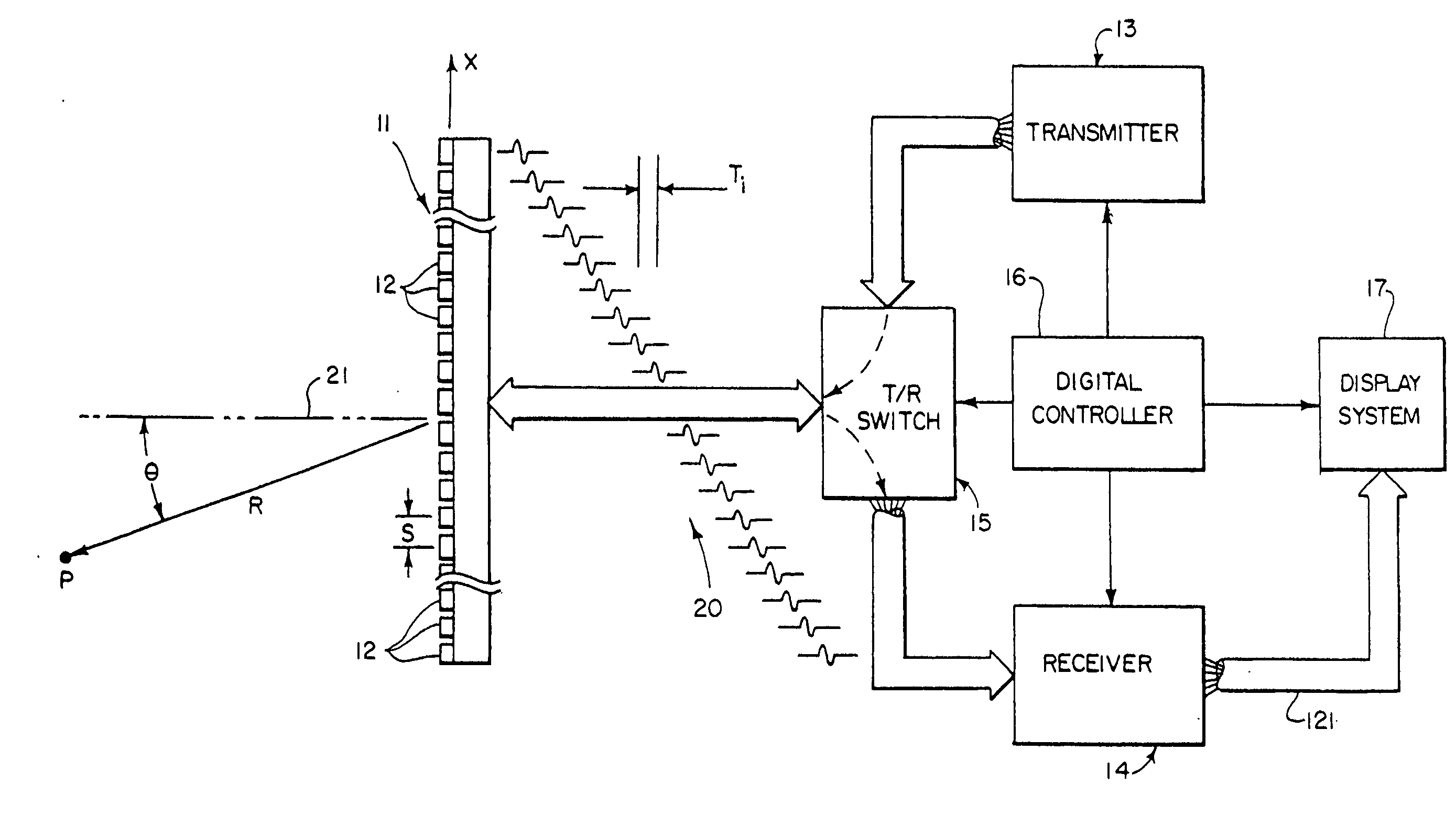
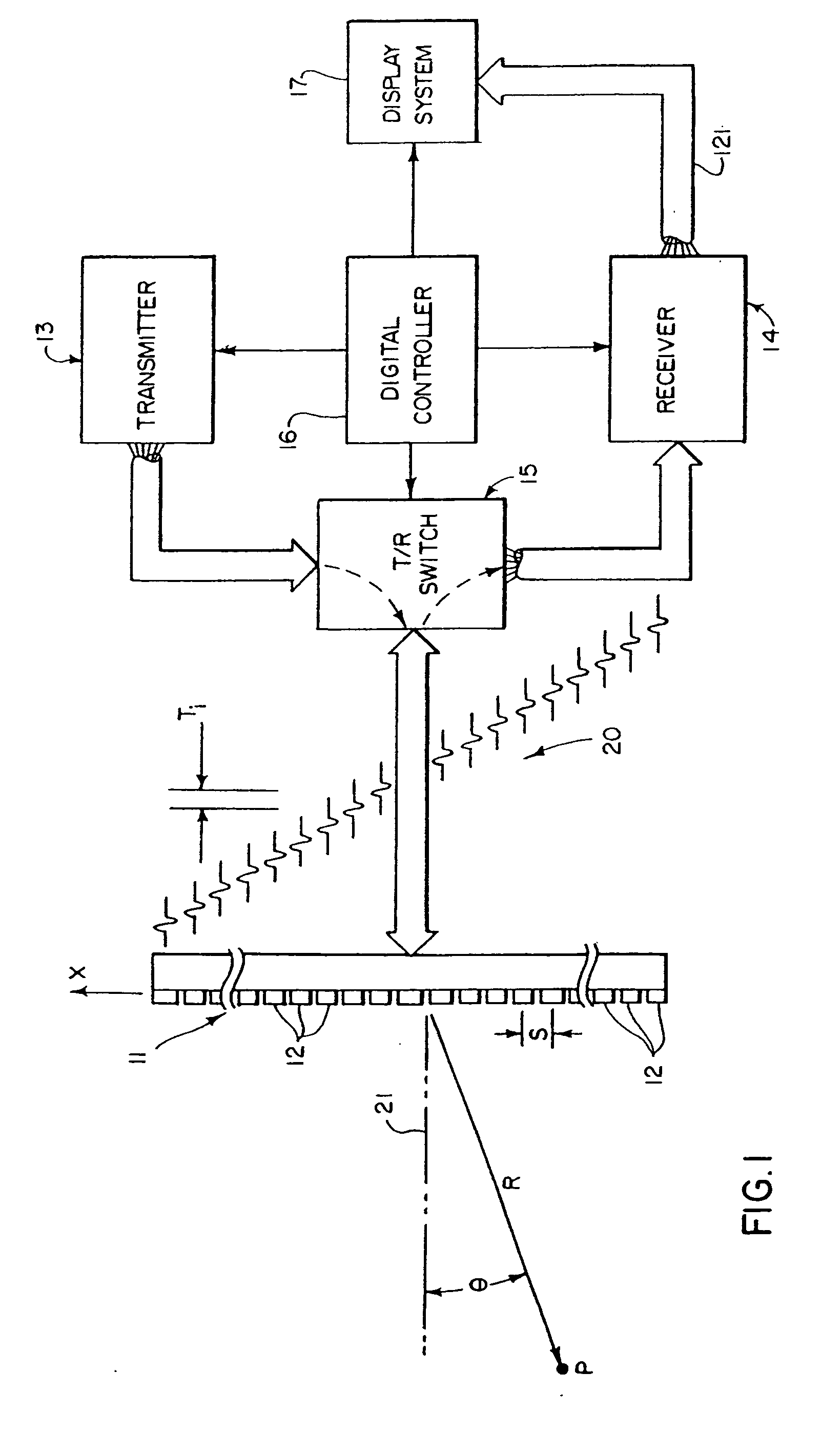
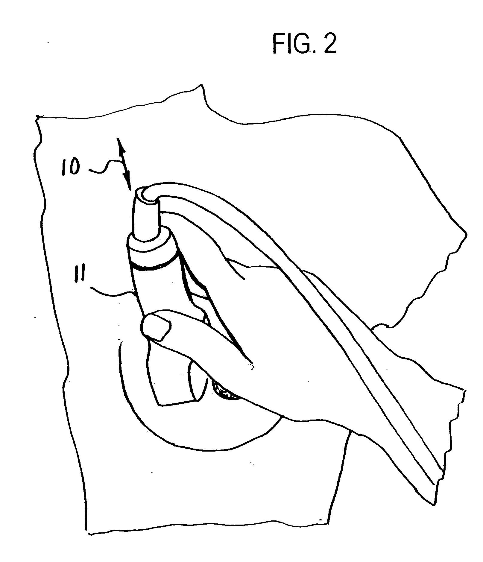
[0015] The present invention is presently implemented using a real-time, freehand strain imaging method on a commercially available ultrasound system (Elegra scanner and 7.5L40 linear array transducer 11 sold by Siemens Medical Solutions, Ultrasound Division) and depicted in FIG. 1. The ultrasound imaging is performed at 7.2 MHz by a sonographer while applying freehand, periodic, gentle axial, loading and unloading to the tissues of interest with the transducer 11 as depicted by the arrow 10 in FIG. 2.

[0016] Referring particularly to FIG. 1, the ultrasonic imaging system includes a transducer array 11 comprised of a plurality of separately driven piezoelectric elements 12 which each produce a burst of ultrasonic energy when energized by a pulse produced by a transmitter 13. The ultrasonic energy reflected back to the transducer array 11 from the subject under study is converted to an electrical signal by each transducer element 12 and applied separately to a receiver 14 through a s...


PUM


Abstract

A series of ultrasound strain images of a breast lesion are acquired along with corresponding B-mode images using a real-time ultrasound strain imaging system and a free-hand technique. A visual assessment of the lesion is made by the sonographer after image acquisition. A conspicuity metric is calculated from the strain images based on the weighted sum of lesion contrast values in each strain image. The weighting of each lesion contrast value is based on observed characteristics of malignant lesions in a series of strain images. Diagnosis is made based on the visual assessment and the conspicuity metric

Description

CROSS-REFERENCE TO RELATED APPLICATIONS [0001] This application claims the benefit of U.S. Provisional patent application Ser. No. 60 / 581,137 filed on Jun. 18, 2004 and entitled “VISUAL AND COMPUTER-AIDED ANALYSIS OF REAL-TIME ULTRASOUND STRAIN IMAGE SEQUENCE FOR DIFFERENTIATION OF BREAST LESIONS”.BACKGROUND OF THE INVENTION [0002] Breast cancer is the most common form of cancer in women in the United States and worldwide. According to the World Health Organization (WHO) more than 1.2 million women were diagnosed with breast cancer in 2000. The American Cancer Society (ACS) estimated that 215,000 cases of invasive breast cancer would be diagnosed in the United States and over 40,000 women would die of this disease in 2004. Mammography and clinical breast examination are used to screen for breast cancer. Because both physical findings and mammographic findings of benign and malignant breast abnormalities overlap, screening for breast cancer results in the detection of many benign bre...

Claims


Application Information

Patent Timeline
Patent Type & Authority Applications(United States)
IPC IPC(8): A61B8/00A61B8/08G06T7/00
CPCA61B8/0825A61B8/485G06T2207/30068G06T7/0012G01S7/52042
Inventor HANGIANDREOU, NICHOLAS J.NORDLAND, MICHELLE R.HESLEY, GINA K.MORTON, MARILYN J.CHARBONEAU, J. WILLIAMMEIXNER, DUANE D.
Owner MAYO FOUND FOR MEDICAL EDUCATION & RES
Why Eureka
Try Eureka