Optical examination method and apparatus particularly useful for real-time discrimination of tumors from normal tissues during surgery

a tumor and optical examination technology, applied in the field of optical examination methods and apparatuses, can solve the problems of limited ability to localize tumor tissue by ultrasound once resection begins, inability to precisely visualize tumors, and complicated tumor removal procedures, so as to enhance the optical detection of target portions

Inactive Publication Date: 2006-01-05
IETMED
View PDF13 Cites 66 Cited by
  • Summary
  • Abstract
  • Description
  • Claims
  • Application Information

AI Technical Summary

Benefits of technology

[0014] An object of the present invention is to provide an improved method and apparatus for enhancing the optical detection of target portions of an object to distinguish same from other portions of the object, particularly for enhancing, in a real-time manner, the optical examination of biological tissue to distinguish cancerous tissue from non-cancerous tissue.

Problems solved by technology

However, during the surgical procedure the tumor cannot always be visualized precisely.
This complicates the tumor removal procedure and makes it totally dependent on the judgment of the surgeon, which is based on his / her experience.
However, the ability to localize tumor tissue by ultrasound once resection commences is limited by signal artifacts caused by blood and surgical trauma at the resection margin (LeRoux et al., Lipson et al., 1961).
Thus intraoperative ultrasound is an unsatisfactory tool for assisting the neurosurgeon in efforts to completely resect primary brain tumors.
However, intraoperative biopsies are hampered by the sampling error, the wait for results, and the difficulty in distinguishing tumor cells from reactive astrocytes [Haglund et al, 1994].
Thus, this method is highly impractical.
However, the intraoperative MRI scanner is not an on-line imaging system.
Also, intraoperative MRI machines are bulky, obstruct and limit the surgical field, and impose special requirements on the neurosurgical facility, such as the need for a non-magnetic environment, special non-ferrous surgical instruments, etc.
The cost of operating and maintaining these liquid-helium cooled machines is extremely high, and as a result, there are very few medical centers that are equipped with intraoperative MRI scanners at present.
However, in the operating room these methods are quite difficult to use [Cheng et al.
They are also unreliable primarily due to contamination of the surgical cavity by blood containing the fluorescent marker and leakage of dyes with accompanying edema into surrounding nontumorous tissue [Stummer et al.
Technical difficulties prevented the development of useful in vivo florescent marking systems.
Despite the appeal of the new tumor contrasting method, its current application remains impractical, primarily because equipment other than the microscope itself is required [Kiroiwa, 1998].
Although almost all neurosurgical operations are performed under an operating microscope, there has been no commercially available microscope with which fluorescence could be observed.

Method used


Image

  • Optical examination method and apparatus particularly useful for real-time discrimination of tumors from normal tissues during surgery
  • Optical examination method and apparatus particularly useful for real-time discrimination of tumors from normal tissues during surgery
  • Optical examination method and apparatus particularly useful for real-time discrimination of tumors from normal tissues during surgery

Examples


Embodiment Construction

[0040] As indicated earlier, the invention relates broadly to a method and apparatus for enhancing the optical detection of target portions of an object, and is particularly useful for enhancing the optical examination of biological tissue in order to distinguish cancerous tissue from non-cancerous tissue. The embodiments of the invention described below are therefore directed to the latter application, namely to enhance the optical examination of biological tissue in order to provide real-time tumor visualization during a surgical procedure, particularly a brain neurosurgical procedure, in order to better enable the surgeon to remove all the cancerous tissue with a minimum of non-cancerous tissue.

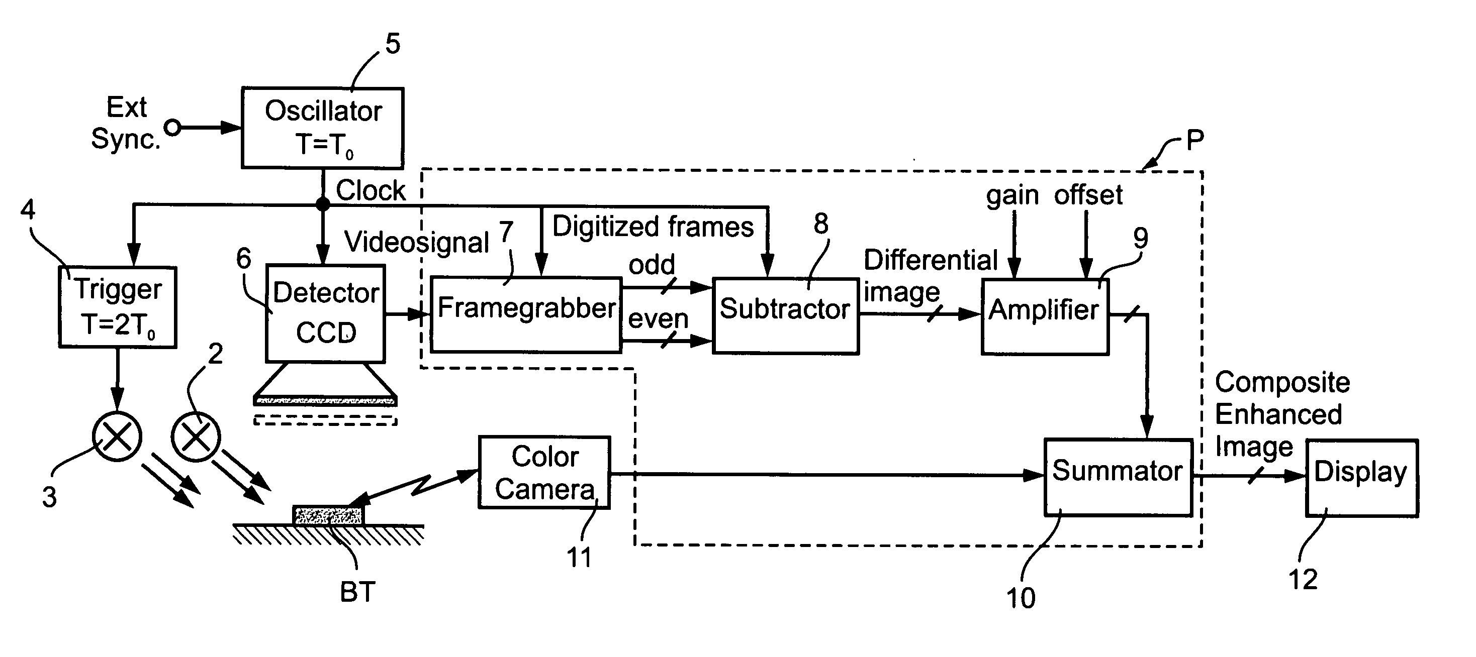
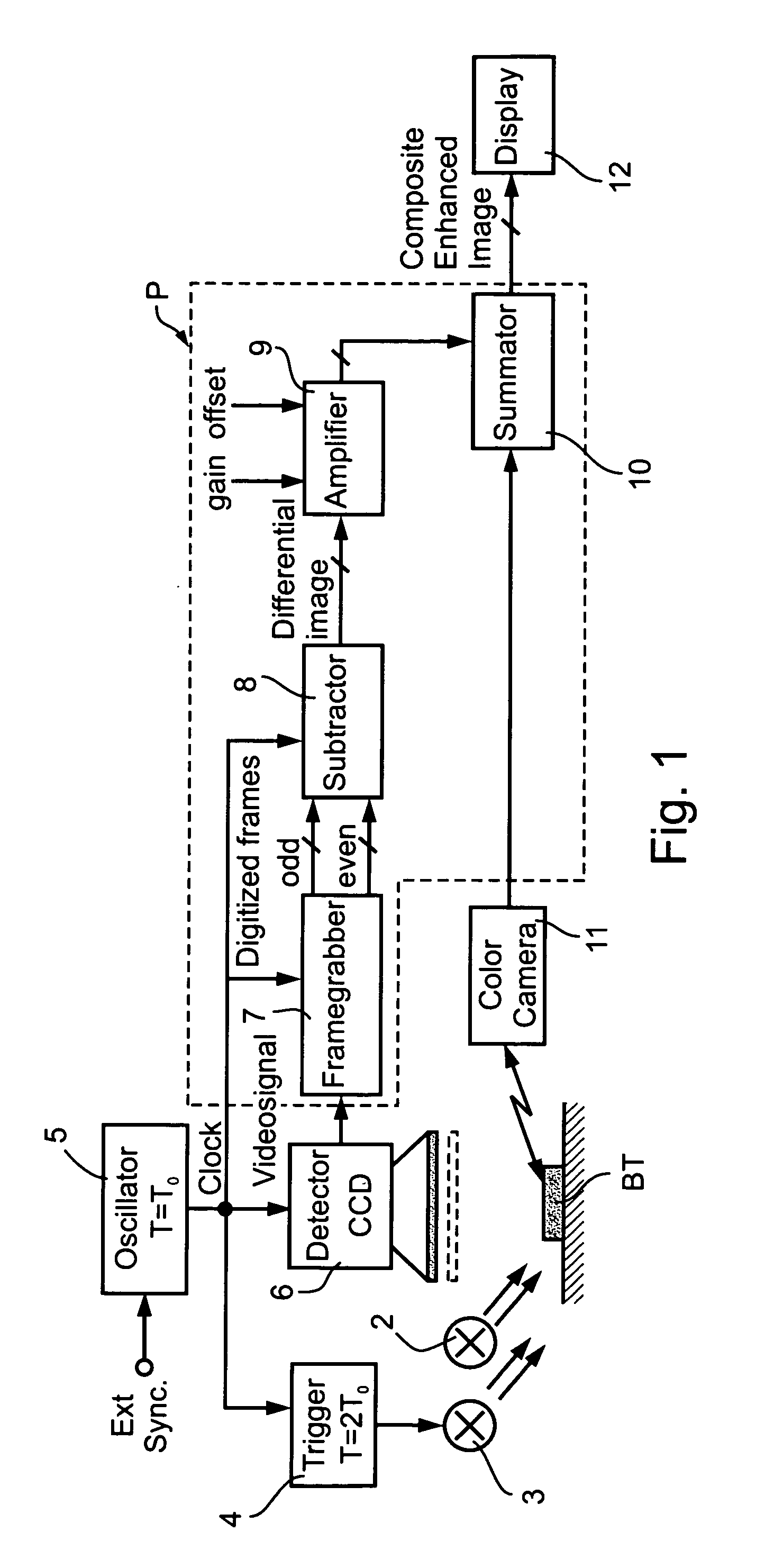
[0041] The apparatus illustrated in FIG. 1 is for use in examining biological tissue BT in a real-time manner while performing a surgical resection procedure. The biological tissue BT is continuously exposed to ambient (e.g., polychromatic or white) light, schematically indicated at 2. In...


PUM


Abstract

A method and apparatus for enhancing the optical detection of target portions of an object particularly useful in the optical examination of biological tissue to distinguish cancerous tissue from non-cancerous tissue. This is accomplished by exposing the object, e.g., biological tissue, to first and second light sources of different spectral contents for short, alternating, first and second time periods; detecting the light received from the object during each of the first and second time periods; and utilizing the light detected in the first and second time periods for producing and displaying a composite image including the image of the object and an enhanced image of the target portions, e.g., the cancerous tissue, overlayed on the image of the object.

Description

RELATED PATENT APPLICATIONS [0001] This is a continuation-in-part of PCT Patent Application No. PCT / IL03 / 01066, filed Dec. 14, 2003, which claims the benefit of U.S. Provisional Patent Application No. 60 / 433,229, filed Dec. 13, 2002. The content of the above applications is incorporated herein by reference in its entirety.FIELD AND BACKGROUND OF THE INVENTION [0002] The present invention relates to a method and apparatus for optically examining objects in order to more clearly discriminate target portions of interest from other portions of the object. The invention is particularly useful for the real-time discrimination of tumors from normal tissues during surgery, especially during brain neurosurgery, and is therefore described below with respect to this application, but it will be appreciated that the invention, or various features thereof, could advantageously be used also in other applications. [0003] The prognosis of patients suffering from malignant gliomas, astrocytomas, meni...

Claims


Application Information

Patent Timeline
Patent Type & Authority Applications(United States)
IPC IPC(8): A61B6/00A61B5/00
CPCA61B5/0071A61B5/4064A61B5/0084A61B5/7425
Inventor BEYLIN, ALEXANDER
Owner IETMED
Why Eureka
Try Eureka