Method, system and storage medium which includes instructions for analyzing anatomical structures

Inactive Publication Date: 2006-02-02
NEW YORK UNIVERSITY
View PDF14 Cites 39 Cited by
  • Summary
  • Abstract
  • Description
  • Claims
  • Application Information

AI Technical Summary

Benefits of technology

[0031] Visual rating of the MTL on MRI scans have shown a good comparability with MRI volume study. Accordingly, visual rating of FDG-PET scans may permit (a) developing a reliable and anatomically valid visual rating scale of MTL hypometabolism on FDG-PET, (b) determining the anatomical validity of the visual MTL rating by comparing the diagnostic capacity of the MTL rating with quantitative metabolic values derived from the gold-standard ROI sampling technique, and (c) comparing the diagnostic accuracy of the MTL rating to the cortical rating in terms of being able to separate clinical groups.
[0032] Generally, the second exemplary embodiment of the method, system and storage medium of the present invention is capable o

Problems solved by technology

The ROI method requires MR scans to guide the PET sampling and time-consuming manual outlining of the hippocampus.
Unfortunately, current implementations typically preserve only the maximum metabolic activity along each projection ray, thus rendering hippocampal hypometabolism invisible to the human observer.
However, few (if any) PET studies using voxel-based or surface projection methods report hippocampal metabolic abnormalities in MCI or AD as compared to controls.
As with the prior discussion of PET studies, such studies rely on estimation of changes in cortical brain metabolism and have not reported data on the hippocampus.
However, the usefulness of cortical hypometabolism in identifying pa

Method used


Image

  • Method, system and storage medium which includes instructions for analyzing anatomical structures
  • Method, system and storage medium which includes instructions for analyzing anatomical structures
  • Method, system and storage medium which includes instructions for analyzing anatomical structures

Examples


first exemplary embodiment

1. First Exemplary Embodiment

Hippocampal Mask (HipMask)

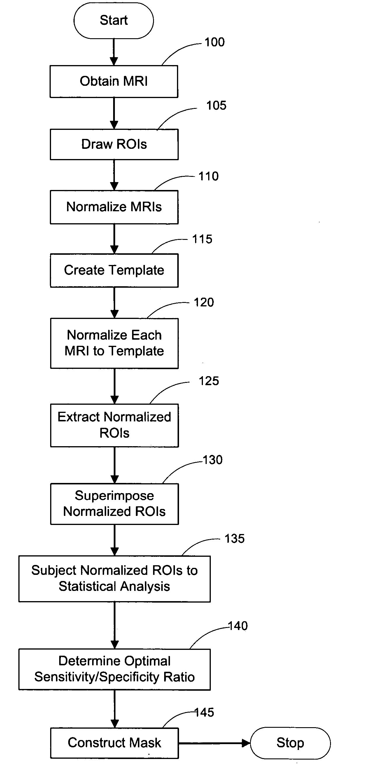
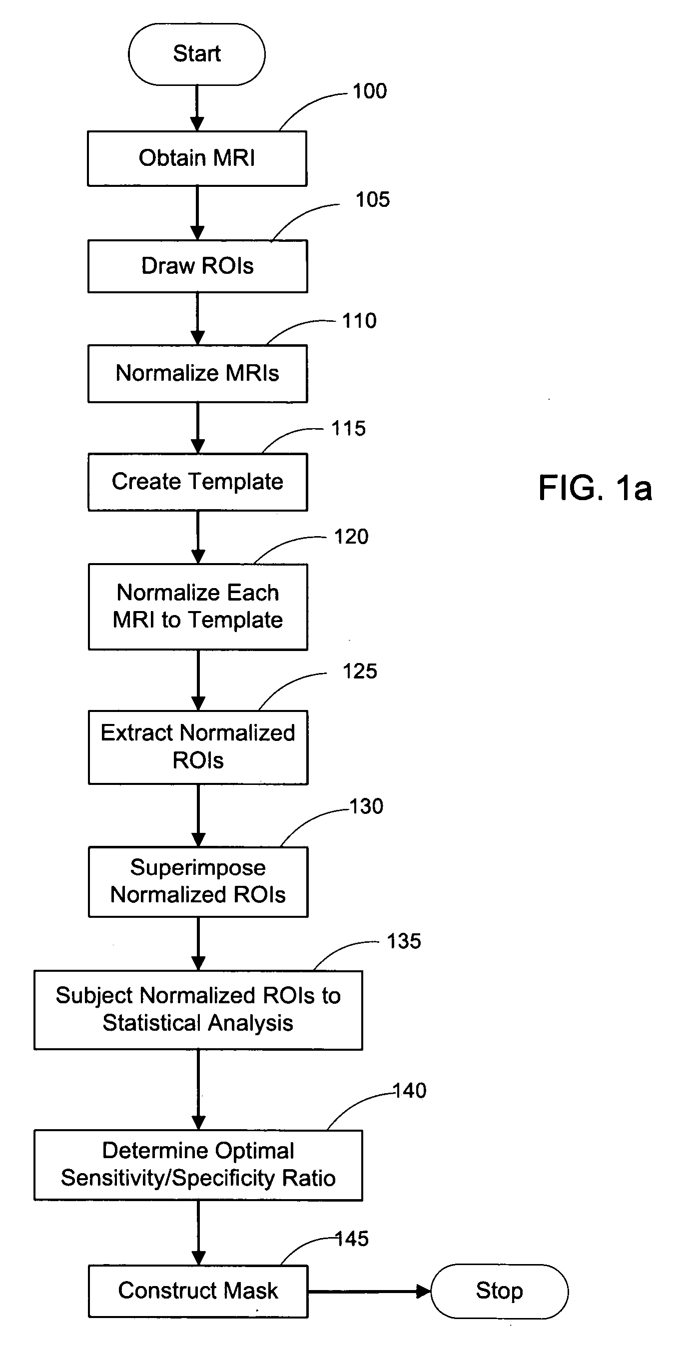
[0053]FIG. 1a depicts a flow diagram of an exemplary embodiment of a method for constructing a HipMask. FIG. 9 depicts an exemplary display of such exemplary method, in which images for the scanned cross-sectional view of a brain is displayed as MRI 10, Hippocampal ROI 20, and HipMask 30. Indeed, the HipMask 30 image can be constructed by utilizing the exemplary method illustrated in the flowchart of FIG. 1. Referencing the exemplary method shown in FIG. 1, after a group of patients of sufficient size are assembled (as discussed below), magnetic resonance images (MRIs) are taken of each patient's brain in operation 100 to create a set of MR scans or MRIs. (The terms “MR scan” and “MRI” are used interchangeably herein.) The MR scanning procedure may employ any scanning parameters that provide a sufficiently precise brain image. Briefly, one embodiment of the present invention may employ MR scans 10 generated by a-1.5 T General E...

second exemplary embodiment

5. Second Exemplary Embodiment

Visual Rating of Medial Temporal Lobes

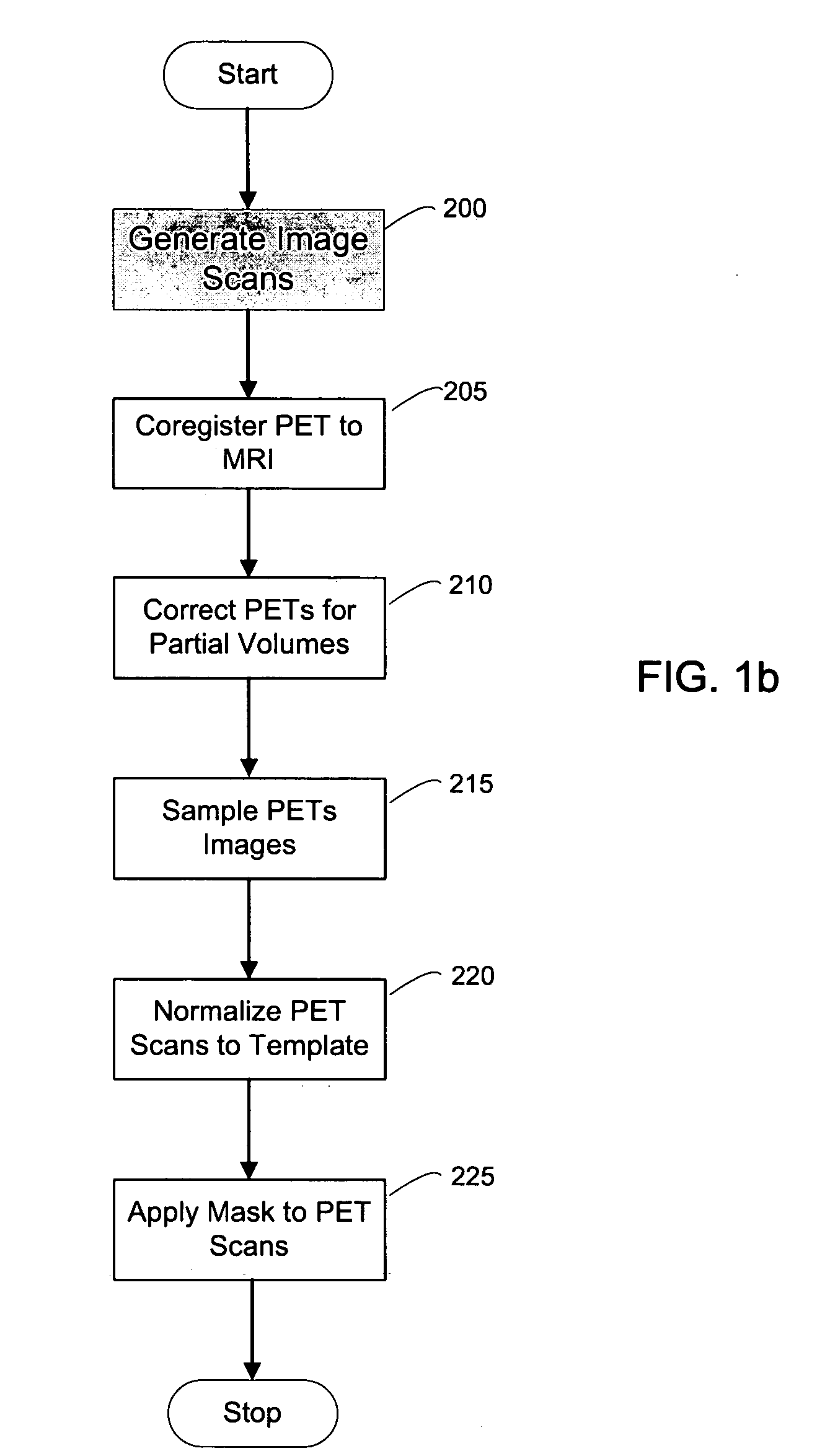
[0131] A second exemplary embodiment of the method, system and storage medium according to the present invention is provide for evaluating and analyzing brain tissue shown on the PET scans 60. Similar to the first exemplary embodiment, the second exemplary embodiment may be broadly applicable to evaluation and analysis of imaged brain tissue, such as that shown in the PET scan 60.

[0132] For example, according to this embodiment, a standardized MRI scan protocol can be conducted on a set of patients to provide a set of MRIs 10. The MRI scanning procedures have been previously described above, as well as in certain references known to those of ordinary skill in the art.

[0133] For example, the MRI scans may be acquired on a 1.5 T General Electric Signa imager (General Electric, Milwaukee, USA) using a T1-weighted fast-gradient-echo sequence with repetition time=35 ms, echo time=9 ms, and flip angle 60°. Images may b...


PUM


Abstract

A method, system and storage arrangement are provided for effectuating an evaluation and analysis of anatomical structures, and creating and/or modifying images associated therewith. In particular, at least two images associated with the anatomical structure can be normalized so as to produce normalized image. A normalized set of regions of interest can be obtained based on the normalized images. Each of the normalized set of regions of interest may be analyzed to provide analysis data. Further, the anatomical structure mask may be created and/or modified based on the analysis data. Another embodiment provides for rating and analyzing anatomical structures.

Description

CROSS-REFERENCE TO RELATED APPLICATION(S) [0001] The present application claims priority from U.S. patent application Ser. No. 60 / 588,647 filed Jul. 16, 2004 and U.S. patent application Ser. No. 60 / 691,715 filed Jun. 16, 2005, the entire disclosures of which are incorporated herein by reference.FIELD OF THE INVENTION [0002] The present invention relates to a method, system and storage medium which includes instructions for analyzing anatomical structures (brain tissue and metabolism). More particularly, this analysis can include sampling and analysis of the hippocampus and medial temporal lobe, as well as an application of the sampling and analysis to assessing the presence of mild cognitive impairment and Alzheimer's disease, including in persons not yet displaying symptoms. BACKGROUND INFORMATION [0003] The following abbreviations will be used throughout the specification as follows: [0004]“AD”—Alzheimer's disease [0005]“MCI”—mild cognitive impairment [0006]“GDS”—global deteriorat...

Claims


Application Information

Patent Timeline
IPC IPC(8): A61B5/05
CPCG06T7/0012G06T7/0022G06T7/0083G06T2207/30016G06T2207/10088G06T2207/10104G06T2207/20104G06T7/0087G06T7/12G06T7/143G06T7/97
Inventor DE LEON, MONY J.MOSCONI, LISATSUI, WAI-HONRUSINEK, HENRY
Owner NEW YORK UNIVERSITY
Why Eureka
Try Eureka