System and method for 3D visualization of lung perfusion or density and statistical analysis thereof

a three-dimensional visualization and density technology, applied in image analysis, image enhancement, instruments, etc., can solve the problems of affecting the quality of life of patients, and putting a strain on the hear

Active Publication Date: 2006-03-16
SIEMENS MEDICAL SOLUTIONS USA INC
View PDF6 Cites 36 Cited by
  • Summary
  • Abstract
  • Description
  • Claims
  • Application Information

AI Technical Summary

Benefits of technology

[0010] The present invention overcomes the foregoing and other problems encountered in the known teachings by providing a system and method for 3D visualization of lung perfusion or de

Problems solved by technology

A pulmonary embolism occurs when a piece of a blood clot from a deep vein thrombosis (DVT) breaks off and travels to an artery in a lung where it blocks the artery, damages the lung and puts a strain on the heart.
This short-term complication is potentially life threatening and occurs in about ten percent of patients with acute DVT events.
However, a large pulmonary embolism can block almost all of the blood flow to a portion of the lungs and cause su

Method used


Image

  • System and method for 3D visualization of lung perfusion or density and statistical analysis thereof
  • System and method for 3D visualization of lung perfusion or density and statistical analysis thereof
  • System and method for 3D visualization of lung perfusion or density and statistical analysis thereof

Examples


Embodiment Construction

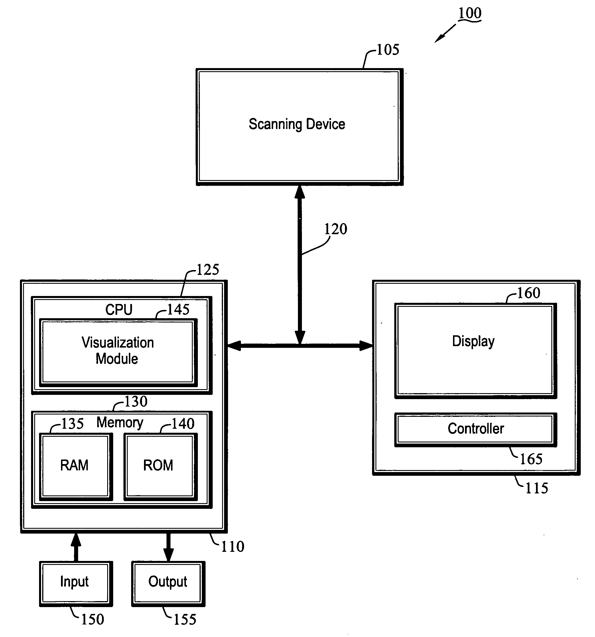
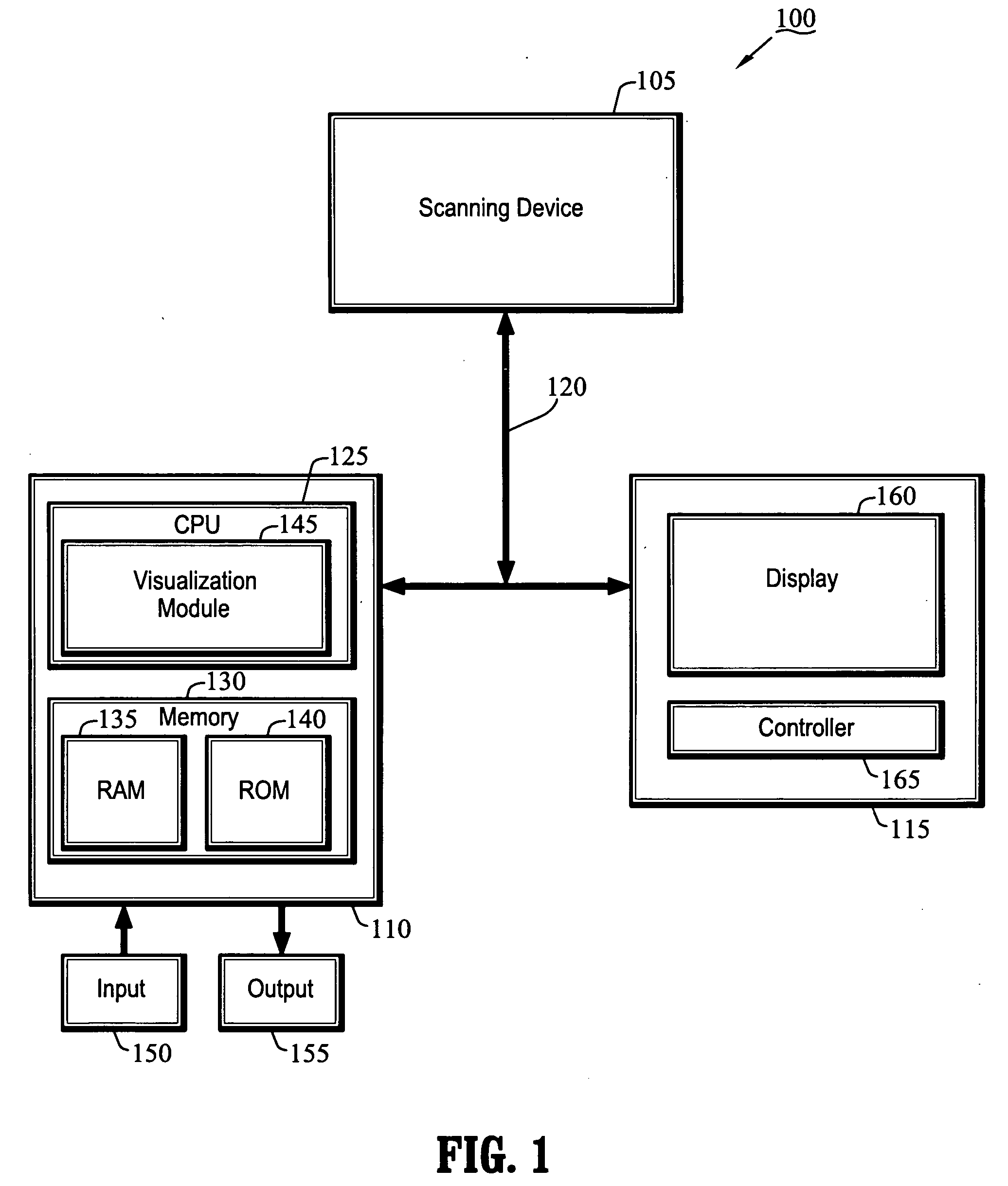
[0038]FIG. 1 is a block diagram of a system 100 for 3D visualization of lung perfusion or density according to an exemplary embodiment of the present invention. As shown in FIG. 1, the system 100 includes, inter alia, a scanning device 105, a personal computer (PC) 110 and an operator's console 115 connected over, for example, an Ethernet network 120. The scanning device 105 may be a magnetic resonance (MR) imaging device, a CT imaging device, a helical CT device, a positron emission tomography (PET) device, a 2D or 3D fluoroscopic imaging device, a 2D, 3D, or four-dimensional (4D) ultrasound imaging device, or an x-ray device. The scanning device 105 may also be a hybrid imaging device capable of CT, MR, PET or other imaging techniques.

[0039] The PC 110, which may be a workstation, portable or laptop computer, a personal digital assistant (PDA), etc., includes a central processing unit (CPU) 125 and a memory 130, which are connected to an input 150 and an output 155. The CPU 125 i...


PUM


Abstract

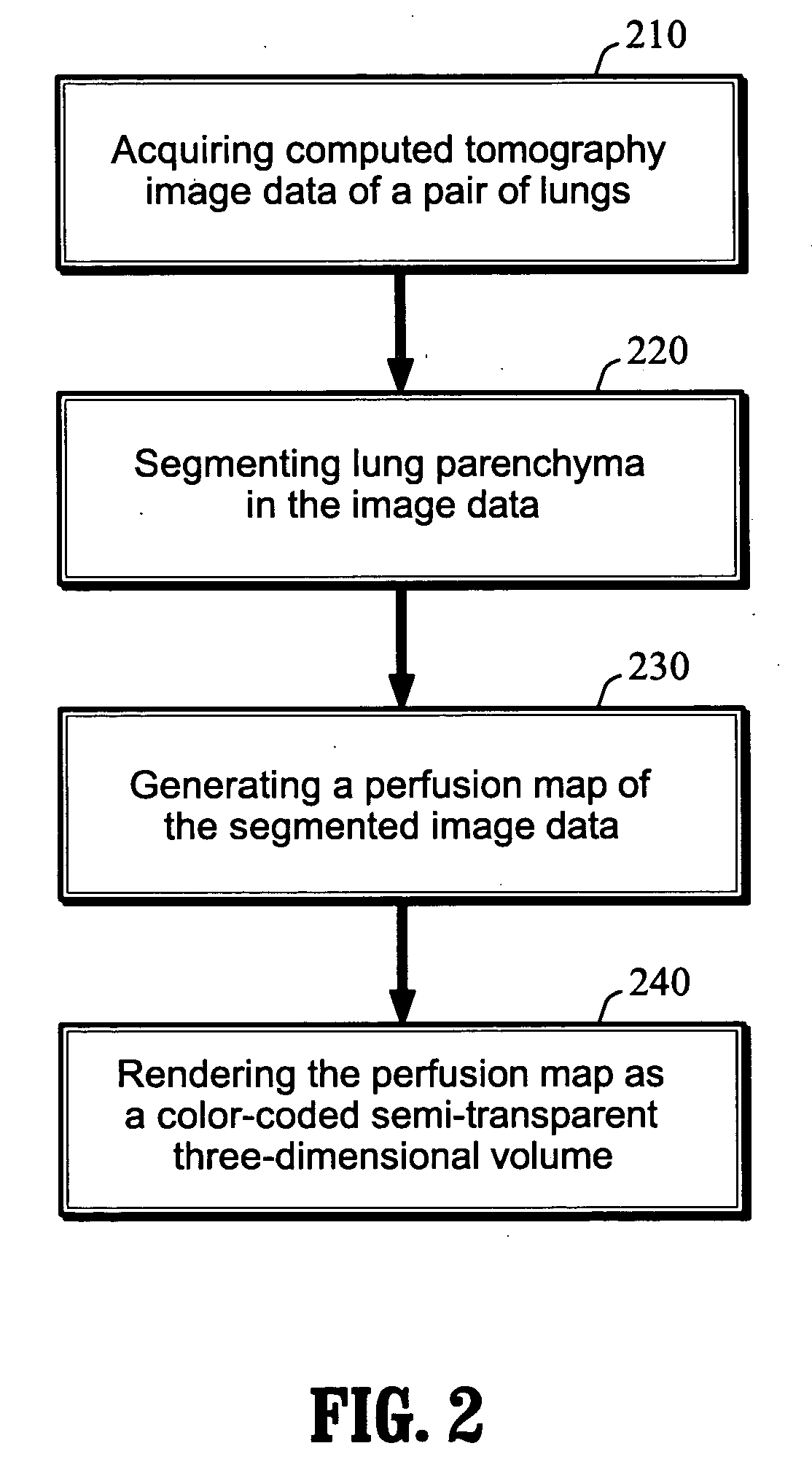
A system and method for 3D visualization of a pair of lungs are provided. The method comprises: segmenting image data of the pair of lungs and lung parenchyma; generating a 3D map as a function of the segmented image data; and rendering the 3D map as a color-coded semi-transparent 3D volume, wherein an opaque region highlights an area of interest.

Description

CROSS-REFERENCE TO RELATED APPLICATION [0001] This application claims the benefit of U.S. Provisional Application No. 60 / 600,257, filed Aug. 10, 2004, a copy of which is herein incorporated by reference.BACKGROUND OF THE INVENTION [0002] 1. Technical Field [0003] The present invention relates to medical image analysis, and more particularly, to a system and method for three-dimensional (3D) visualization of lung perfusion or density and a method for analyzing lung perfusion or density distribution in patients for diagnosis. [0004] 2. Discussion of the Related Art [0005] A pulmonary embolism occurs when a piece of a blood clot from a deep vein thrombosis (DVT) breaks off and travels to an artery in a lung where it blocks the artery, damages the lung and puts a strain on the heart. This short-term complication is potentially life threatening and occurs in about ten percent of patients with acute DVT events. It may be even more common than generally realized because a majority of embol...

Claims


Application Information

Patent Timeline
IPC IPC(8): G06K9/34G06T15/08
CPCG06T7/0012G06T7/0081G06T7/0091A61B6/507G06T2207/10072G06T2207/20148G06T2207/30061G06T15/08G06T7/11G06T7/136G06T7/155
Inventor VAZ, MICHAELKIRALY, ATILLA PETERNOVAK, CAROL L.
Owner SIEMENS MEDICAL SOLUTIONS USA INC
Why Eureka
Try Eureka