Method for electrically detecting oligo-nucleotides with nano-particles

a nano-particle and nucleotide technology, applied in the field of electrically detecting oligo-nucleotides with nano-particles and electrodes, can solve the problems of increasing experiment time and further tasks, slowing the detection process, etc., and achieves the effect of short time and high sensitivity

Inactive Publication Date: 2006-03-16
THINKFAR NANOTECH CORP
View PDF2 Cites 167 Cited by
  • Summary
  • Abstract
  • Description
  • Claims
  • Application Information

AI Technical Summary

Benefits of technology

[0009] The present invention provides a method for electrically detecting oligo-nucleotides with nano-particles, by which a small scale of samples can be used for detecting with high sensitivity in a short time, and the present invention also provides a portable device for detecting oligo-nucleotides.

Problems solved by technology

However, the procedure aforecited raises further tasks before a simple detection can be undertaken, and these will slow the detection process.
Nevertheless, nano particles and portions of nucleotides have to be pre-treated in the conventional use, and this increases experiment time and further tasks.

Method used


Image

  • Method for electrically detecting oligo-nucleotides with nano-particles
  • Method for electrically detecting oligo-nucleotides with nano-particles
  • Method for electrically detecting oligo-nucleotides with nano-particles

Examples


example 1

Substrate Activation and Monolayer of Gold Particles Formation

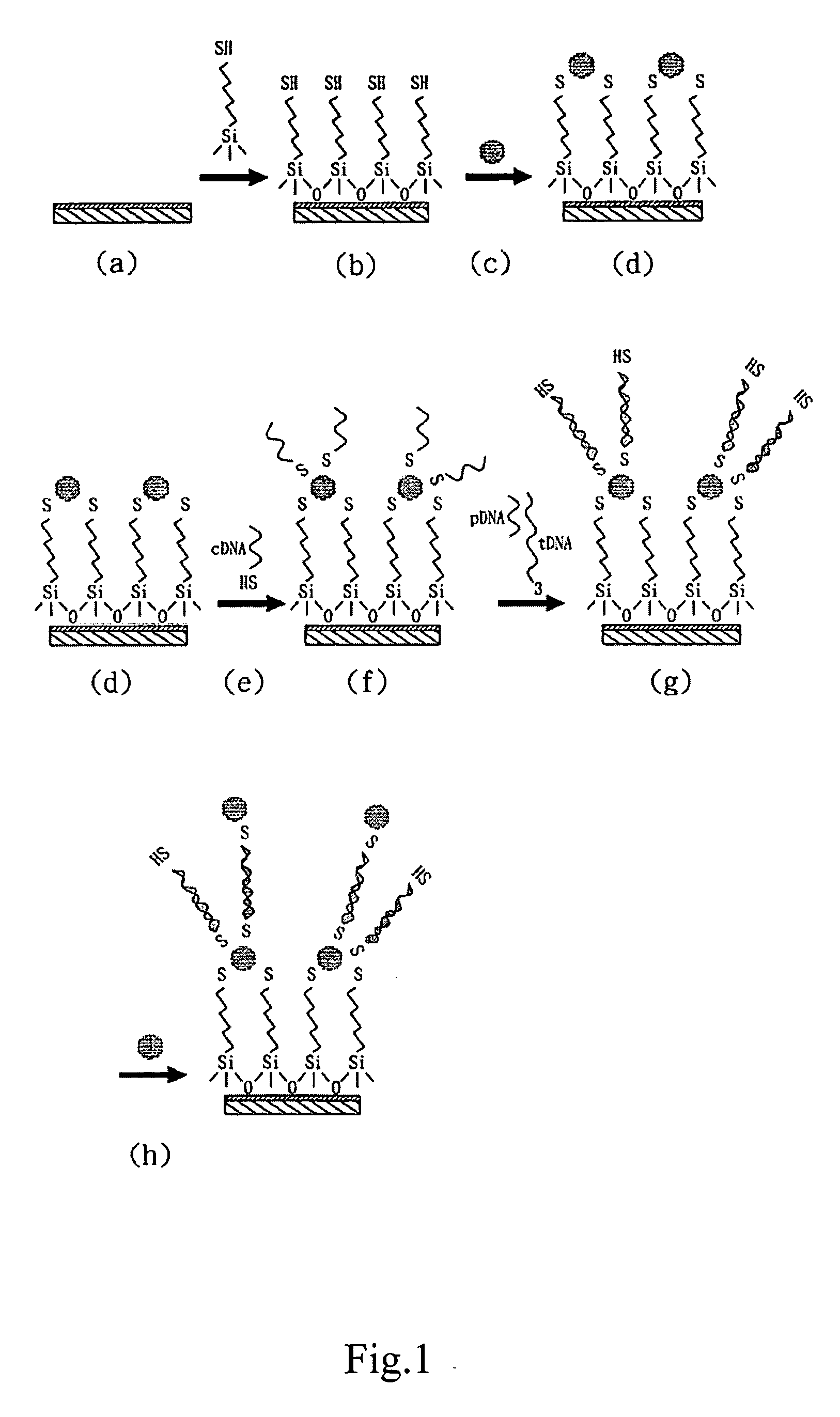
[0027] In reference to FIG. 1(a), a substrate mounted with two electrodes is provided, and the gap between the two electrodes is 300-600 nm.

[0028] Immerse the substrate into a solution with equal volume of concentrate sulfuric acid and methanol for 30 minutes, and wash the substrate. Rinse the substrate with non-ionic water (over 18 ΩW cm). Immerse the substrate into concentrate sulfuric acid for 5 minutes and wash it with water again. Place the substrate in boiled non-ionic water for several minutes and begin the procedures of substrate activation.

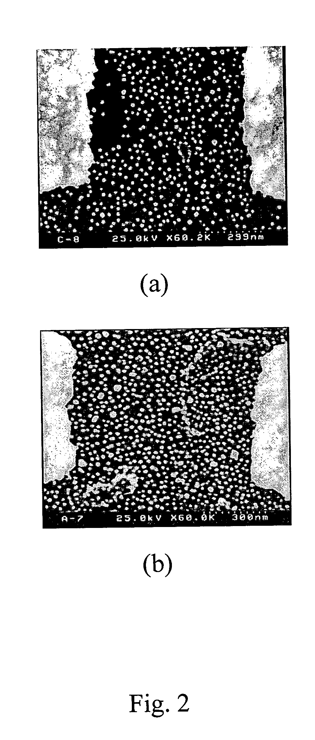
[0029] Prepare 1 mM solution of 3-Mercaptopropyl trimethoxysilane (Sigma Chemical Co.) with DMSO. Immerse the substrate into the solution for at least 2 hours at room temperature, rinse the substrate with DMSO and dry in the environment of nitrogen (FIG. 1(b)). Immerse the activated substrate into nano-gold particles solution (AuNPs) for 8-12 hours (FIG. 1(c)), wash the subs...

example 2

Immobilizaton and Hybridizaiton of Oligo-Nucleotides

[0031] Four oligo-nucleotide fragments are designed for the example: capture oligonucleotide (cDNA) as seen in SEQ ID NO. 1; target oligonucleotide (tDNA) as seen in SEQ ID NO. 2; probe oligonucleotide (PDNA) as seen in SEQ ID NO. 3; and single base mismatched target oligonucleotide (m-tDNA) as seen in SEQ ID NO. 4. Wherein, cDNA fragment and pDNA fragment both have a portion which is complementary to a different portion of tDNA.

[0032] In reference to table 1, four fragments are shown with sequences. Nucleotide bases with underlines indicate a complementary portion, and the single base with frame indicates a single base mutant, which leads to mis-match.

TABLE 1Oligo-nucleotidesSequencecDNA3′-HS-A10-CCT AAT AAC AAT-5′tDNA5′GGA TTA TG TTA AAT ATT GAT AAGGAT-3′m-tDNA5′GGA TTA TG TTA AAT ATT GAT AAGGAT-3′pDNA3′-TTA TAA CTA TTC CTA-A10-SH-5′

[0033] In reference to FIG. 1(e), 100 μl of 1 μM cDNA is deaerated with pH 6.6 HEPES(4-(2-hydr...

example 3

Results

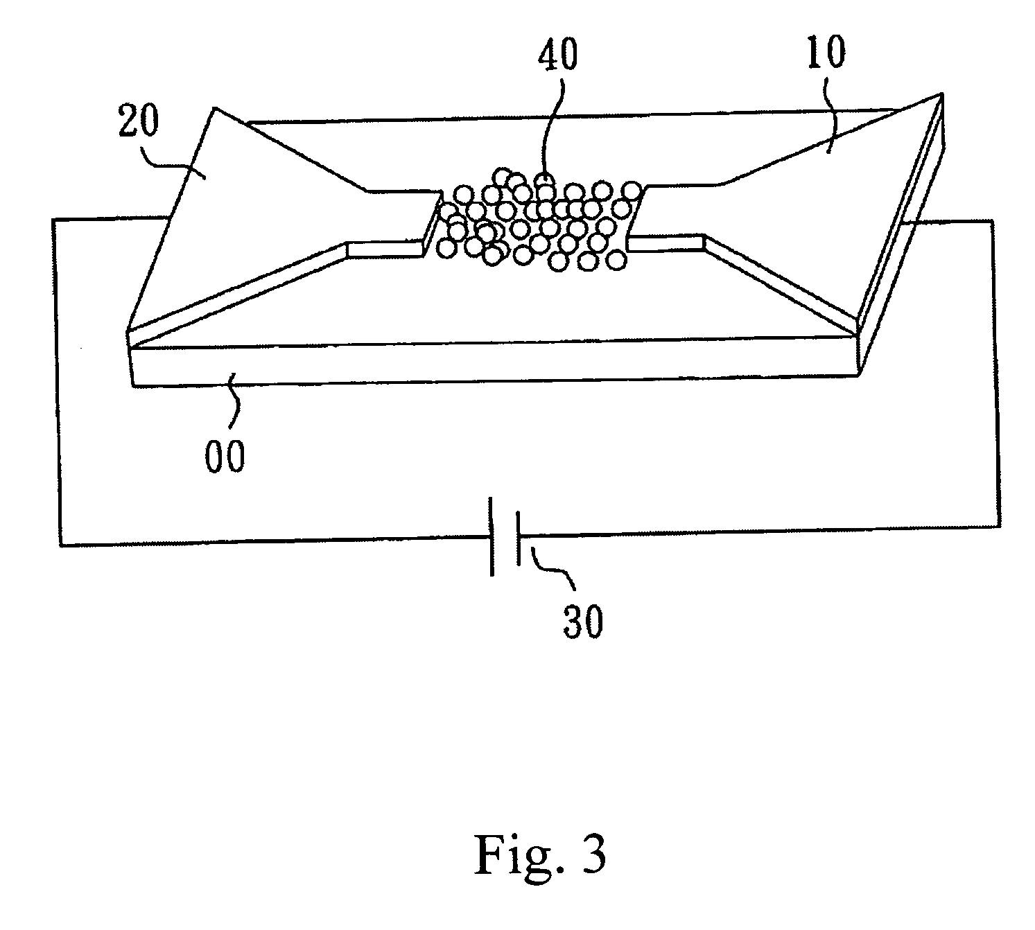
[0037]FIG. 3 shows the device to measure the electrical characteristics on the substrate. A source 10, a drain 20 and a voltage generator 30 are on the substrate 00. When the voltage generator 30 provides a voltage, AuNPs 40 with hybridized oligo-nucleotides gather in the nano-gap of two electrodes 10, 20. Because of the conductivity of gold, the changing electrical behavior is measured. Based on the results, when no AuNPs 40 attach to the nano-gap of two electrodes 10, 20, the current measured is less than 50 mA.

[0038]FIG. 4 (a) shows the current-voltage curves for monolayer of gold particles, scan rate 10 mV / s; (b) shows current-voltage curves for multilayer (hybridization successful) of gold particles, scan rate 10 mV / s. Electrons tunnel more readily through the junction when enough energy is supplied. The linear curve is typical of ohmic devices.

[0039]FIG. 5 shows the I-V curves of the nano-gap electrode measured by using four different concentrations of tDNA: (A) 0.1 ...


PUM

PropertyMeasurementUnit
diameteraaaaaaaaaa
diameteraaaaaaaaaa
currentaaaaaaaaaa

Abstract

A method for detecting a target oligo-nucleotides includes the following steps: providing a substrate mounted with at least a pair of detecting electrodes separated with a gap; coating a surface activation agent on the substrate; providing a plurality of nano-particles and immobilizing them between the two detecting electrodes on the substrate; providing a plurality of capturing oligo-nucleotides; providing a plurality of target oligo-nucleotides and a plurality of probe oligo-nucleotides in order, wherein a portion of the capturing oligo-nucleotides is complementary to the first portion of the sequence of the target oligo-nucleotides; and a portion of the probe oligo-nucleotides is complementary to the second portion of the sequence of the target oligo-nucleotides; and adding a plurality of nano-particles to the gap between the two detecting electrodes.

Description

BACKGROUND OF THE INVENTION [0001] 1. Field of the Invention [0002] The present invention relates to a method for detecting nucleotides with nano particles and electrodes, and more particularly, to a method for minor DNA or RNA sample sequencing and detecting. [0003] 2. Description of Related Art [0004] Biochemical reactions or the detection of human diseases are performed in the molecule level, and the molecules as minor as DNA or RNA have to be purified before the biochemical detection can proceed. Polymerase chain reaction (PCR) is the major equipment currently used to amplify nucleotide molecules, and enlarge samples for detecting with high sensitivity. However, the procedure aforecited raises further tasks before a simple detection can be undertaken, and these will slow the detection process. [0005] A nanoparticle probes with electrical DNA sequencing detector is used to measure electrical characteristics such as resistance values, capability values, current values, frequencies...

Claims


Application Information

Patent Timeline
Patent Type & Authority Applications(United States)
IPC IPC(8): C12Q1/68
CPCC12Q1/6825C12Q2563/155
Inventor CHEN, CHEI-CHIANGTSAI, CHIEN-YING
Owner THINKFAR NANOTECH CORP
Why Eureka
Try Eureka