Because
cancer varies from person to person, no single treatment may be effective for all patients.
Surgery, however, is not a viable option for many patients because of the location and / or type of cancer.
Surgical treatments may also result in complications with
anesthesia or infection, and surgical treatments may have long, painful
recovery periods.
Chemotherapy is not a desirable option for several types of cancers and it can also have many complications.
Increasing the
radiation dose, however, also increases the potential for complications to healthy tissues.
One difficulty of
radiation therapy is that the target often moves within the patient either during or between radiation sessions.
This is not a desirable solution because larger treatment margins generally result in irradiating larger volumes of
normal tissue.
These initial CT images are often not sufficient for carrying out radiation treatments because they do not address the internal motion of the tumor.
For example,
CT scanners are very expensive machines that require dedicated rooms because they use an ionizing energy for imaging the tumor and the gold fiducials.
Additionally, the
CT scanners for obtaining the initial images are typically different than the
CT scanners that are used in the
simulation procedures such that two separate dedicated areas with very expensive machines are required in these applications.
This requires additional time and expense for skilled personnel, and it is also subject to
human error.
This may result in under utilization of the expensive equipment, facilities, and personnel.
Conventional setup procedures for aligning the tumor with the
isocenter of the
radiation beam are also an area of concern because they can be time-consuming and subject to error.
Conventional setup procedures using external marks may be inadequate because the target may move relative to the external marks between the patient planning procedure and the treatment session and / or during the treatment session.
Conventional setup procedures using internal gold fiducials are also generally inadequate because this is a time-consuming process that may produce inaccurate results.
This process is time-consuming and may be unpleasant for the patient because the
technician must vacate the radiation vault while the X-rays are acquired.
This procedure may also be inaccurate because the patient may inadvertently move after taking the stereotactic X-rays such that the tumor is not at the location in the images.
The potential inaccuracy of this process may be exacerbated because a person typically determines the offset by subjectively interpolating the CT images.
Therefore, conventional setup procedures using gold fiducials tie up expensive linear accelerators in the radiation vault for extensive periods of time just to position patients for treatment, and conventional setup procedures may be inaccurate.
Such conventional
verification procedures, however, are subject to errors because the tumor is assumed to be at the
machine isocenter throughout the radiation fraction.
In conventional
radiation therapy systems, the changes in shape or trajectory of the tumor can be determined using additional CT scans, but this requires additional time and use of expensive CT scanners and personnel.
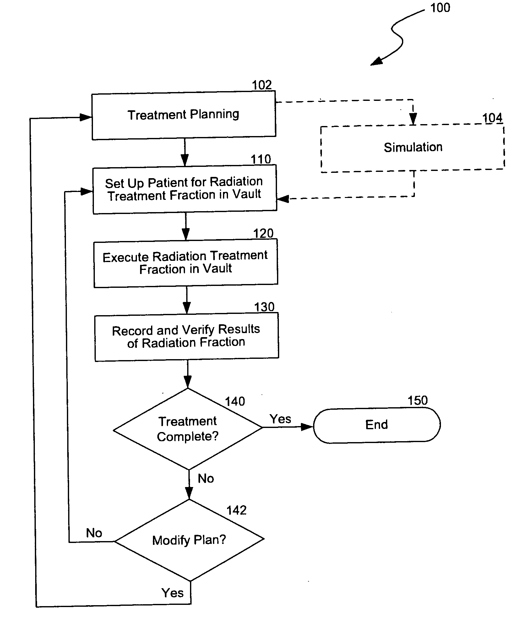
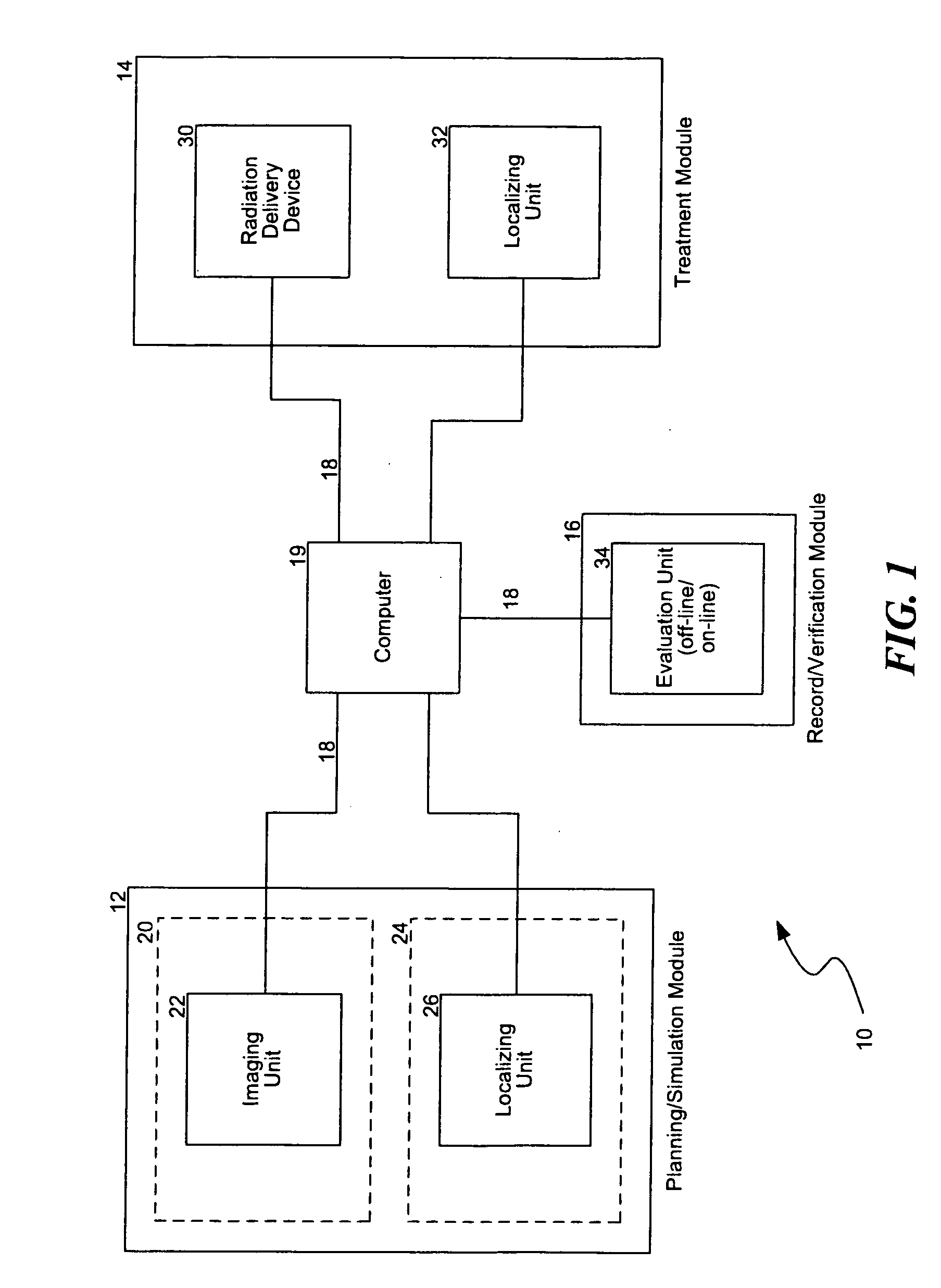
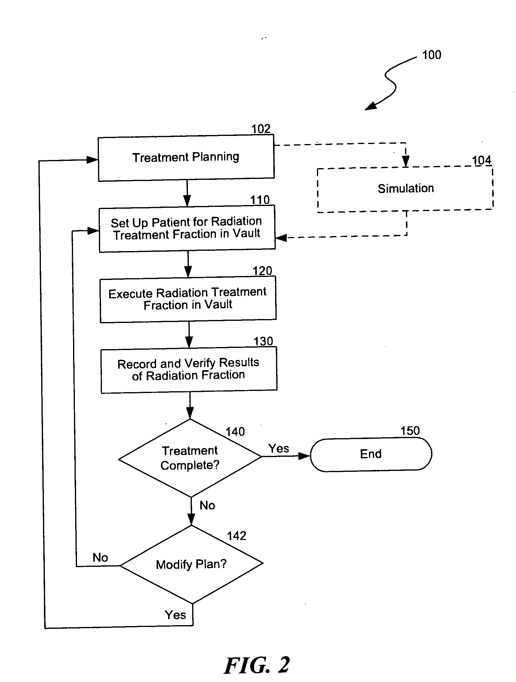
Another challenge of providing
radiation therapy is that the information from the planning,
simulation, setup, treatment, and
verification procedures is typically generated from different equipment in various formats.
This is inefficient because managing the data from the different procedures in a coherent, integrated manner may be difficult.
As such, expensive equipment and highly trained technicians are often under utilized because information is not readily available.

