

System and method for forming a non-ablative cardiac conduction block
a non-ablative, cardiac technology, applied in the direction of depsipeptides, powder delivery, peptide/protein ingredients, etc., can solve the problems of collagen shrinkage, thrombosis, and each is associated with certain adverse consequences, and achieve the effect of enhancing the retention of living cells
- Summary
- Abstract
- Description
- Claims
- Application Information
AI Technical Summary
Benefits of technology
Problems solved by technology
Method used
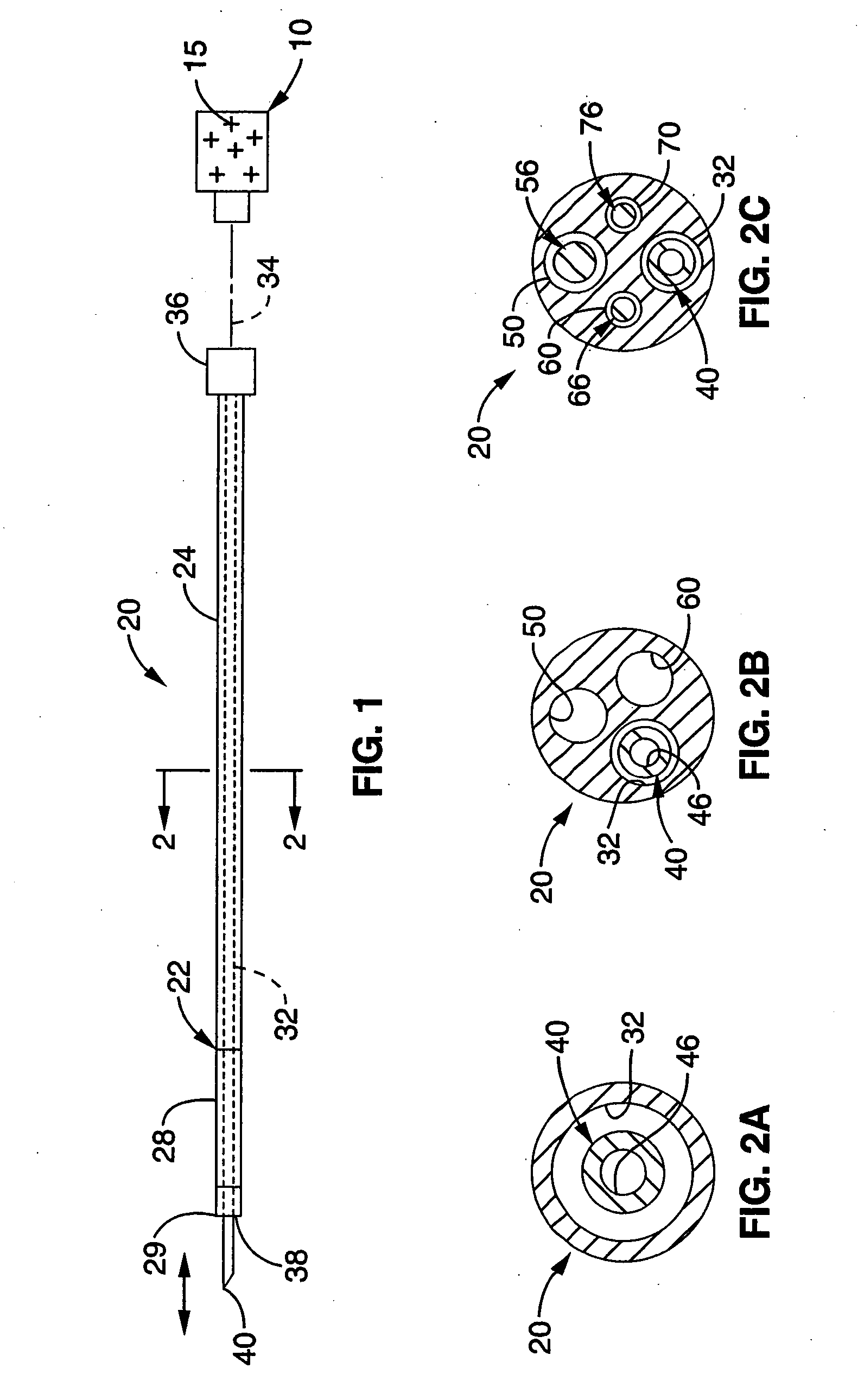
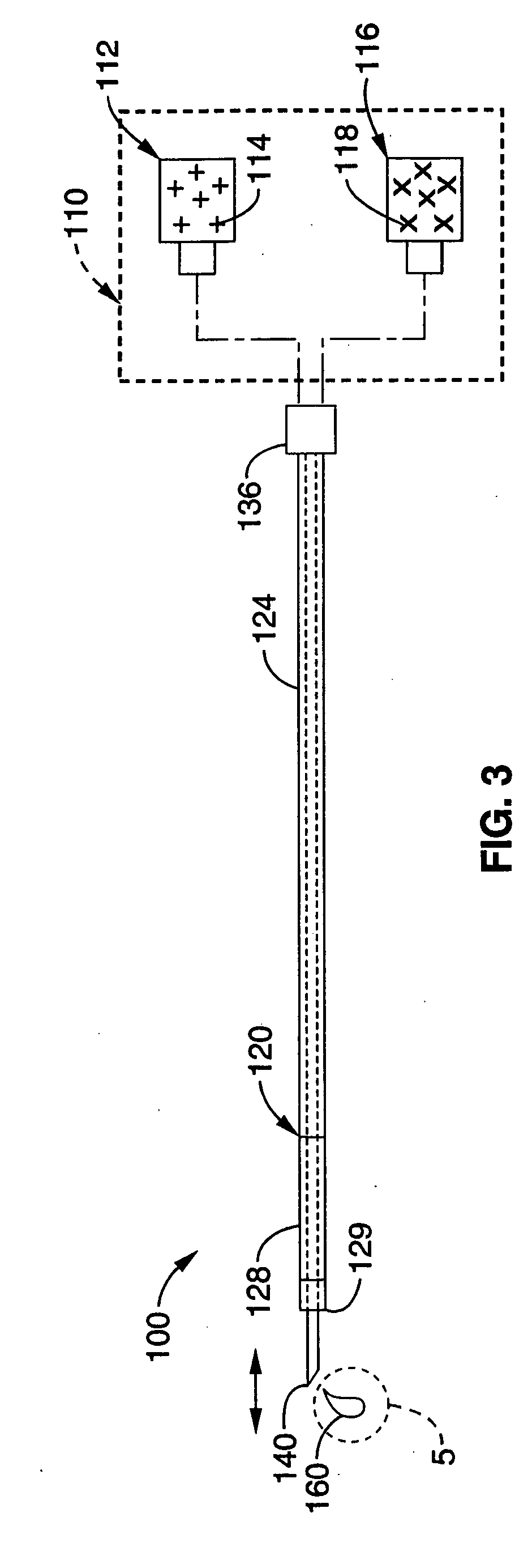
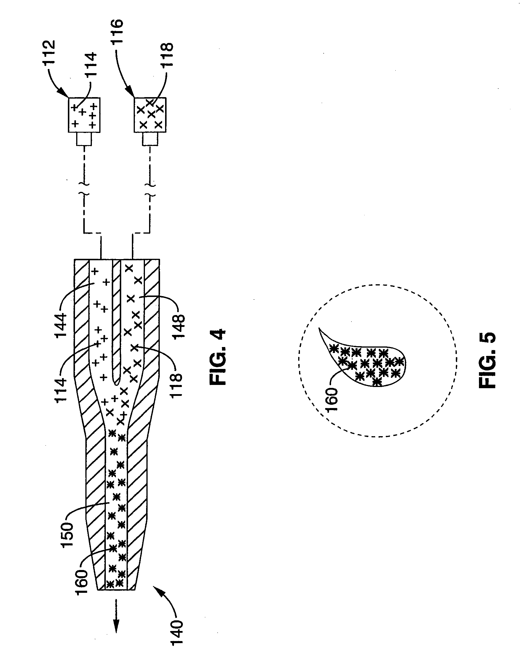
Image

Examples
example 1
[0154] Coupling requirements for successful impulse propagation with skeletal myocytes transplanted in myocardium have been determined by computer modeling as follows in order to determine whether transplanted myocytes can propagate electrical impulses within the myocardium.
[0155] The methods according to this example use computer modeling, which constructed theoretical strands of skeletal and mixed skeletal and ventricular myocytes. The ventricular cells were an adaptation of the dynamic Luo Rudy ventricular cell formulation.
[0156] Results according to this computer modeling study were as follows. In the mixed strand model, cardiac to skeletal coupling requirements were similar to cardiac-cardiac requirements. In contrast, skeletal to cardiac propagation failed at 300 nS, consistent with the need for a high degree of coupling. According to these results, conditions which decrease intercellular coupling appear to have a marked decrease on transmission between transplanted skeletal...
example 2
[0158] To assess the electrophysiologic consequences of skeletal muscle transplantation into the myocardium, an in vivo model was used to assess cardiac conduction. The feasibility of gene transfer to specific areas of the cardiac conduction system has been previously demonstrated (Lee et al. 1198 PACE 21-II: 606; Gallinghouse et al. November 1996 Am Heart Assoc.; U.S. Pat. No. 6,059,726). For example, the highly efficient and specifically localized expression of recombinant beta galactosidase in the AV node of rats and pigs has been described. The accuracy and reproducibility of AV nodal injections has been validated by the production of AV block in rats (Lee et al. 1998 J Appl Physiol. 85(2): 758-763). As an electrically insulated conduit for electrical transmission between the atrium and the ventricle, the AV conduction axis is in a strategic position for the study of cardiac electrophysiology.
[0159] To observe the effect of skeletal muscle transplantation on conduction and in p...
example 3
[0164] In this study skeletal muscle was chosen as a test form of cell therapy for transplantation into the myocardium in arrhythmic animals to observe for antiarrhythmic effects.
[0165] The materials and methods used according to this study were as follows. Neonatal skeletal myoblasts were isolated as previously described by enzymatic dispersion from 2-5 days old Sprague Dawley neonatal rats and cultured as previously described (Rando, T., and Blau, H. M. (1994), J. Cell Biol. 125, 1275-1287). After isolation, cells were cultured with growth medium (GM) (80% F-10 medium (GIBCO BRL), 20% FBS (HyClone Laboratories, Inc.), penicillin G 100 U / ml and streptomycin 100 ug / ml,bFGF 2.5 ng / ml(human, Promega Corp)). Skeletal myoblasts were maintained in GM medium in humidified 95% air and 5% CO2 until used for transplantation.
[0166] Sprague-Dawley rats underwent 30 minutes of left coronary artery occlusion and 2 hours of reperfusion. One week following the creation of a myocardial infarction...
PUM
| Property | Measurement | Unit |
|---|---|---|
| temperatures | aaaaa | aaaaa |
| temperatures | aaaaa | aaaaa |
| time | aaaaa | aaaaa |
Abstract
Description
Claims
Application Information