Volume rendering apparatus and method

a volume rendering and volume technology, applied in the field of volume rendering, can solve the problems of high computational intensity of medical volume rendering, difficulty in implementing a practical gpu-based medical image renderer, and insufficient processing power of modern general purpose computers, so as to achieve low additional cost and increase the speed of rendering

Inactive Publication Date: 2006-06-01
TOSHIBA MEDICAL VISUALIZATION SYST EURO
View PDF2 Cites 39 Cited by
  • Summary
  • Abstract
  • Description
  • Claims
  • Application Information

AI Technical Summary

Benefits of technology

[0013] By conceptually dividing the volume data into blocks, the processing power of the GPU can be employed to render MPR images notwithstanding the modest memory available to a typical GPU. Furthermore, by scheduling the transfer of blocks which the CPU predicts are likely to be needed in the future, the effects of inconsistent performance associated with irregular bus traffic with known schemes for GPU-based rendering of medical image volume data are reduced and more rapid rendering of sequences of images can be performed.
[0036] A cache of volume data blocks can be maintained on the graphics processor to accelerate rendering of subsequent cross-sectional images.

Problems solved by technology

Medical volume rendering is thus highly computationally intensive and the processing power of a modern general purpose computer's CPU is often inadequate for performing the task at an acceptable speed.
However, while a GPU might have sufficient raw computing power to perform medical image rendering, it is nonetheless a difficult task to implement a practical GPU-based medical image renderer.
Difficulties arise because the medical image volume data is typically larger than the memory available on the graphics card supporting the GPU, and because of the limited bandwidth available for the transfer of data from the system memory associated with the CPU to the graphics card.
Further difficulties arise because volume data are generally stored linearly in system memory.
This difficulty is especially important in MPR volume rendering because MPR rendering frequently requires access to voxels arranged in an arbitrarily oriented plane which includes voxels spread throughout system memory and not in a contiguous series which would be more easy to access.
Although for a given MPR view it is possible to duplicate the volume data in system memory in a more appropriate order, this is generally undesirable because of the cost in memory overheads.
This can lead to stilted and jerky performance, especially during real-time cine.

Method used


Image

  • Volume rendering apparatus and method
  • Volume rendering apparatus and method
  • Volume rendering apparatus and method

Examples


Embodiment Construction

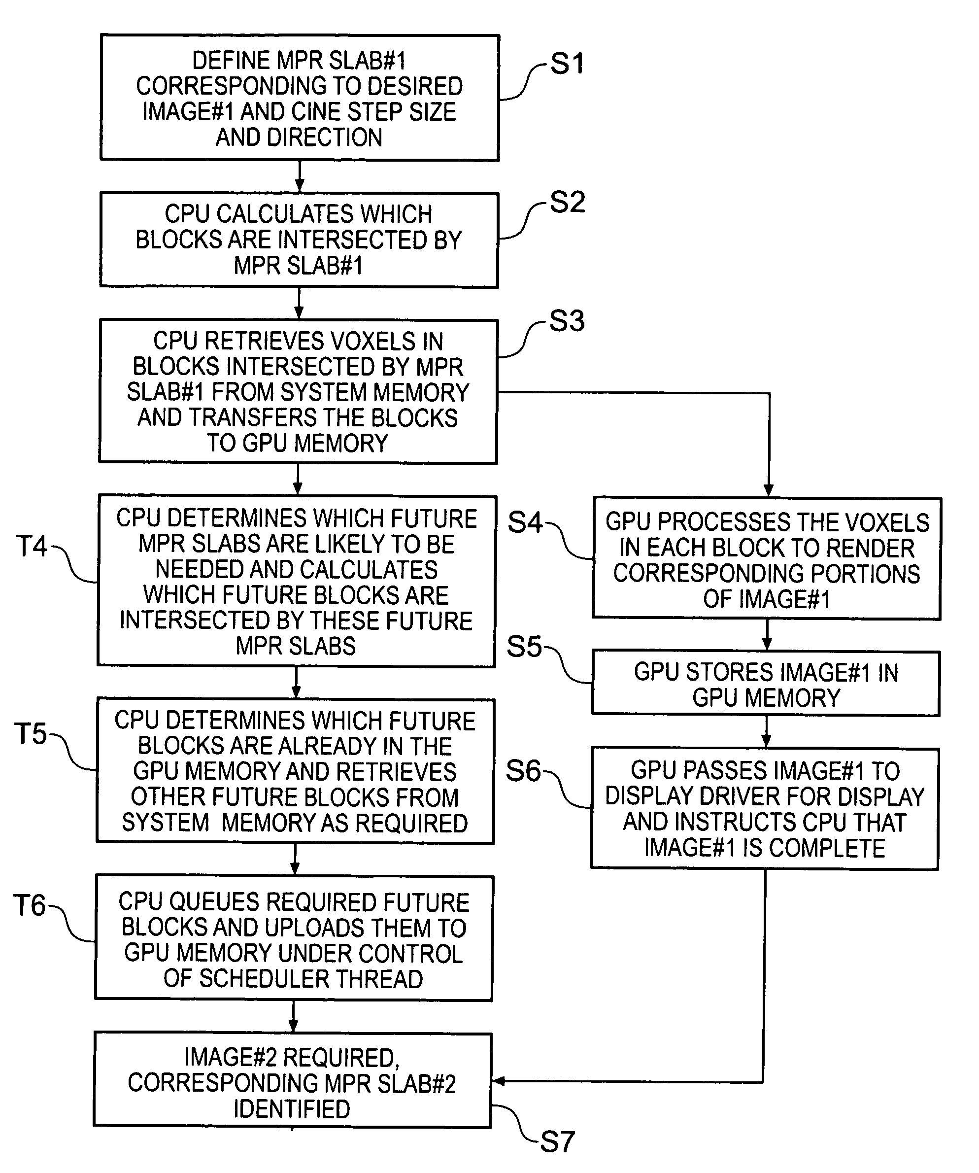
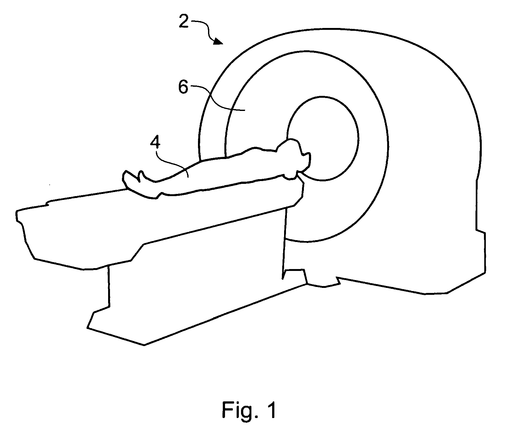
[0058]FIG. 1 is a schematic perspective view of a generic MR scanner 2 for obtaining a 3D scan of a region of a patient 4. An anatomical feature of interest (in this case a head) is placed within a circular opening 6 of the NMR scanner 2 and a series of image slices through the patient is taken. Raw image data are derived from the MR scanner and could comprise a collection of one thousand 2D 512×512 data subsets, for example. These data subsets, each representing a slice of the region of the patient being studied, are combined to produce volume data. The volume data comprise a collection of voxels each of which corresponds to a pixel in one of the slices. Thus the volume data are a 3D representation of the feature imaged and various user-selected 2D projections (output images) of the 3D representation can be displayed (typically on a computer monitor).

[0059] Different imaging modalities (e.g. CT, MR, PET, ultrasound) typically provide 15 different image resolutions (i.e. voxel size...


PUM


Abstract

An apparatus and method for rendering multiplanar reformatting (MPR) images of volume data to be displayed to a user. The apparatus may comprise a conventional personal computer system having a central processing unit (CPU) coupled to a system memory for storing the volume data and a graphics processing unit (GPU) having a GPU memory connected to the computer bus. The computer system CPU is configured to predict an MPR image which may be required for display at a future time and to identify blocks of voxels comprising the volume data which will be needed to render the predicted MPR image. The CPU is further operable to retrieve these blocks from the system memory and to queue them for transfer to the GPU memory. The transfer of blocks from the queue to the GPU memory is controlled by a scheduler such that at least some of the queued blocks are transferred to the GPU memory prior to the predicted MPR image becoming required for display. The GPU retrieves blocks from the GPU memory and renders corresponding image parts for assembly into the predicted MPR image should it become required for display.

Description

BACKGROUND OF THE INVENTION [0001] The invention relates to volume rendering, in particular to multi-planar reformatting (MPR) using a computer system that includes a graphics processing unit (GPU). [0002] MPR volume rendering is a standard method of displaying two-dimensional (2D) representations of three-dimensional (3D) data sets collected by medical imaging equipment, such as computer-assisted tomography (CT) scanners, magnetic resonance (MR) scanners, ultrasound scanners and positron-emission-tomography (PET) systems. These 3D data sets are sometimes referred to as volume data. In the early days of medical imaging, rendering of volume data was performed on vendor-specific software and hardware associated with the scanner. However, for a number of years, application software to implement volume rendering on general purpose computers, for example standard personal computers and workstations, and which does not utilize any bespoke hardware has been well known. [0003] Medical image...

Claims


Application Information

Patent Timeline
Patent Type & Authority Applications(United States)
IPC IPC(8): G06T17/00G06T15/08
CPCG06T15/08G06T2200/28G06T2210/41
Inventor DAY, TIMPAPAGEORGIOU, PAVLOSCRAYFORD, DOMINIC
Owner TOSHIBA MEDICAL VISUALIZATION SYST EURO
Why Eureka
Try Eureka