
Apparatus and methods for noninvasive monitoring of cancerous cells

a cancerous cell and non-invasive technology, applied in the field of apparatus and methods for non-invasive monitoring of cancerous cells, can solve the problems of increasing morbidity and mortality, increasing intrapericardial pressure, increasing cardiac output, etc., to reduce the risk of improper implantation, reduce the risk of power consumption, and prolong the effect of us

Active Publication Date: 2017-06-13
SEQUANA MEDICAL AG
View PDF66 Cites 13 Cited by
  • Summary
  • Abstract
  • Description
  • Claims
  • Application Information

AI Technical Summary

Benefits of technology

Enables continuous, noninvasive monitoring of cancerous cells, reducing the risk of infection and complications, improving patient outcomes by allowing for timely treatment adjustments and reducing the need for costly treatments and invasive procedures.

Problems solved by technology

Such cancers may cause chronic ascites, pleural effusion, or pericardial effusion, where chronic fluid collections persist and result in increased morbidity and mortality.
If left untreated, the fluid accumulation may interfere with proper lung function, significantly increasing morbidity and mortality.
In pericardial effusion, fluid accumulates in the pericardial sac and may lead to increased intrapericardial pressure and reduced cardiac output.
Untreated ascites can result in respiratory compromise, compression of the inferior vena cava (a vital blood vessel) and spontaneous bacterial peritonitis (a life-threatening condition).
Such methods suffer from a variety of drawbacks including high cost, continuous visits to a physician, ineffectiveness, and risk of serious complications.
A biopsy often must be sent to a remote laboratory for the analysis, delaying the physician's ability to efficiently and effectively treat the cancer.

Method used


Image

  • Apparatus and methods for noninvasive monitoring of cancerous cells
  • Apparatus and methods for noninvasive monitoring of cancerous cells
  • Apparatus and methods for noninvasive monitoring of cancerous cells

Examples


first embodiment

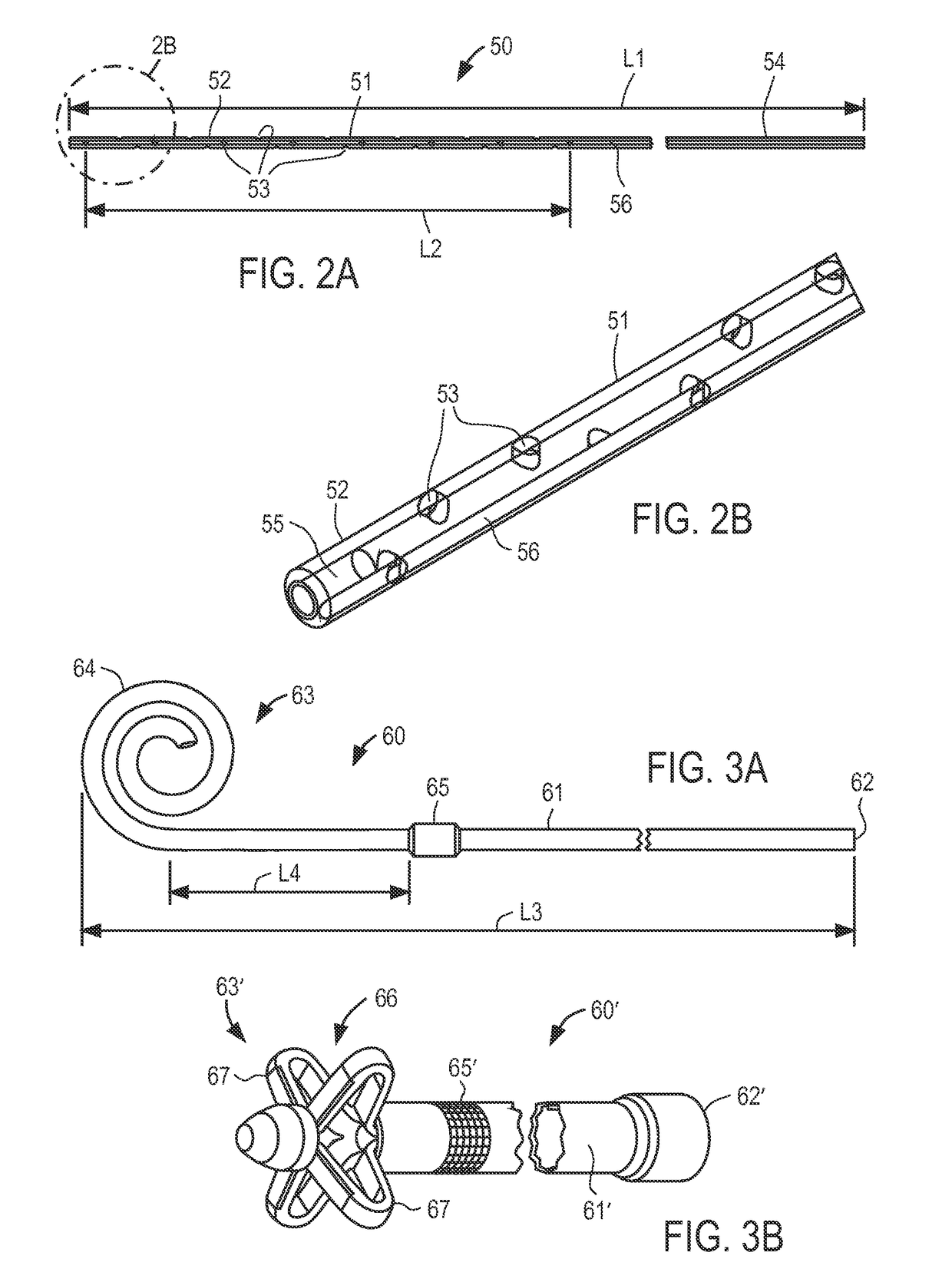
[0052]With respect to FIG. 3A, outflow catheter 60 of the present invention is described, corresponding to bladder catheter 25 of FIG. 1. Outflow catheter 60 preferably comprises tube 61 of medical-grade silicone having inlet end 62 and outlet end 63 including spiral structure 64, and polyester ingrowth cuff 65. Outflow catheter 60 includes a single internal lumen that extends from inlet end 62 to a single outlet at the tip of spiral structure 64, commonly referred to as a “pigtail” design. Inlet end 62 may include a connector for securing the inlet end of the outflow catheter to implantable device 20, or may have a length that can be trimmed to fit a particular patient.

[0053]When configured for use as the outflow catheter in an ascites treatment system, outflow catheter may have length L3 of about 45 cm, with cuff 65 placed length L4 of about 5 to 6 cm from spiral structure 64. Outflow catheter 60 may be loaded onto a stylet with spiral structure 64 straightened, and implanted usin...

second embodiment

[0057]With respect to FIG. 3B, an outflow catheter of the present invention is described, in which similar components are identified with like-primed numbers. Outflow catheter 60′ preferably comprises tube 61′ of medical-grade silicone having inlet end 62′, outlet end 63′ and polyester ingrowth cuff 65′. In accordance with this embodiment, outlet end 63′ includes malecot structure 66, illustratively comprising four resilient wings 67 that expand laterally away from the axis of the catheter to reduce the risk that outlet end 63′ of the catheter will be inadvertently pulled loose after placement. Inlet end 62′ may include a connector for securing the inlet end of the outflow catheter to implantable device 20, or may have a length that can be trimmed to fit a particular patient.

[0058]Malecot structure 66 preferably is constructed so that wings 67 deform to a substantially flattened configuration when a stylet is inserted through the lumen of the catheter. In this manner, outflow cathet...


PUM

PropertyMeasurementUnit
flow ratesaaaaaaaaaa
length L2aaaaaaaaaa
length L2aaaaaaaaaa

Abstract

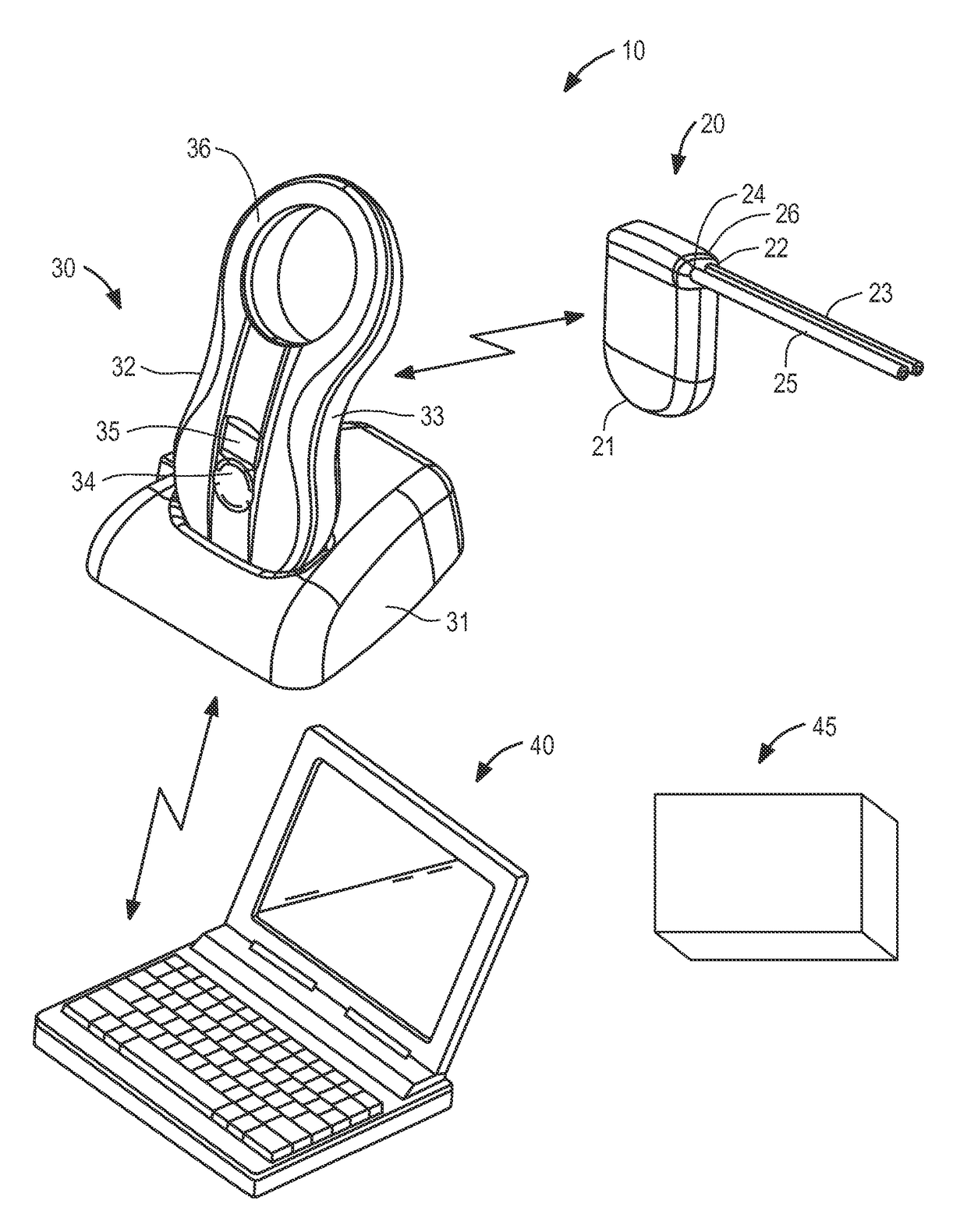
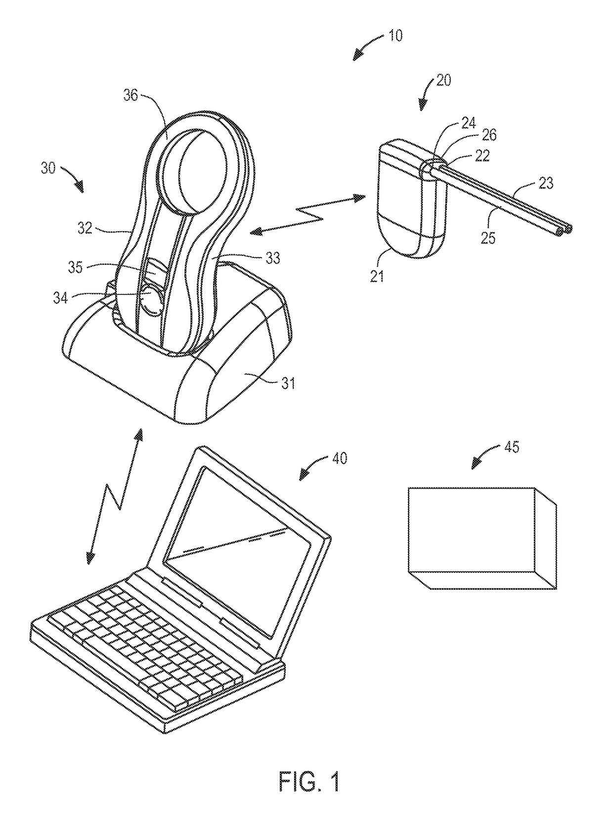
A system for noninvasively monitoring cancerous cells within bodily fluid accumulated due to ascites, pleural effusion or pericardial effusion is provided including an implantable pump having control circuitry; a charging and communication system configured to periodically charge and communicate with the implantable pump to retrieve performance data; and monitoring and control software, suitable for use with conventional personal computers, for configuring and controlling operation of the implantable pump and charging and communication system. The implantable pump is configured to move fluid containing cancerous cells from a body cavity to the bladder such that the fluid may be excreted during urination, collected and analyzed with an analysis station to assess progress of the cancer or efficacy of a cancer treatment program.

Description

I. CROSS-REFERENCE TO RELATED APPLICATIONS[0001]This application is a continuation-in-part application of U.S. patent application Ser. No. 13 / 397,509, filed Feb. 15, 2012, and a continuation-in-part application of U.S. patent application Ser. No. 13 / 397,523, filed Feb. 15, 2012, both of which claim priority to U.S. Provisional Patent Application No. 61 / 443,668, filed Feb. 16, 2011, the entire contents of each of which are incorporated herein by reference.II. FIELD OF THE INVENTION[0002]This application relates to apparatus and methods for noninvasive monitoring of cancerous cells in intracorporeal fluid accumulations.III. BACKGROUND OF THE INVENTION[0003]Certain cancers can result in pathologic chronic collection of bodily fluids within cavities such as the peritoneum, pleura, or pericardial sac. Such cancers may cause chronic ascites, pleural effusion, or pericardial effusion, where chronic fluid collections persist and result in increased morbidity and mortality.[0004]In pleural e...

Claims


Application Information

Patent Timeline
Patent Type & Authority Patents(United States)
IPC IPC(8): A61M1/00A61B10/00A61M5/142G01N33/493
CPCA61B10/007A61M5/14236A61M5/14276G01N33/493A61M2205/3507A61M2205/3569A61M2205/8243
Inventor JOHNSON, NOEL L.WHITTAKER, STEVE
Owner SEQUANA MEDICAL AG
Learn More