Nucleic acids coding for adhesion factor of group b streptococcus, adhesion factors of group b streptococcus and further uses thereof
a technology of adhesion factor and nucleic acid, which is applied in the field of nucleic acids coding for adhesion factor of group b streptococcus, and further uses thereo
- Summary
- Abstract
- Description
- Claims
- Application Information
AI Technical Summary
Benefits of technology
Problems solved by technology
Method used
Image
Examples
example 2
Identification of a Novel S. agalactiae Adhesion by a Signal Peptide Tagging Screen
[0380] Results
[0381] GBS strain 6313, belonging to serotype III, was tested in binding experiments for its interaction with radiolabelled human vitronectin, laminin, fibronectin, fibrinogen, and IgG. Strain 6313 accumulated about 50% of the total fibrinogen on its surface. Of the other proteins tested, none interacted in significant amounts (>5%) with GBS 6313. Treatment of the bacteria with either trypsin or pronase reduced the amount of bound fibrinogen to levels below 5%, indicating a proteinacious nature of the fibrinogen-binding structures of GBS 6313.
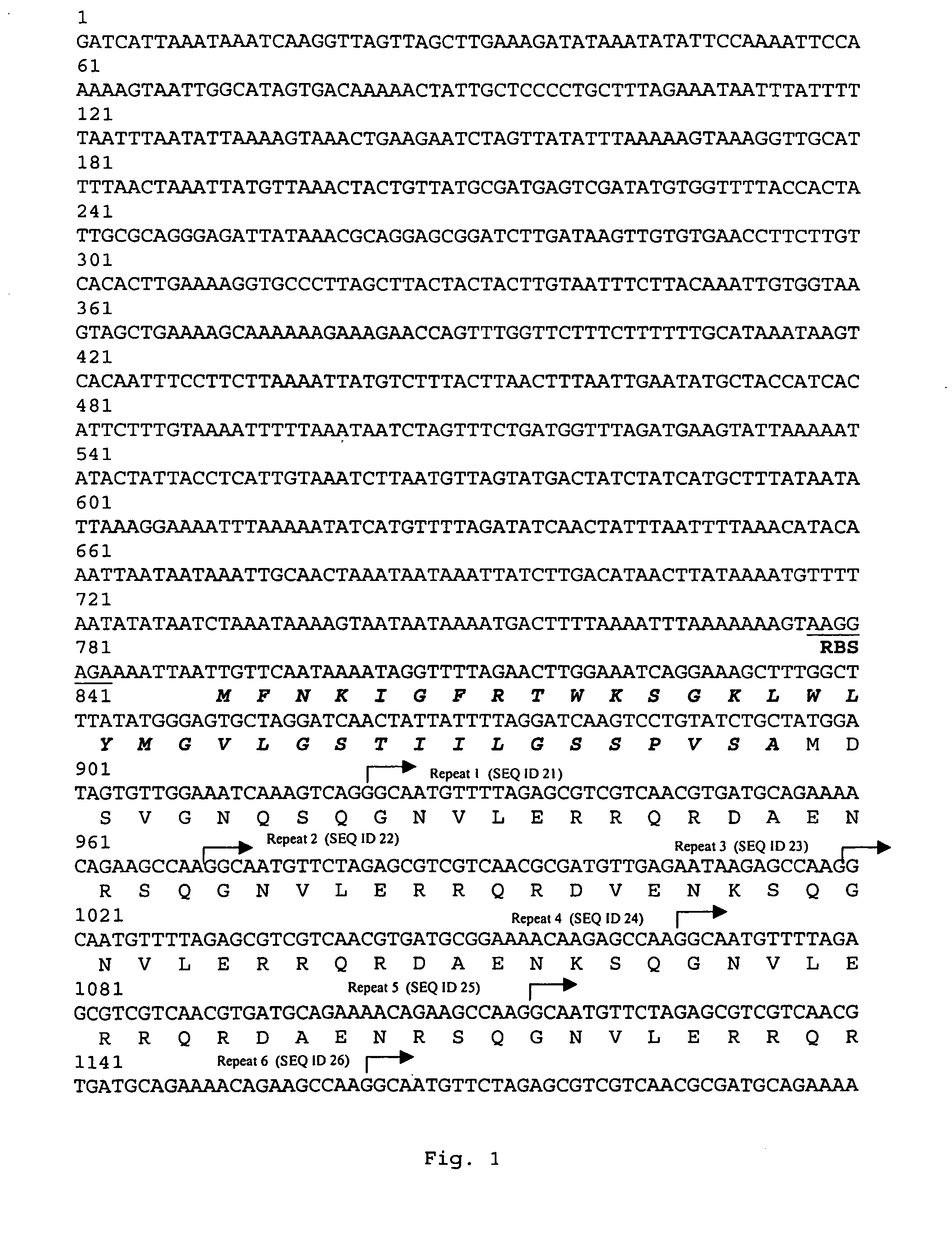
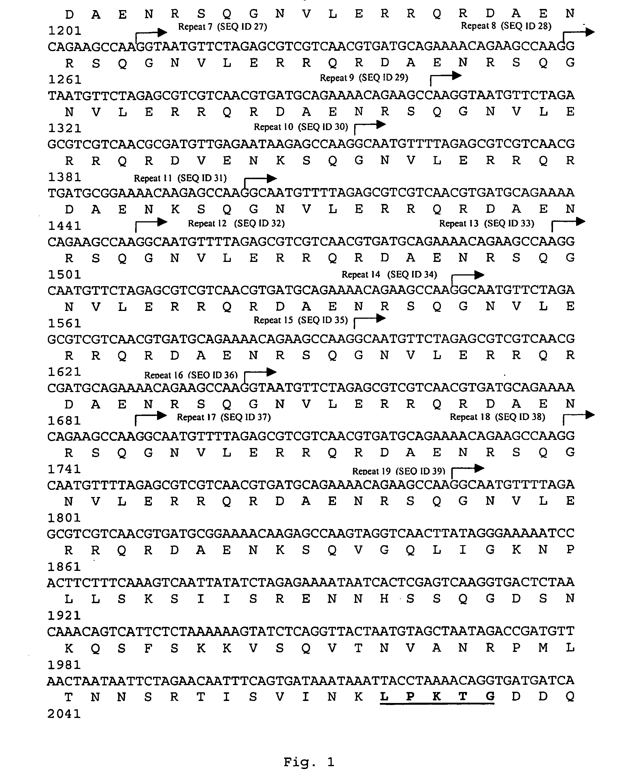
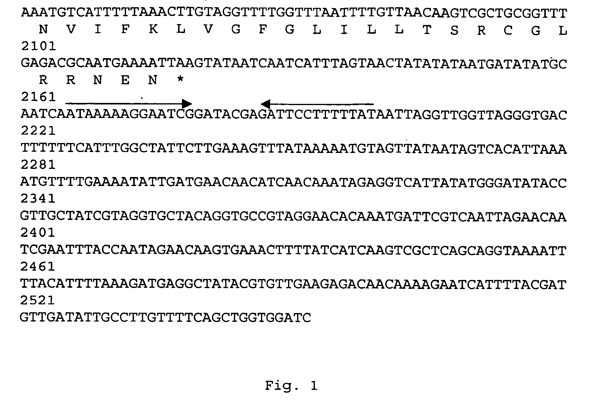
[0382] An Escherichia coli cosmid gene library of GBS 6313 was screened by colony blotting for the presence of fibrinogen-binding E. coli clones, resulting in the identification of a clone that revealed strong interaction with human fibrinogen. Partial digestion of its cosmid with Sau3A and subcloning of fragments in the range of 2-3 kb in plasmi...
example 3
FbsA is the Fibrinogen Receptor of Streptococcus agalactiae
[0384] Results
[0385] For functional analysis of FbsA, a truncated FbsA polypeptide (FbsA-19), devoid of a signal peptide and a membrane-spanning region was synthesized as a hexa-histidyl fusion protein in E. coli BL21 and purified by affinity chromatography. In Western blot experiments FbsA-19 revealed binding to human fibrinogen (FIG. 9), confirming FbsA as a fibrinogen receptor from GBS. To localize the fibrinogen-binding region in the FbsA protein, the N-terminal and the C-terminal regions of FbsA were synthesized as FbsA-N and FbsA-C fusion proteins and tested for fibrinogen binding. As shown in FIG. 9, fibrinogen binding was observed for FbsA-N but not for FbsA-C, indicating that the N-terminal repeats of FbsA mediates fibrinogen binding.
[0386] In competitive inhibition experiments with 125I-labelled fibrinogen, different proteins were tested for their capability to interfere with the binding of radiolabelled fibrino...
example 4
FbsA Contributes to Adherence and Invasion of Epithelial Cells and Inhibits Opsonophagocytosis
[0391] Results
[0392] To analyse the importance of FbsA for protecting GBS from opsonophagocytosis, the GBS strains 6313 and 6313ΔfbsA were tested for survival in a classical bactericidal assay in whole human blood. After inoculation of heparinized human blood with 10±30 colony forming units (cfu) of either of the two strains, both strains revealed growth, however, after three hours of incubation, strain 6313 grew to 2500±500 cfu / assay while strain 6313ΔfbsA grew only to 800±100 cfu / assay. This finding indicates a role of fbsA in preventing opsonization.
[0393] The GBS strains 6313, 706 S2 and O90R, and their respective fbsA deletion mutants were also tested for their ability to adhere to and invade the human lung epithelial cell line A549. As shown in FIG. 14A, the adhesion of the fbsA deletion mutants to A549 cells was significantly impaired compared to their parental strains. Similarly,...
PUM
| Property | Measurement | Unit |
|---|---|---|
| Fraction | aaaaa | aaaaa |
| Fraction | aaaaa | aaaaa |
| Fraction | aaaaa | aaaaa |
Abstract
Description
Claims
Application Information
 - R&D
- Intellectual Property
- Life Sciences
- Materials
- Tech Scout
- Unparalleled Data Quality
- Higher Quality Content
- 60% Fewer Hallucinations
Browse by: Latest US Patents, China's latest patents, Technical Efficacy Thesaurus, Application Domain, Technology Topic, Popular Technical Reports.
© 2025 PatSnap. All rights reserved.Legal|Privacy policy|Modern Slavery Act Transparency Statement|Sitemap|About US| Contact US: help@patsnap.com


