
Apparatus and method for rapid separation of cells without using density gradient and antibodies
a cell separation and density gradient technology, applied in the field of apparatus and a method for rapid cell separation without density gradient and antibody, can solve the problems of difficult operation and time consumption, cell separation medium could be toxic to cells to be separated, affecting the quality of cells, etc., and achieves simple and rapid operation procedure, the effect of reducing the cost involved
- Summary
- Abstract
- Description
- Claims
- Application Information
AI Technical Summary
Benefits of technology
Problems solved by technology
Method used
Image

Examples
example 1
Interaction of Cell and Chemical
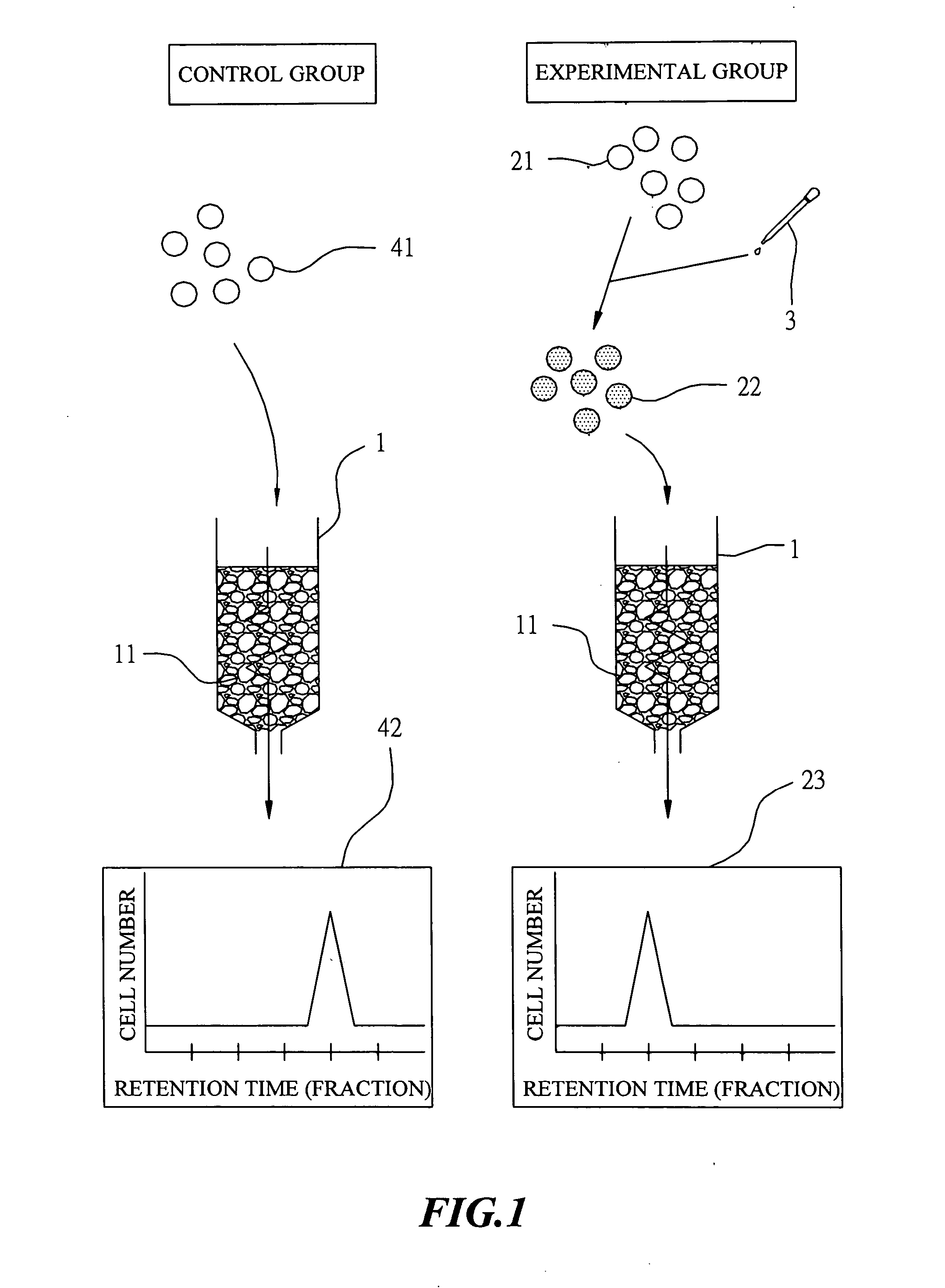
[0027] In this example, the column used for separating cell has an inner diameter of 6 mm, a length of 180 mm and a volume of 5 ml. This column was packed with resin particles and was washed first and thereafter, filled with a phosphate buffered saline (PBS).
The Control Group
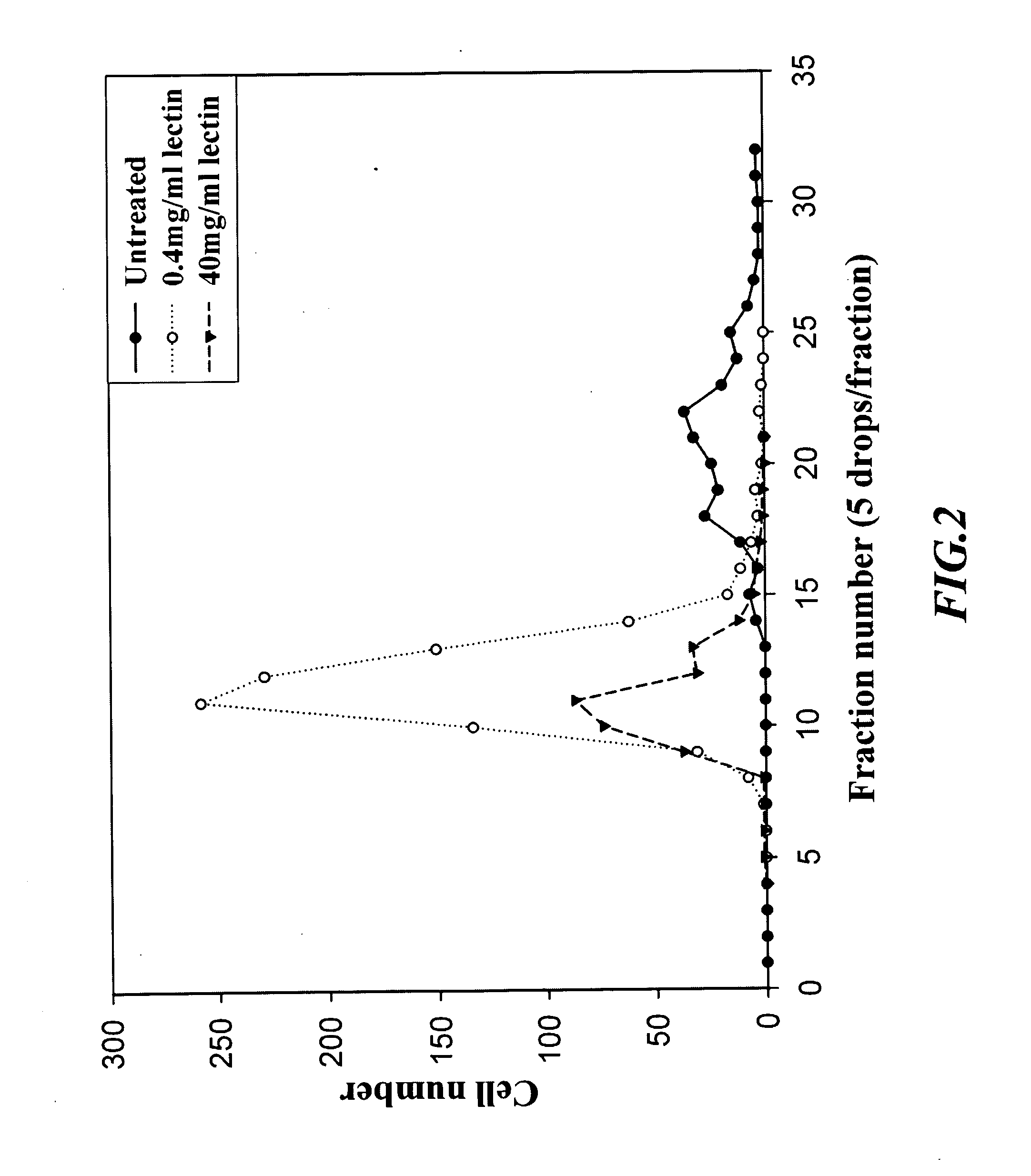
[0028] Blood sample was separated first by centrifugation using dextran cell separation medium (Ficoll) to yield peripheral blood mononuclear cell (PBMC). The thus-obtained PBMC was diluted with phosphate buffered saline (PBS) into a cell suspension at concentration of 1×10⁶ cells / ml. This cell suspension was loaded then on the upper frontier of the above-described column. After adding PBS to a level of a pre-determined height, the column was eluted with PBS at a flow rate of 3 ml / min and cell fractions were collected in test tubes, respectively, in a manner that each test tube collected 5 drops of cell suspension eluent. Thereafter, the eluted cell suspension in each test tube...
example 2
Separation of Sub-Population of Mononuclear Cells
EXAMPLE 2(A)
[0031] In example 2(A), the column used for separating cell has an inner diameter of 6 mm, a length of 180 mm and a volume of 5 ml. This column was packed with resin particles and was washed first and thereafter, filled with a phosphate buffered saline (PBS).
[0032] A concentrated mononuclear cells suspension was diluted with PBS to a cell suspension at a concentration of 1×10⁶ mononuclear cells / ml, containing still a certain amount of erythrocytes. This cell suspension was loaded then on the above-described column. The column was eluted subsequently with PBS at a flow rate of 3 ml / min and cell fractions were collected in test tubes, respectively, in a manner that each test tube collected 5 drops of cell suspension eluent. Thereafter, the eluted cell suspension in each test tube was examined under an optical microscope and numbers of erythrocytes and leukocytes were counted by a cell counter, respectively. The results we...
example 2 (
EXAMPLE 2(B)
[0034] In example 2(B), the column used for separating cell was changed and has an inner diameter of 8 mm, a length of 200 mm and a volume of 10 ml. The procedure of example 2(A) was repeated, and the column was filled at a flow rate of 1.2 ml / min. The result was shown in FIG. 6 and 7. FIG. 6 shows the number of mononuclear cells, while FIG. 7 shows the relative percentage of each sub-population of mononuclear cells.
[0035] The results obtained above suggest that the column could not achieve any separation effect against a single population of erythrocyte as shown in FIG. 3. For mononuclear cells in a same sample, several bands were eluted successively, as shown in FIG. 4 and 6. After analyzing further by fluorescence immuno-staining, the column provided a partition effect with respect to various sub-populations of mononuclear cells such as, T lymphocyte, B lymphocyte and monocyte, and revealed a significantly difference variation, as shown in FIG. 5 and 7.
[0036] The si...
PUM
 Abstract
Description
Claims
Application Information