Method and system for determining platelet-mediated clot formation

a platelet-mediated clot and platelet technology, applied in the field of thrombosis and hemostasis, can solve the problems of excess clot formation, thrombophilia, and slow blood flow in the vessel, and achieve the effect of preventing thrombophilia and preventing thrombophilia

Inactive Publication Date: 2006-06-29
VARON DAVID +1
View PDF16 Cites 6 Cited by
  • Summary
  • Abstract
  • Description
  • Claims
  • Application Information

AI Technical Summary

Benefits of technology

[0039] ii) an insignificant increase in fibrin clots formed at said surface as compared with a referenc

Problems solved by technology

First, the blood vessels slow the flow of blood past the wound site.
Disorders in either system can yield conditions that cause either too much or too little clotting.
If the clotting system can not adequately form clots, then the result is a bleeding disorder (hemophilia); if the clotting system forms clots too easily, then the result is formation of excess clots (thrombophilia).
Having too few platelet or platelets that do not function well (for example, aspirin use) can lead to a bleeding tendency.
Likewise, too many platelets can predispose to a t

Method used


Image

  • Method and system for determining platelet-mediated clot formation
  • Method and system for determining platelet-mediated clot formation
  • Method and system for determining platelet-mediated clot formation

Examples


example 1

Factor Xa Mediated Clot Formation

[0137] Bovine factor Xa was dissolved in normal saline containing 0.5% BSA to yield a stock solution of 7Units / ml. The stock solution was stored at −20° C. in small aliquots. Before use, the aliquots were thawed at 37° C. and the factor Xa was diluted 1:100 in the saline BSA solution to obtain a solution containing 0.07 Units / ml. One μl of the diluted solution contained 7×10⁻⁵ unit of factor Xa.

[0138] On the upper cap of a CPA well set attached to a magnetic stirrer were placed 1.5 μl of 1M CaCl2 solution and 2 μl of the diluted factor Xa solution (1.5×10⁻⁵ U). As control, no factor Xa or CaCl2 were added to the same blood sample.

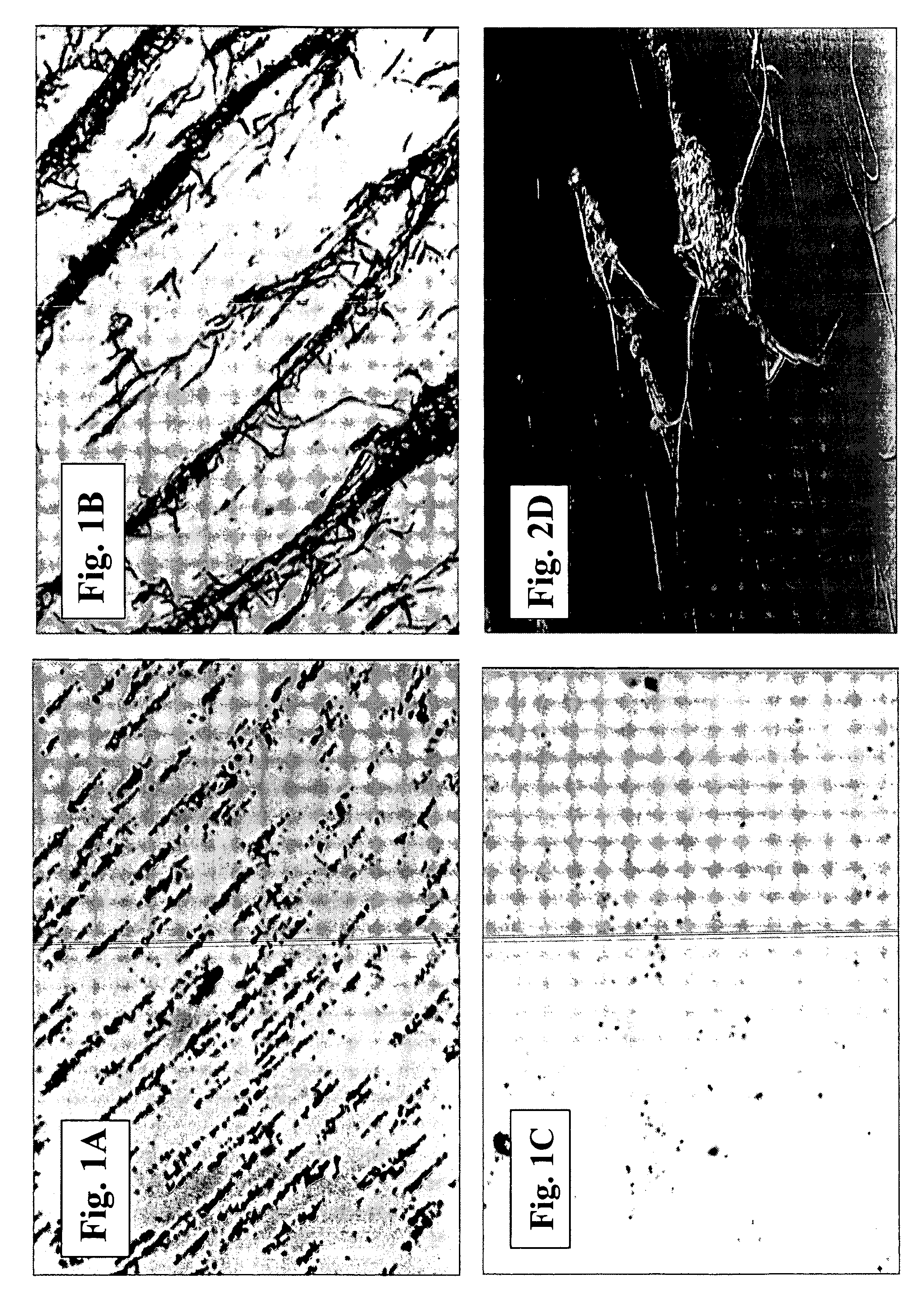
[0139] The stirrer assembly was placed in a CPA well containing 130 μl of whole blood obtained from a healthy subject and spinning at about 750 rpm started immediately. After 2 minutes the spinning was stopped, the magnet and cap were removed from the well and the well was washed with tap water to remove all residual bloo...

example 2

Determining Correlation Between Surface Coverage / Clot Size and Concentration of Clot Initiator

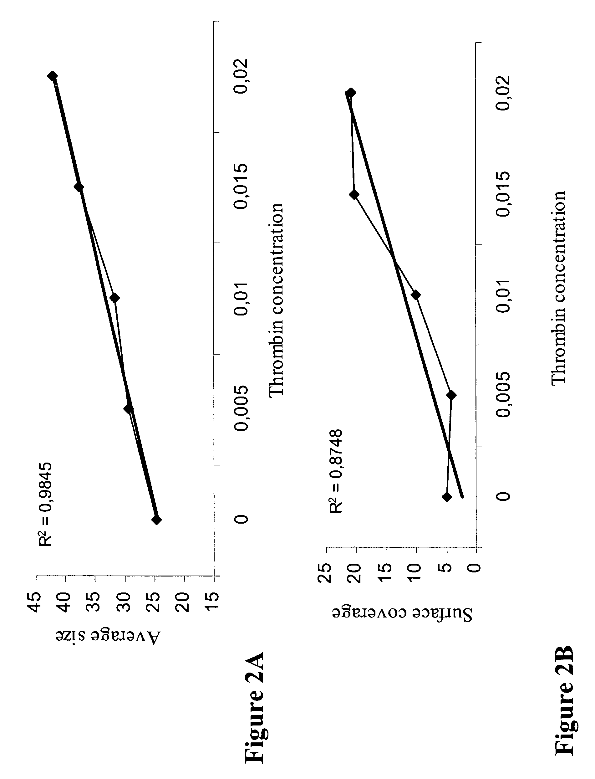
[0143] In this assay, increasing concentrations of thrombin, which promotes fibrin clot formation, were applied to normal blood samples. In particular, thrombin was placed on the tip of the rotating conical disc, in an amount yielding final concentration according to the points indicated in FIG. 3A and FIG. 3B. The conical disc was then applied in the well containing 130 μL of citrated whole blood, rotated at 720 rpm for two minutes followed by washing and staining of the well. For quantitative determination of fibrin clot formation image analysis (using Impact-R image analysis system, DiaMed, Switzerland) was performed.

[0144] Image analysis of the stained fibrin clots was performed expressing the amount of clot in terms of % of surface covered (SC, %), and the average size (AS, μm2) of the stained clots.

[0145]FIG. 2A shows that there is a significant correlation (R2=0.9845) between the ...

example 3

Use of the aPTT Reaction to Determine Factor VIII Deficiency

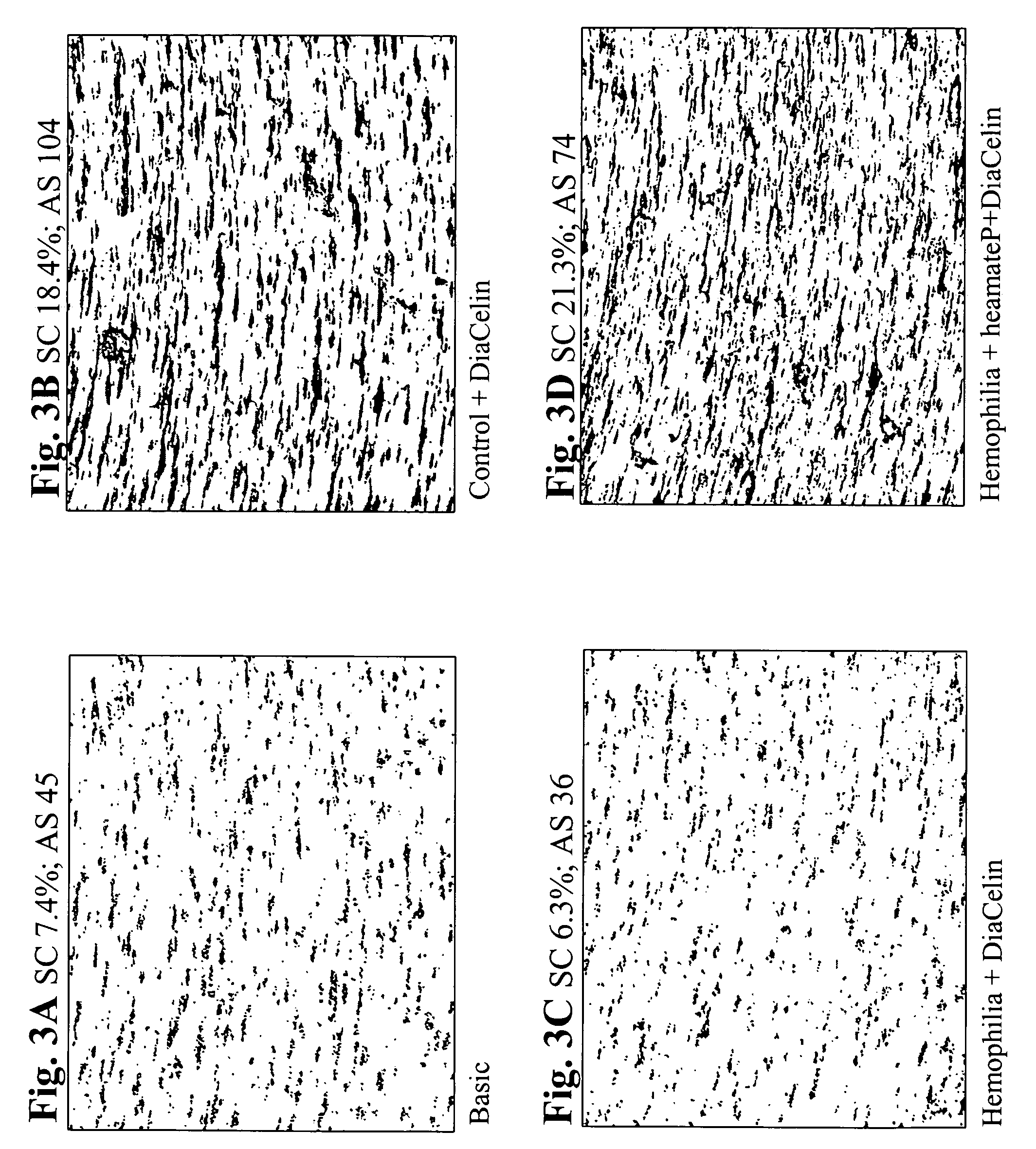
[0147] In the following assay undiluted aPTT reagent Diacelin (Daimed, Switzerland) was used to initiate clot formation. In particular, on the upper cap of a CPA well set attached to a magnetic stirrer 1.6 μl of 1M CaCl2 and 3 μl of Diacelin were placed. The stirrer assembly was placed in a well containing 130 μl of whole blood and spinning at 720 rpm started immediately.

[0148] After 2.5 min. the spinning was stopped, the magnet and cap were removed from the well and the well washed with tap water to remove all residual blood. The washed wells were then stained using May-Gruenwald stain (Sigma) and clot % surface coverage and clot average size was quantitatively determined using an image analysis system (Impact-R).

[0149]FIG. 3A shows the results of the assay when no aPTT reagent or CaCl2 were added to the blood. As a result, only single adsorbed platelets and platelet aggregates are shown, with no fibrin clots formation....


PUM


Abstract

The present invention concerns methods for determining platelet medicated clot formation. Specifically, the invention provides a method for determining platelet-medicated clot formation in a blood sample comprising: (i) obtaining sample of blood, and optionally mixing same with an anti-coagulant in an amount effective to inhibit clot formation; (ii) in a vessel, mixing the sample with a minute amount of an initiator to obtain a mixed sample, the amount being effective to initiate coagulation; (iii) rotating the mixed sample inside the vessel, whereby shear forces are developed at the surface of said vessel in a manner and for a time sufficient to allow adhesion of platelets at the surface of said vessel; (iv) determining clot formation at the surface of said adherent platelets. The method has different applications, among others, for determining a coagulation disorder in a subject having a coagulation factor dysfunction.

Description

FIELD OF THE INVENTION [0001] The present invention concerns thrombosis and hemostasis and more specifically methods of diagnosing and treating coagulation disorders. BACKGROUND OF THE INVENTION [0002] When a wound occurs, several changes take place to minimize blood loss, referred to as the hemostatic response. First, the blood vessels slow the flow of blood past the wound site. Next, platelets that circulate in the blood are collected at the wound site to form a plug. Finally, a second system (the coagulation cascade), based upon the action of multiple proteins (called clotting factors) that act in concert to produce a fibrin clot is activated. These two systems (also known by the terms “primary hemostasis” and “secondary hemostasis”) work in concert to form a clot. Disorders in either system can yield conditions that cause either too much or too little clotting. [0003] Platelets serve three primary functions. When a break in a blood vessel occurs, substances are exposed that norm...

Claims


Application Information

Patent Timeline
IPC IPC(8): G01N33/48
CPCG01N33/4905
Inventor GITEL, SANFORD N.
Owner VARON DAVID
Why Eureka
Try Eureka