[0006] Therefore, a need has arisen to provide apparatus for recording electrophysiological signals associated with a subject and methods of ascertaining and recording such electrophysiological signals which overcome the above-described and other shortcomings of the related art. One of the advantages of the present invention is that electrophysiological signals may be determined and recorded in a MRI environment. Specifically, various noises associated with subject movements can be removed from data associated with intrinsic voltages measured from the subject in order to generate a display of electrophysiological signals. Another
advantage of the present invention is that the noise associated with a ballistocardiogram motion within the subject and the noise associated with a
blood flow motion within the subject also can be removed from the data associated with the intrinsic voltages to generate the display of electrophysiological signals.
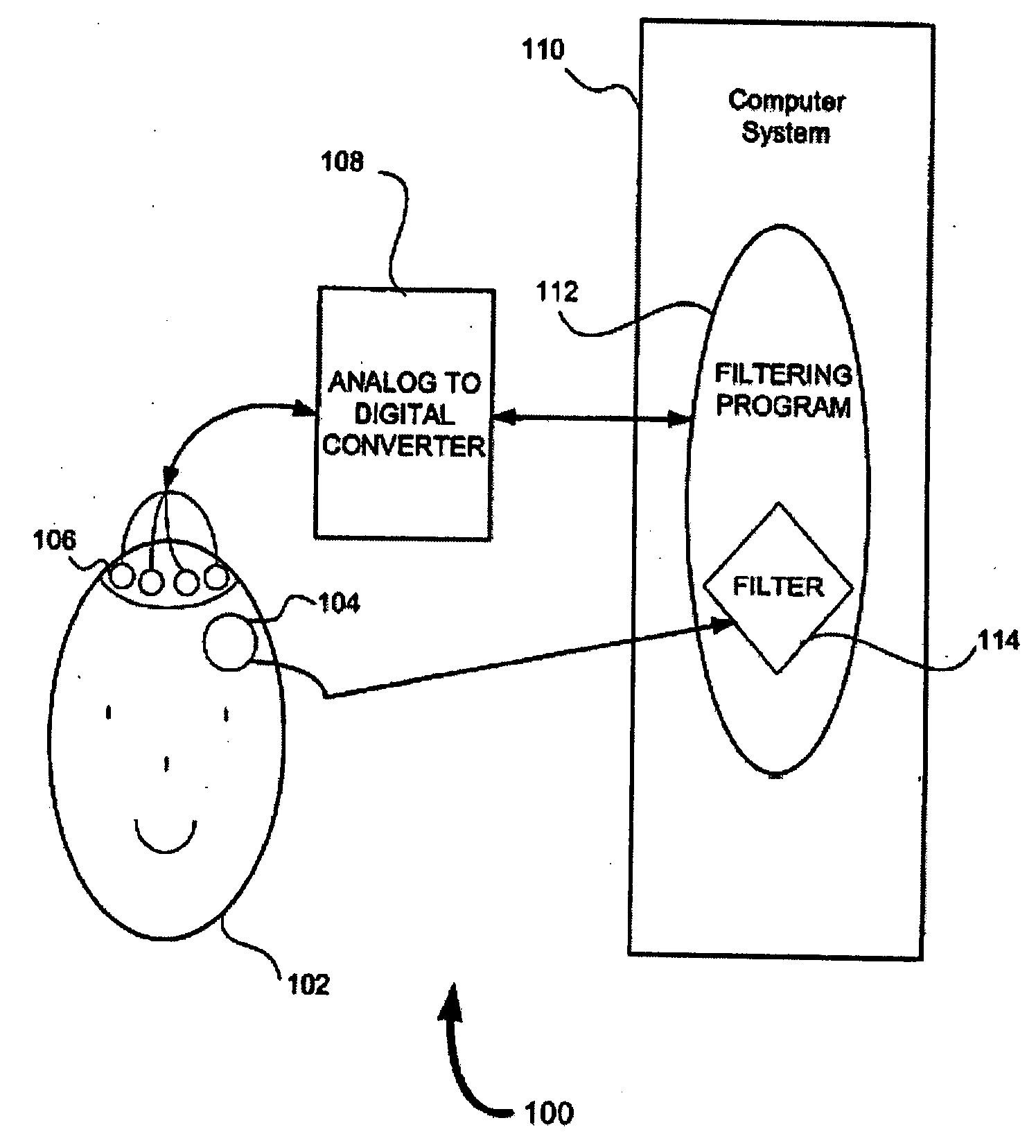
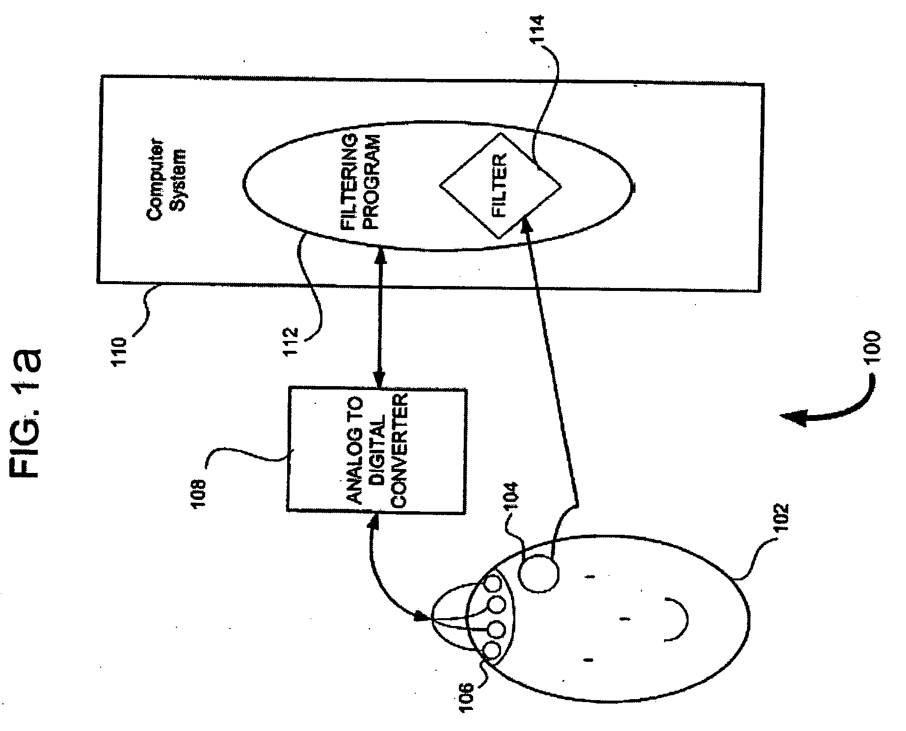
[0007] According to an exemplary embodiment of the present invention, an arrangement and method for ascertaining and / or recording electrophysiological signals (e.g.,
electroencephalography (EEG) signals, electromyogram (EMG) signals, single and / or multi-
cell signals, evoked potentials (EP) any other behavioral event
signal, etc.) associated with a subject are provided. In particular, a
processing system in the arrangement (e.g., a
processing system associated with a computer
system or a
processing system associated with an EEG system) may be adapted to execute a filtering program. When the filtering program is executed, the processing system may be adapted (e.g., configured) to receive first data associated with a movement of the subject from one or more
motion sensors (e.g., directly from the one or more
motion sensors or indirectly from the one or more sensors via an
analog to digital converter). Such movements may include
head movements by the subject,
swallowing by the subject, etc., and the first data may be amplified and then
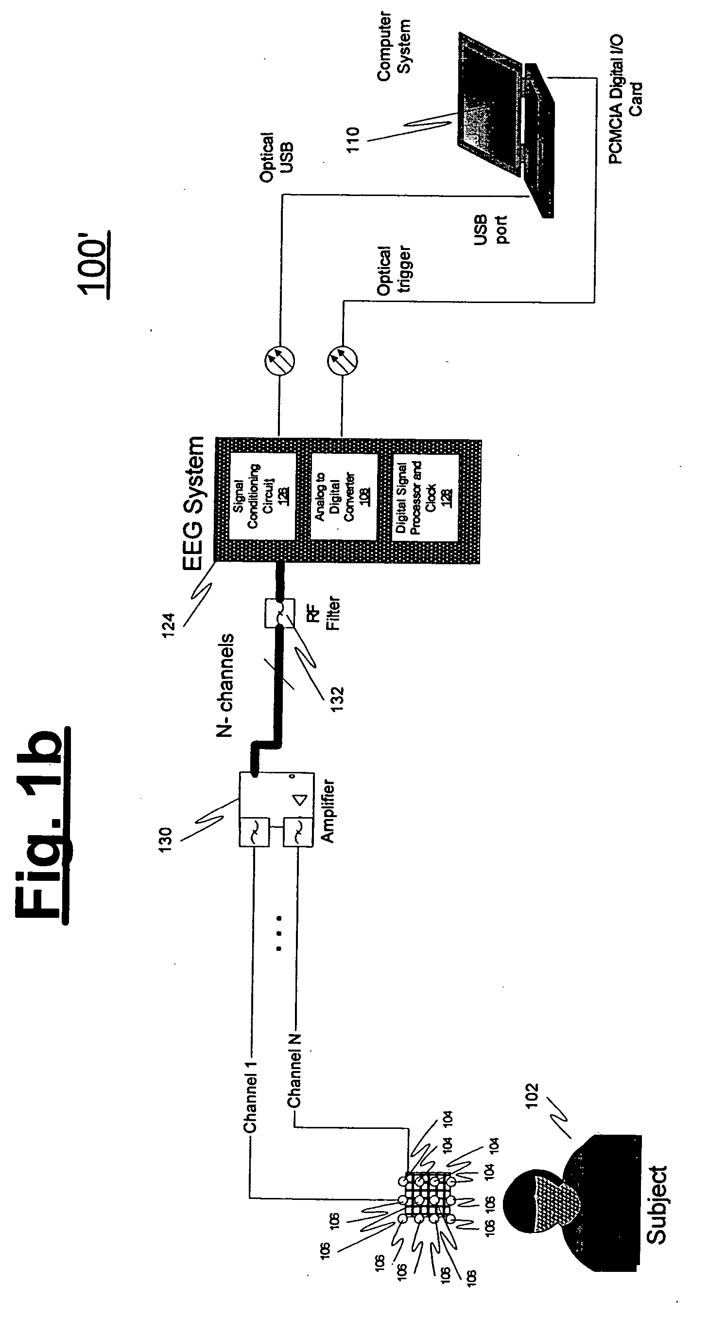
radio frequency (RF) filtered before processing system receives the first data. The motion data also can include noise associated with a
blood flow motion within the subject, noise associated with a ballistocardiac motion within the subject, etc. The processing system also may be adapted to receive second data associated with intrinsic voltages measured from the subject (e.g., after the second data is amplified and RF filtered), and to calculate result data based on the first and second data, with the result data being associated with the electrophysiological signal. For example, the filtering program can include a filtering routine which receives the first data from the motion sensor, and generates an output which is subtracted from of the second data to generate the result data. Moreover, the processing system may further be adapted to generate a continuous,
real time display of electrophysiological signals associated with the result data.
[0008] According to another exemplary embodiment of the present invention, a plurality of electrodes are positioned on at least one portion of the subject. An analog to digital (A / D) converter (e.g., a twenty-four (24) bit A / D converter) may be provided to be coupled to each of the electrodes. For example, the EEG system may include the A / D converter. The processing system can be coupled to the A / D converter. For example, the electrodes can be positioned on the
scalp of the subject. The filter routine can be an
adaptive filter routine, such as a Kalman-type
adaptive filter routine. Moreover, the A / D converter is preferably adapted to measure intrinsic voltages associated with the subject, and to transmit the second data (which is associated with the intrinsic voltages) to the filtering program. In yet another exemplary embodiment of the present invention, the A / D converter can be positioned inside a MRI environment, and the processing system can be positioned outside the MRI environment. Alternatively, when the processing system is associated with the EEG system, the processing system can be positioned inside the MRI environment, and the computer system can be positioned outside the MRI environment. The motion sensor (e.g., a piezoelectric
transducer) can provide signals and information to the filter routine (e.g., directly or via the A / D converter). For example, the motion sensor may be positioned adjacent to the subject, or on a portion of the subject (e.g., on a temporal
artery of the subject). Further, at least one portion of the motion sensor may be filled with an acoustic dampening material, such as
silicon, and can be adapted to measure the first data.

