Method of measurement of nucleated red blood cells

a measurement method technology, applied in the field of nucleated red blood cell measurement, can solve the problems of complex fluorescence measurement, high detection cost, and difficult to differentiate nucleated red blood cell only based on electronic or optical properties

Active Publication Date: 2006-07-20
BECKMAN COULTER INC
View PDF25 Cites 39 Cited by
  • Summary
  • Abstract
  • Description
  • Claims
  • Application Information

AI Technical Summary

Benefits of technology

[0018] In one embodiment, the present invention is directed to a method of measuring nucleated red blood cells in a blood cell sample. The method comprises the steps of exposing a blood cell sample to a reagent system to lyse mature red blood cells and to form a sample mixture; analyzing the sample mixture in a flow cell by measuring axial light loss, DC impedance and medium angle light scatter signals; differentiating nucleated red blood cells from other cell types using obtained axial light loss, DC impedance and medium angle light scatter signals; and reporting nucleated red blood cells in the blood cell sample. The medium angle light scatter signals are measured in a range from about 15° to about 70°, and preferably in a range from about 20° to about 45°. The differentiation of nucleated red blood cells from other cell types can use one or more functions including functions of the medium angle light scatter and DC impedance signals, functions of the medium angle light scatter and axial light loss signals, functions of the axial light loss and DC impedance signals, or combinations thereof.
[0019] In a further embodiment, the present invention is directed to a method of measuring nucleated red blood cells in a blood cell sample. The method comprises the steps of exposing a blood cell sample to a reagent system to lyse mature red blood cells and to form a sample mixture; analyzing the sample mixture in a flow cell by measuring axial light loss, low angle light scatter and medium angle light scatter signals; differentiating nucleated red blood cells from other cell types using obtained axial light loss, low angle light scatter and medium angle light scatter signals; and reporting nucleated red blood cells in the blood cell sample. The low angle light scatter signals are measured less than 10°, preferably in a range from about 1° to about 7°. Differentiation of nucleated red blood cells from other cell types can use one or more functions including functions of the medium angle light scatter and low angle light scatter signals, functions of the medium angle light scatter and axial light loss signals, functions of the axial light loss and low angle light scatter signals, or combinations thereof.
[0020] In another embodiment, the present invention is directed to a method of measuring nucleated red blood cells in a blood sample. The method comprises the steps of exposing a blood cell sample to a reagent system to lyse mature red blood cells and to form a sample mixture; analyzing the sample mixture in a flow cell by measuring axial light loss, DC impedance, low angle light scatter and medium angle light scatter signals; differentiating nucleated red blood cells from other cell types using obtained axial light loss, DC impedance, low angle light scatter and medium angle light scatter signals; and reporting nucleated red blood cells in the blood cell sample. Differentiation of nucleated red blood cells from other cell types can use one or more functions including functions of the axial light loss and DC impedance signals, functions of the axial light loss and low angle light scatter signals, functions of the low angle light scatter and DC impedance signals, functions of the medium angle light scatter and DC impedance signals, functions of the medium angle light scatter and low angle light scatter signals, functions of the medium angle light scatter and axial light loss signals, functions of the axial light loss, DC impedance and low angle light scatter signals, functions of the axial light loss, DC impedance and medium angle light scatter signals, functions of the axial light loss, low angle light scatter and medium angle light scatter signals, or combinations thereof.
[0021] In a further embodiment, the present invention is directed to a differential analysis method for differentiating nucleated red blood cells from other cell types. Upon obtaining DC impedance, axial light loss, low angle light scatter and medium angle light scatter signals, the cell debris is first differentiated from the nucleated red blood cells and the white blood cells using the medium angle light scatter signals, in conjunction with one or more other measured signals, or functions thereof; and the nucleated red blood cells are differentiated from the white blood cells using the low angle light scatter, axial light loss and DC impedance signals. The cell debris includes platelets, large platelets, giant platelets, platelet clumps, sickle cells or unlysed red blood cell membrane. The method further includes obtaining and reporting a corrected white blood cell count of the blood sample.
[0022] As will be better appreciated from the ensuing Detailed Description of Preferred Embodiments, the invention is particularly advantageous compared to the prior art in that it provides differentiation of nucleated red blood cells utilizing DC impedance, axial light loss, low angle and medium angle light scatter measurements without nuclear staining and the use of costly fluorescence detection methods. An additional feature of the present method is that it utilizes the medium angle light scatter measurement, in conjunction with DC impedance, low angle light scatter, or axial light loss measurement to further differentiate nucleated red blood cells from various interference materials. The invention will be better understood from the ensuing description of preferred embodiments.

Problems solved by technology

This approach is extremely time-consuming as well as being subjective to the interpretation of the individual analyzing the slide.
These methods utilize nuclear specific staining techniques to distinguish nucleated red blood cells from other cell types because it is difficult to differentiate nucleated red blood cells only based on their electronic or optical properties.
However, fluorescence measurements are complex and expensive detection methods.
It is known that differentiation of nucleated red blood cells from other cell types, particularly white blood cells, is technically challenging because of the potential overlapping signals from other cell types, when measured by their size, and light scatter and fluorescence properties.
Furthermore, it is known that certain cell types and / or cellular materials tend to interfere with the nucleated red blood cell measurement.
For example, when fluorescence measurement is used for measuring the nucleated red blood cells, aged white blood cells tend to interfere with the measurement, because they tend to be lysed by the reagent used for preparing the sample mixture, and the nuclei of the lysed white blood cells tend to overlap with nucleated red blood cells.
It has been found that when using light scatter and impedance measurements for measuring nucleated red blood cells, giant platelets, platelet clumps and sickle cells can cause interference, because these materials can overlap with nucleated red blood cells in one or more of the measurements.

Method used


Image

  • Method of measurement of nucleated red blood cells
  • Method of measurement of nucleated red blood cells
  • Method of measurement of nucleated red blood cells

Examples


example 1

[0085] 14 μL of an anticoagulated whole blood sample, also referred to as peripheral blood, was diluted by 614 μL of an isotonic blood diluent, Isoton® 3E (product of Beckman Coulter, Inc. Miami, Fla.), and mixed with 125 μL of a lytic reagent in a mixing chamber on an experimental hematology analyzer. About nine seconds after the addition of the lytic reagent the sample mixture was delivered to a flow cell with a sheath fluid, Isoton® 3E, for differential analysis of nucleated red blood cells. The lytic reagent was an aqueous solution containing active components for lysing red blood cells and analysis of nucleated red blood cells: 36 g / L dodecyltrimethylammonium chloride (50% solution), 3.6 g / L tetradecyl-trimethylammnonium bromide, and has a pH about 4.

[0086] An experimental hematology analyzer was equipped with a detection system for detecting DC impedance, low angle light scatter and axial light loss signals generated when a cell in the sample mixture passed through the flow c...

example 2

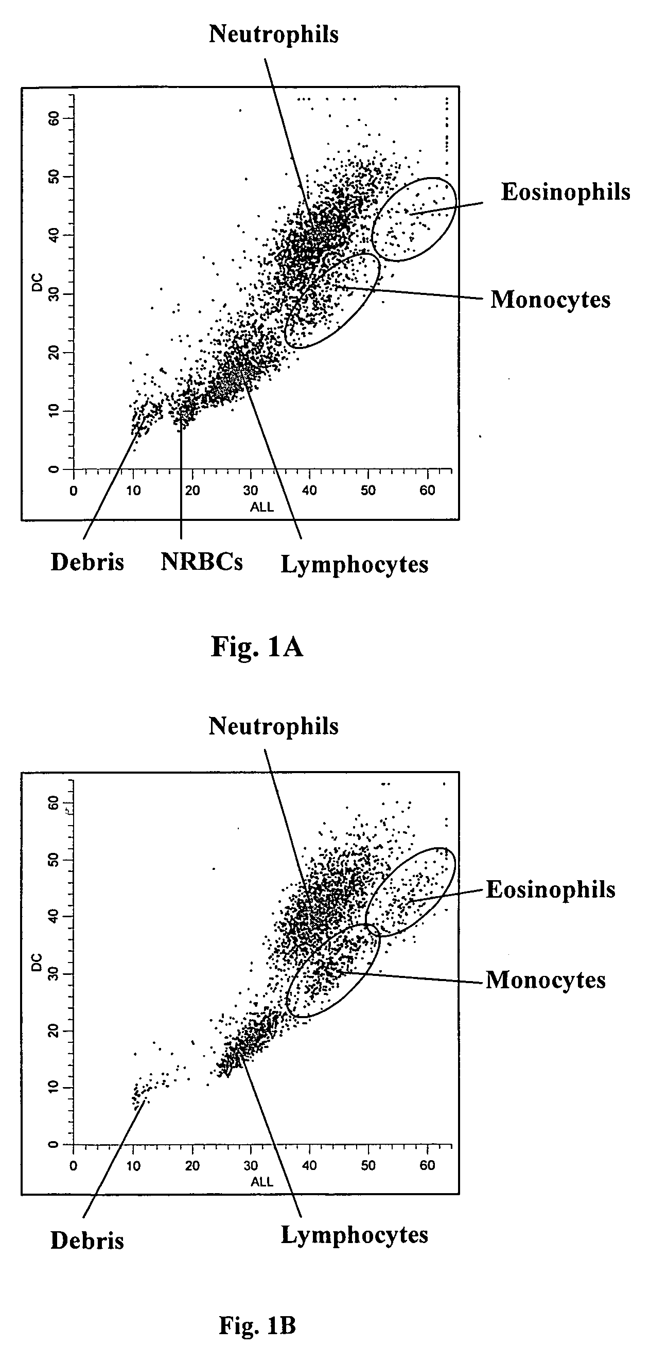
[0089] A fresh normal whole blood sample was analyzed using the same reagents and procedure described in Example 1. FIG. 1B shows an obtained DC vs. ALL scattergram of a normal blood sample. As shown, no cell population was present in the region where nucleated red blood cells reside.

[0090] Furthermore, as shown in FIGS. 1A and 1B the white blood cells were further differentiated into four subpopulations, i.e., lymphocytes, monocytes, neutrophils and eosinophils.

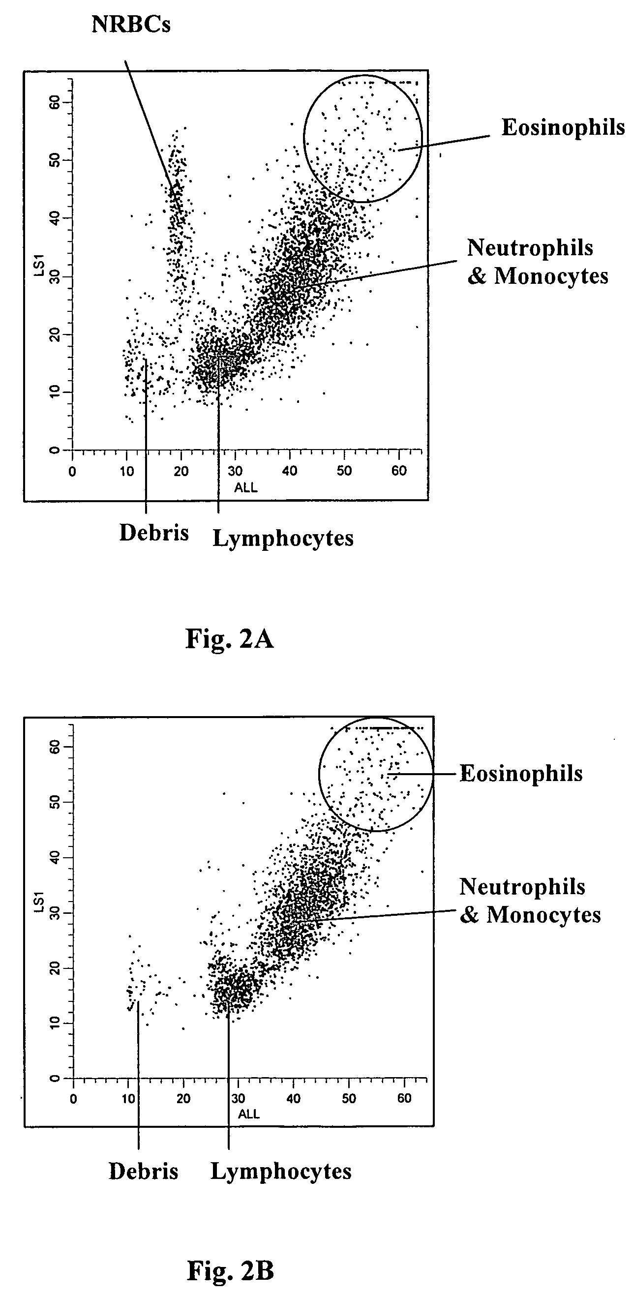
[0091]FIG. 2B shows an obtained LS1 vs. ALL scattergram of the same normal whole blood sample shown in FIG. 1B. Again, no cluster was present in the region where nucleated red blood cells reside.

[0092] Also, as shown in FIGS. 2A and 2B the white blood cells were further differentiated into three subpopulations, i.e., lymphocytes, a sum of monocytes and neutrophils, and eosinophils.

example 3

[0093] A clinical whole blood sample containing nucleated red blood cells and a fresh normal whole blood sample were analyzed using the process described in Example 1 on another experimental hematology analyzer which was equipped with a detection system equivalent to that described in Example 1, but the data was acquired with a 14-bit ADC resolution. The lytic reagent and the diluent used were Lyse S® 4 and Isoton® 4 (products of Beckman Coulter, Inc. Miami, Fla.).

[0094]FIGS. 5A thru 8B show the obtained scattergrams. As shown in FIGS. 5A, 6A, 7A and 8A, the nucleated red blood cells in the clinical sample clearly separate from white blood cells and from cell debris in DC vs. ALL, LS1 vs. ALL, F1 vs. ALL and F2 vs. ALL scattergrams, respectively. As shown in FIGS. 5B, 6B, 7B and 8B obtained from the normal blood sample, no cell population was present in the region where nucleated red blood cells reside. It is noted that the axis scales of the scattergrams in FIGS. 5A thru 8B are di...


PUM

PropertyMeasurementUnit
angleaaaaaaaaaa
angleaaaaaaaaaa
angleaaaaaaaaaa

Abstract

Methods are provided for measurement of nucleated red blood cells in a blood sample. The methods include exposing a blood cell sample to a reagent system to lyse mature red blood cells, subsequently analyzing nucleated red blood cells in a flow cell by axial light loss, DC impedance, and medium angle light scatter measurements; by axial light loss, low angle light scatter, and medium angle light scatter measurements; or by axial light loss, DC impedance, low angle light scatter, and medium angle light scatter measurements; and then differentiating nucleated red blood cells from other cell types by using measured signals and/or functions thereof.

Description

CROSS REFERENCE TO RELATED APPLICATION [0001] This application is a continuation-in-part of application Ser. No. 11 / 048,086, filed on Feb. 1, 2005, which claims the benefit under 35 USC 119 (e) of the provisional patent application Ser. No. 60 / 543,162, filed on Feb. 10, 2004. All parent applications are hereby incorporated by reference in their entirety.FIELD OF THE INVENTION [0002] The present invention relates to a method for the measurement of nucleated red blood cells by optical and impedance measurements. BACKGROUND OF THE INVENTION [0003] Normal peripheral blood contains mature red blood cells which are free of nucleus. Nucleated red blood cells (NRBC), or erythroblasts, are immature red blood cells. They normally occur in the bone marrow but not in peripheral blood. However, in certain diseases such as anemia and leukemia, nucleated red blood cells also occur in peripheral blood. Therefore, it is of clinical importance to measure NRBC. Traditionally, differentiation and enume...

Claims


Application Information

Patent Timeline
Patent Type & Authority Applications(United States)
IPC IPC(8): G01N33/50G01N15/00G01N15/10G01N15/12G01N15/14G01N33/48G01N33/80
CPCG01N15/1031G01N15/12G01N15/1459G01N33/5094G01N33/80Y10T436/101666G01N2015/008G01N2015/1037G01N2015/1062G01N2015/1486Y10T436/25125G01N2015/0073
Inventor LOPEZ, LIDICE L.PEREZ, CARLOS A.WELLS, MARK A.CAREAGA, DIANA B.HUO, ZILINGQIAN, CHENGTORKE, NANCY J.
Owner BECKMAN COULTER INC
Why Eureka
Try Eureka