Methods and compositions for differentiating tissues for cell types using epigenetic markers
a cell type and epigenetic technology, applied in the field of molecular diagnostic markers, can solve the problems of lack of quantitative methods in the prior art, and achieve the effect of accurate and efficient manner
- Summary
- Abstract
- Description
- Claims
- Application Information
AI Technical Summary
Benefits of technology
Problems solved by technology
Method used
Image
Examples
example 1
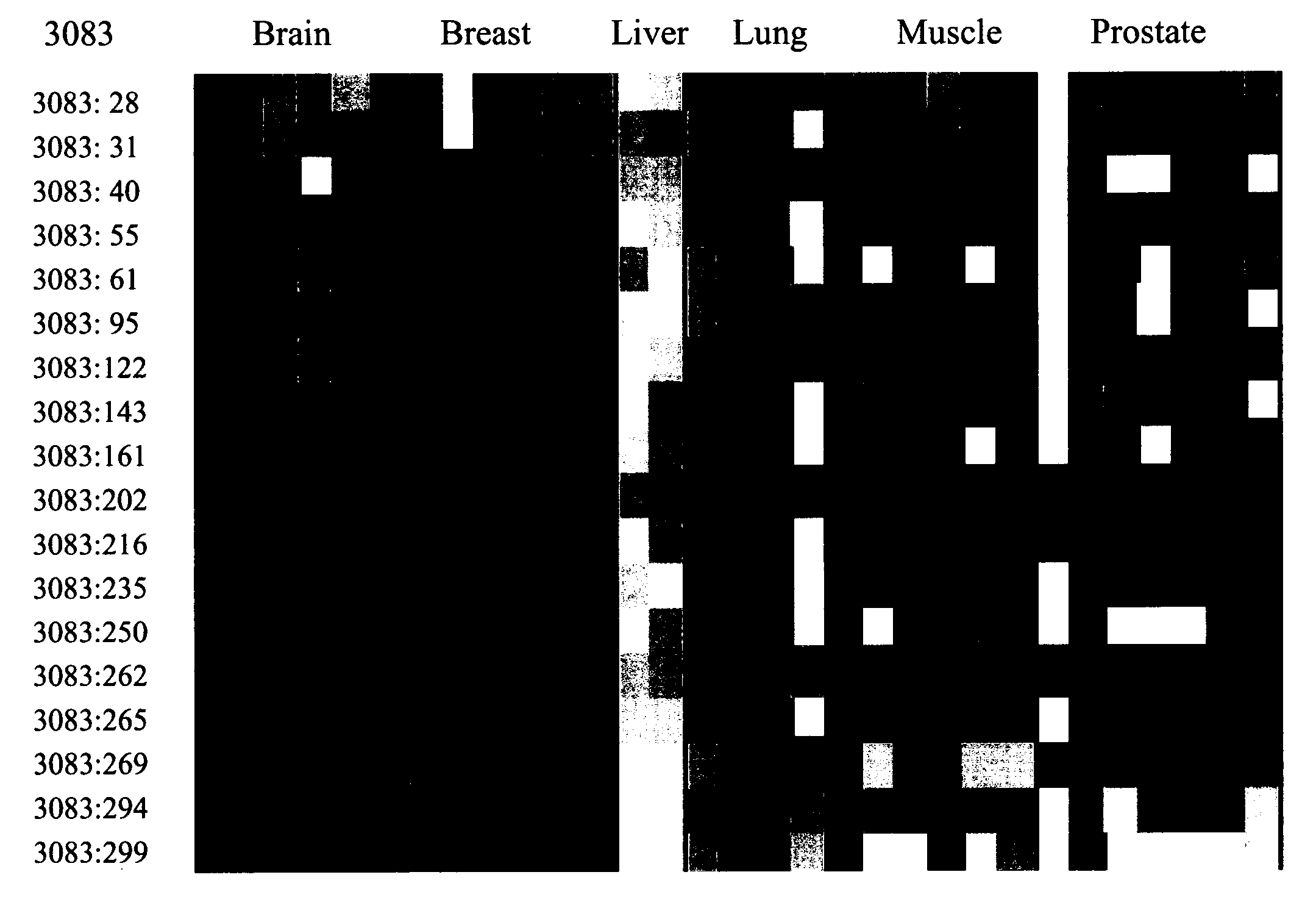
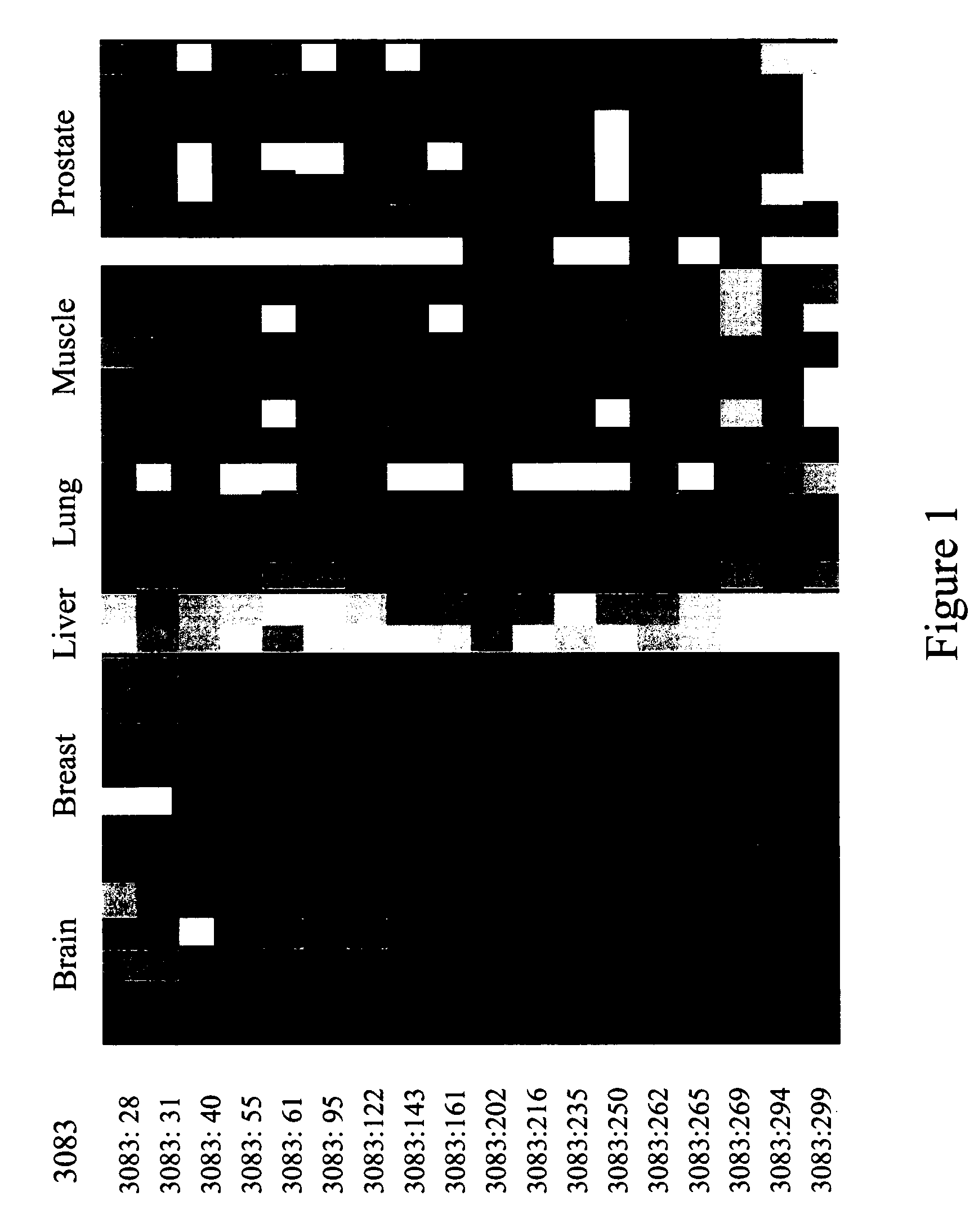
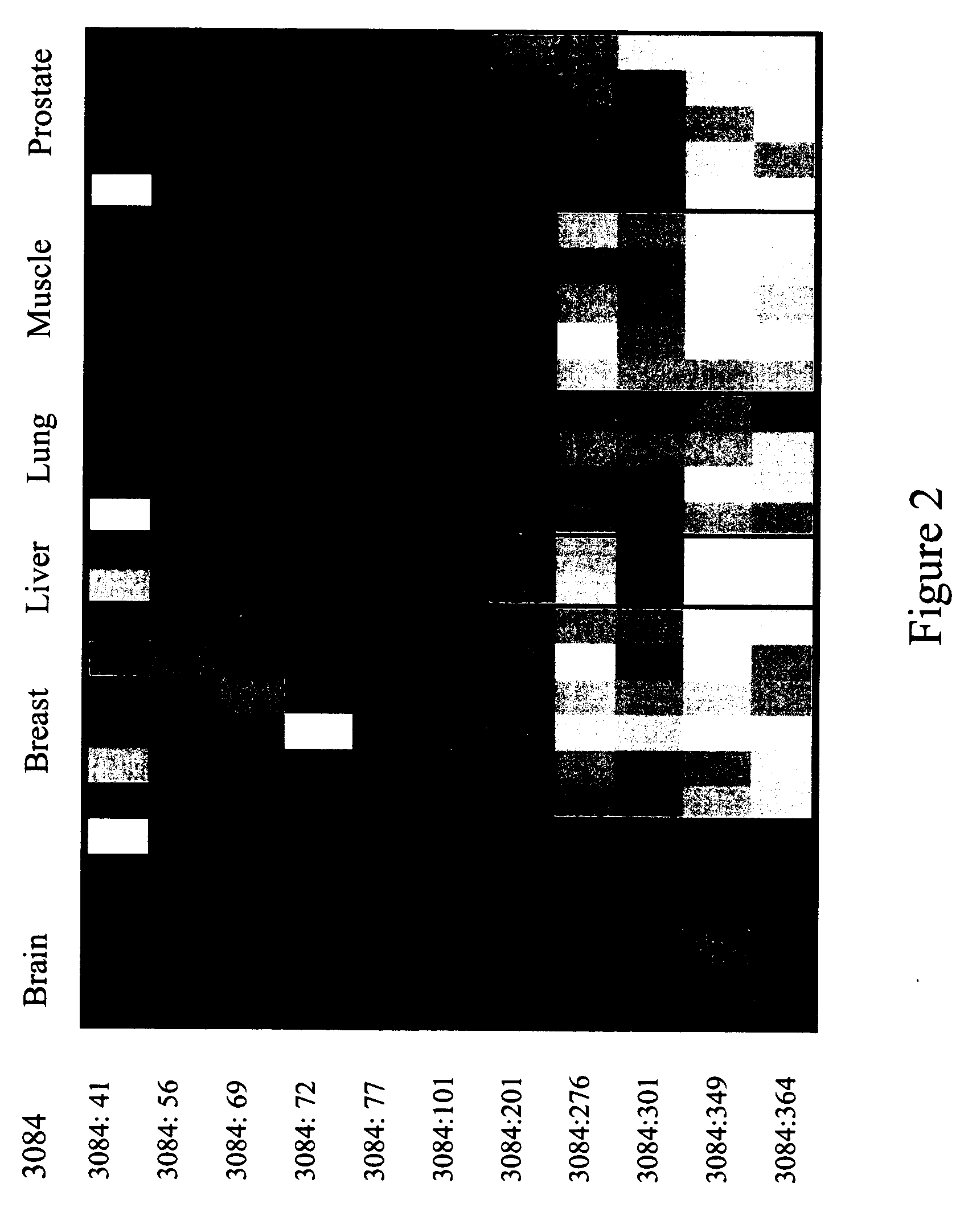
MVPs and Markers Comprising Multiple MVPs in the Major Histocompatability Complex (MHC) were Identified According to Methods of the Present Invention
[0271] The selected loci of this example are all located within the major histocompatability complex (MHC), as is disclosed in SEQ ID NO:205.
[0272] Cloned DNA cannot be used for sequencing for present purposes, because the methylation information is lost during cloning. Therefore, protocols for the design of primers and for the generation of amplificates of genes within the MHC were developed. Available sequence information from the MHC was used for this purpose, and specific primer-sets were designed to be used to amplify (gene-derived) fragments or regions comprising putative variable methylation information. The amplificates were obtained in multiplex PCR experiments, and the primer molecules designed therefore (see herein above) are listed in Table 1, and referring to the sequence protocol SEQ ID NO:137 through SEQ ID NO:204 (see ...
example 2
The Marker ROI 3083 and the Attendant Epigenetic Map is Used to Identify Liver Tissue as the Source of Origin of a Sample Containing Genomic DNA. A HeavyMethyl™ Assay is Used for Differentiation of Liver Tissue Amongst Other Tissues
[0362] The experiments of the following example occur in the setting of a diagnostic laboratory where two tubes, each containing isolated genomic DNA from one of two different tissue samples, are accidentally randomized. It is known, however, that one sample is obtained from a liver biopsy (intended for use in a molecular cancer test), whereas the other sample is derived from muscle cells of a dead body (intended for use with a SNP-based test for forensic studies). A lack of sufficient tissue material to repeat the extraction (DNA isolation) leads to a decision to quickly test each DNA for its source of origin using one of the inventive liver markers out of a group of several, as disclosed herein above according to the present invention.
[0363] According...
example 3
The Marker ROI 3105 and the Attendant Epigenetic Map is Used in a Sensitive Detection Assay for Unambiguous Identification of Breast Tissue as the Source of Origin of Genomic DNA. A HeavyMethyl™ Assay is Used for Differentiation of Breast Tissue Amongst Other Tissues
[0376] The experiments of this example are in the context of a diagnostic laboratory, where two tubes arrive at the same day from the same practitioner, who has sent in biopsy samples from two of his female patients both named Smith. No other description is deciphered, but it is known that one sample is taken from a breast biopsy (to monitor the clearance of tumor cells after surgical removal and radiation therapy), whereas the other sample comes from a lung biopsy. The genomic DNA is already isolated when the ambiguity is noticed, so that a visual differentiation is no longer possible.
[0377] According to the present invention, only a quick test employing one of the breast markers disclosed herein is required to determ...
PUM
| Property | Measurement | Unit |
|---|---|---|
| length | aaaaa | aaaaa |
| nucleic acid sequencing | aaaaa | aaaaa |
Abstract
Description
Claims
Application Information
 - R&D
- Intellectual Property
- Life Sciences
- Materials
- Tech Scout
- Unparalleled Data Quality
- Higher Quality Content
- 60% Fewer Hallucinations
Browse by: Latest US Patents, China's latest patents, Technical Efficacy Thesaurus, Application Domain, Technology Topic, Popular Technical Reports.
© 2025 PatSnap. All rights reserved.Legal|Privacy policy|Modern Slavery Act Transparency Statement|Sitemap|About US| Contact US: help@patsnap.com


