Method and apparatus for percutaneous reduction of anterior-posterior diameter of mitral valve

a technology of mitral valve and anterior-posterior diameter, which is applied in the field of mitral valve repair, can solve the problems of mechanical valve carrying the risk of thromboembolism, biological prosthesis suffering from limited durability, and mitral regurgitation, and achieve the effect of promoting coaptation of mitral leaflets

Inactive Publication Date: 2006-08-17
KARDIUM
View PDF56 Cites 245 Cited by
  • Summary
  • Abstract
  • Description
  • Claims
  • Application Information

AI Technical Summary

Benefits of technology

[0016] In general, it is an object of the present invention to provide a method and a device which is deployed in the coronary sinus and right atrium for effecting a 5-10 mm approximation of the septal annulus and lateral annulus of the mitral valve and promote coaptation of the mitral leaflets and dynamic function of the mitral valve annulus. Key to the method of the invention is appreciation that the anterior leaflet of mitral valve is not in same plane as tricuspid valve but sits close to the base of a heart and can be compressed from the right atrial side by applying pressure on the atrial septum in certain particular locations.
[0017] Some aspects of the invention relate to a device system for treating mitral regurgitation comprising an elongate element having a first end member and an opposite second end member, wherein the first end member is deployed in a coronary sinus and the second end member is deployed in a right atrium sized and configured for effecting an approximation of a septal annulus and a lateral annulus of the mitral valve. In one embodiment, the approximation is between about 1 and 20 mm, preferably between about 5 and 10 mm.
[0018] In one embodiment, the first end member of the elongate element is configured bendable that enables anchoring the first end member in the coronary sinus. In another embodiment, the first member is connected to the second end member of the elongate element by an adjustment system that is co

Problems solved by technology

Improper function of any one of these structures or in combination can lead to mitral regurgitation.
The mechanical valve carries the risk of thromboembolism and requires anticoagulation with all of its potential hazards, whereas the biological prosthesis suffers from limited durability.
Another hazard with replacement is the risk of endocarditis.
While these methods have been able to successfully treat mitral regurgitation, they have not been without problems and potential adverse consequences.
Such a procedure is expensive, requires considerable time, and is associated with high morbidity and mortality.
Due to the risks associated with this procedure, many of the sickest patients are denied the potential benefits of surgical correction of mitral regurgitation.
In addition, patients with moderate, symptomatic mitral regurgitation are denied early intervention and undergo surgical correction only after the development of cardiac dysfunction.
Furthermore, the effectiveness of such procedures is difficult to assess during the

Method used


Image

  • Method and apparatus for percutaneous reduction of anterior-posterior diameter of mitral valve
  • Method and apparatus for percutaneous reduction of anterior-posterior diameter of mitral valve
  • Method and apparatus for percutaneous reduction of anterior-posterior diameter of mitral valve

Examples


Embodiment Construction

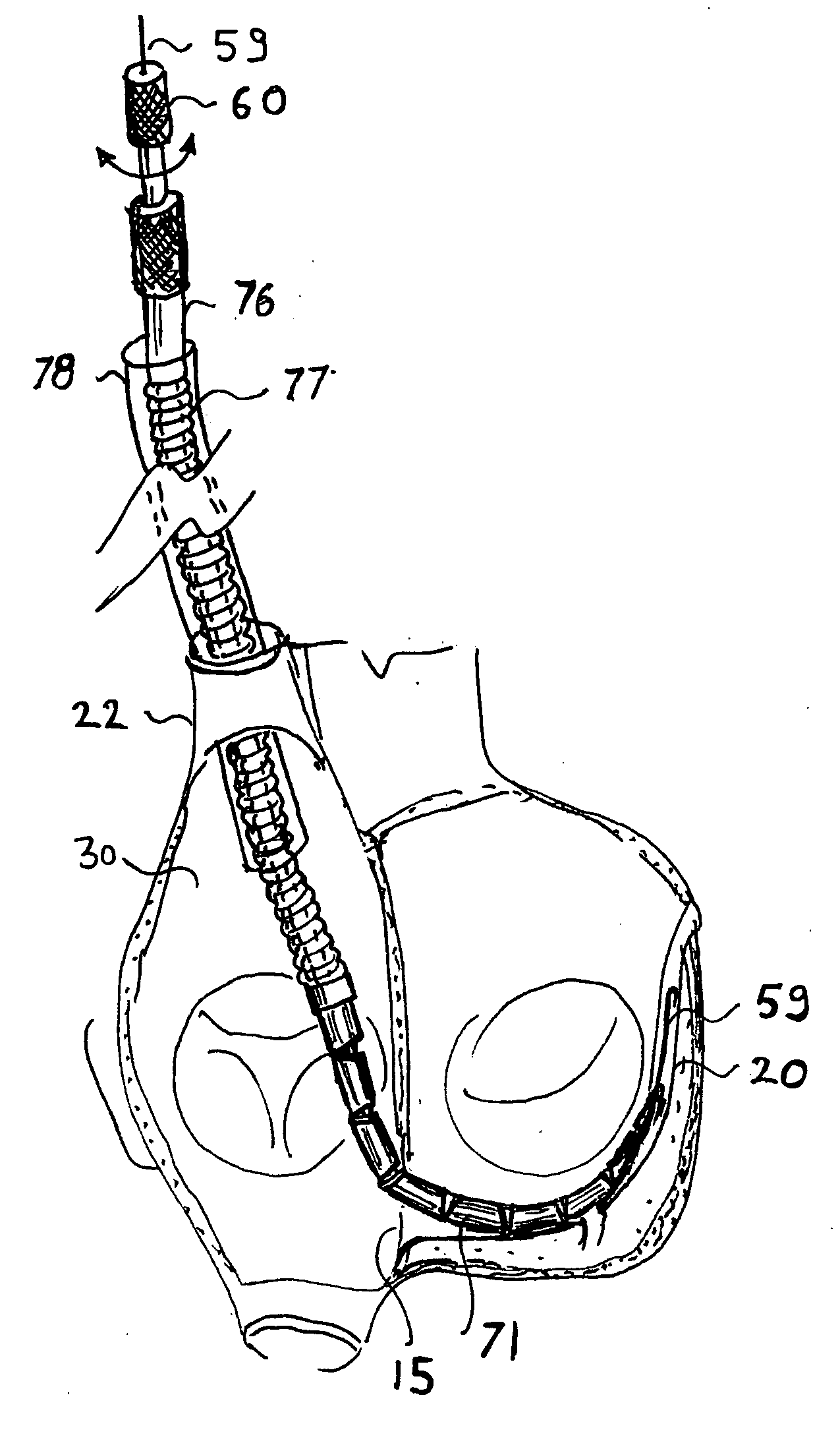
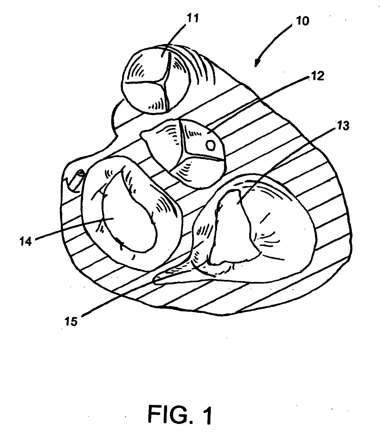
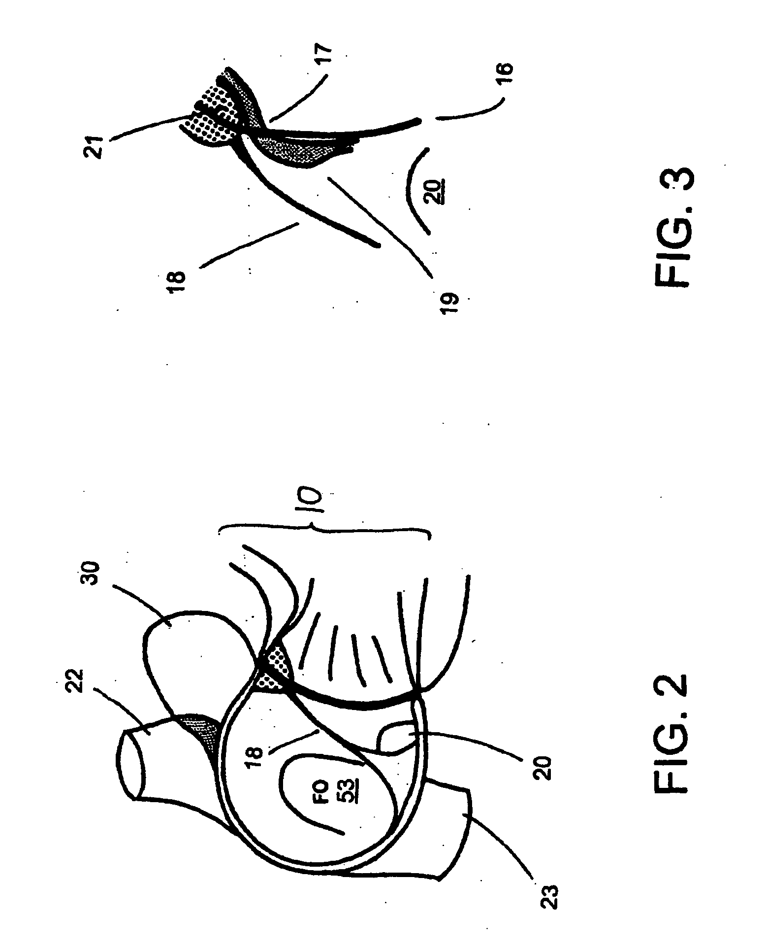
[0038]FIGS. 1-15 show a device system and methods for treating mitral regurgitation by approximating the septal and lateral (clinically referred to as anterior and posterior) annuli of the mitral valve. While the description sets forth various embodiment specific details, it will be appreciated that the description is illustrative only and should not be construed in any way as limiting the invention. Furthermore, various applications of the invention, and modifications thereto, which may occur to those who are skilled in the art, are also encompassed by the general concepts described below.

[0039] The present invention provides an improved apparatus and method to treat mitral regurgitation. Of particular importance and a salient aspect of the present invention allows mitral regurgitation to be treated without resorting to open heart surgery. This is rendered possible not only by the realization that the coronary sinus of a heart is near to and at least partially encircles the latera...


PUM


Abstract

A method and apparatus for treating mitral regurgitation by approximating the septal and lateral (clinically referred to as anterior and posterior) annulus of the mitral valve. The distal end of the device is inserted into the coronary sinus of the heart and the proximal end of the device rests within the right atrium along the tendon of Todaro and extends to at least the membranous septum of the tricuspid valve. Because the coronary sinus approximates the lateral (posterior) annulus of the mitral valve and the tendon of Todaro approximates the septal (anterior) annulus of the mitral valve, the device encircles approximately one half of the mitral valve annulus. The apparatus is then adapted to deform the underlying structures i.e. the septal annulus and lateral annulus of the mitral valve in order to move the posterior leaflet anteriorly and the anterior leaflet posteriorly and thereby improve leaflet coaptation and eliminate mitral regurgitation.

Description

FIELD OF THE INVENTION [0001] The present invention generally relates to cardiac surgery, and in particular to mitral valve repair. BACKGROUND OF THE INVENTION [0002] Mitral regurgitation with structurally normal leaflets is generally caused by ischemic heart disease and dilated cardiomyopathy. The mitral apparatus is made up of four major structural components and includes the annulus, the two leaflets, the chordae and the papillary muscles. Improper function of any one of these structures or in combination can lead to mitral regurgitation. It is generally believed that acute mitral regurgitation due to myocardial ischemia results from discordant function of the papillary muscles. Annular dilation is a major component in the pathology of mitral regurgitation regardless of causes and is manifested in mitral regurgitation related to dilated cardiomyopathy and chronic mitral regurgitation due to ischemia. [0003] The mitral valve is intended to prevent the regurgitation of blood from t...

Claims


Application Information

Patent Timeline
IPC IPC(8): A61F2/24
CPCA61B17/00234A61B2017/00243A61B2017/0496A61F2/2451A61F2/2466
Inventor LICHTENSTEIN, SAMUEL
Owner KARDIUM
Why Eureka
Try Eureka