Use of GFAP for identification of intracerebral hemorrhage

a technology of intracerebral hemorrhage and gfap, which is applied in the field of intracerebral hemorrhage identification using gfap, can solve the problems of simultaneous expansion of cells in the penumbra, physical obstruction of arterial blood supply to the brain, and blockage or severe reduction of occlusion supply to the brain with oxygen and nutrients

Inactive Publication Date: 2006-10-26
FOERCH CHRISTIAN +1
View PDF9 Cites 23 Cited by
  • Summary
  • Abstract
  • Description
  • Claims
  • Application Information

AI Technical Summary

Benefits of technology

[0031] The present invention also relates to a test kit for performing the analysis for GFAP as well as to marker panels comprising the marker GFAP and one or more additional markers used in the differentiation of intracerebral hemorrhage from other types of stroke.

Problems solved by technology

Thrombi or emboli can result from atherosclerosis or other disorders, for example, arteritis, and lead to physical obstruction of arterial blood supply to the brain.
As a consequence of vessel occlusion supply with oxygen and nutrients to the brain is blocked or severely reduced.
However, as ischemia or bleeding from hemorrhage continues, the core of dead cells can expand from the site of insult, resulting in a concurrent expansion of cells in the penumbra.
The zone immediately beyond the infarct soon lacks suitable blood concentrations of the nutrients essential for cell survival.
Hemorrhagic stroke can induce cell death by direct trauma, elevation in intracranial pressure, and the release of damaging biochemical substances in blood.
Current diagnostic methods for stroke include costly, time-consuming or invasive procedures such as noncontrast computed tomography (CT) scan, magnetic resonance imaging (MRI), or intra-arterial angiography.
Determining the immediate cause of stroke and differentiating ischemic from hemorrhagic stroke is difficult.
MRI may be more effective than CT scan in early detection of ischemic stroke, but it is less accurate at differentiating ischemic from hemorrhagic stroke, and is not widely available.
It is, however, an invasive procedure that is also limited by cost and availability.
Moreover, delays in the identification of stroke type limit the number of patients that may benefit from early intervention and appropriate therapy.
A marker, which is useful in differentiation of intracerebral hemorrhage from other types of stroke or TIAs, has to be very specific for intracerebral hemorrhage, because false classification might result in a not adequate treatment.
Recent reports from researchers investigating serum GFAP levels and their associatiation with stroke are severely limited by the methods used, and have produced controversial results.

Method used


Image

  • Use of GFAP for identification of intracerebral hemorrhage
  • Use of GFAP for identification of intracerebral hemorrhage

Examples


specific embodiments

EXAMPLE 1

Antibodies / Antibody Derivatives Used For Measurement of GFAP

[0082] With the exception of the blocking antibodies, all immunological reagents have been modified according to one of the following procedures. All antibodies used are commercially available, details / sources are given in detail below.

a) Biotinylation of Monoclonal Antibody MAB4A11 (Research Diagnostics Inc., Catalogue Number GFAPabm-411)

[0083] The purified antibody preparation comprising MAB4A11 was dialysed against the biotinylation buffer (50 mM KPO4, 100 mM NaCl, pH 8.5) and afterwards the solution was adjusted to a protein concentration of 0.5 mg / ml. D-biotinoyl-aminocaproic acid-N-hydroxysuccinimide ester was dissolved in DMSO and added to the antibody solution in a molar ratio of 1:5. The reaction was stopped by adding L-lysine and the surplus of the labelling reagent was removed by dialysis.

b) Ruthenylation of PABR-IgG, (DAKO-Cytomation, Catalogue-nr. ZO334)

[0084] The antibodies were dialysed agai...

example 2

Measurement of GFAP in a Patient Sample

[0086] Two assay formats have successfully been used in measurement of GFAP. Details are given below.

a) Microtiter Plate Assay

[0087] Wells of streptavidin-coated microtiter plates (RDG, catalogue nr. 148 705 1001) were incubated with 100 μl / well phosphate buffered saline with 0.05% Tween-20 (PBS-T) and with 1% bovine serum albumin (BSA) containing 1 μg / ml biotinylated IgG of MAB 4A11 (see Example 1a). Incubation was performed for 60 min at room temperature under shaking.

[0088] Wells were washed 4 times with the washing buffer PBS-T.

[0089] The incubation with native antigen in patient plasma, diluted in PBS-T buffer, was carried out with 100 μl / well for 1 hour at room temperature under shaking.

[0090] Wells were washed 4 times with the washing buffer PBS-T.

[0091] The incubation with PABR-IgG-digoxigenylated (cf. Example 1c) was performed with 100 μl / well PBS-T containing 8 μg / ml digoxigenylated PAB and 1% BSA for 1 hour at room temperatu...

example 3

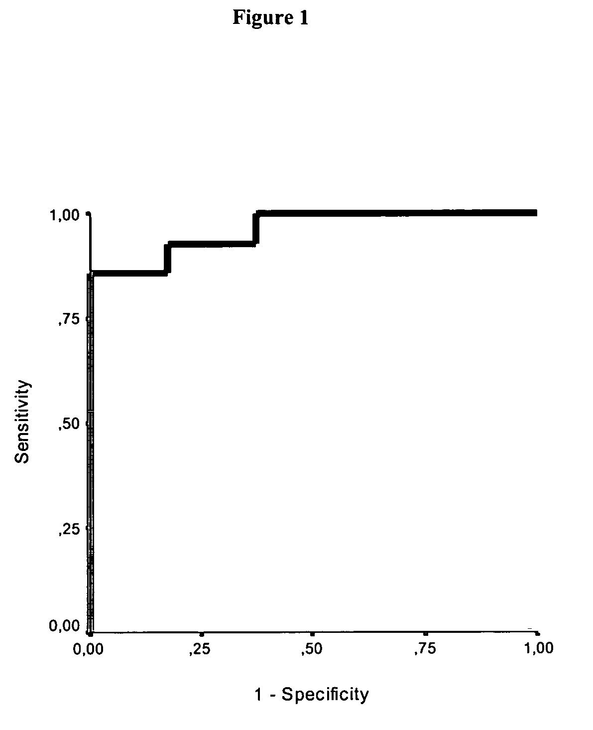
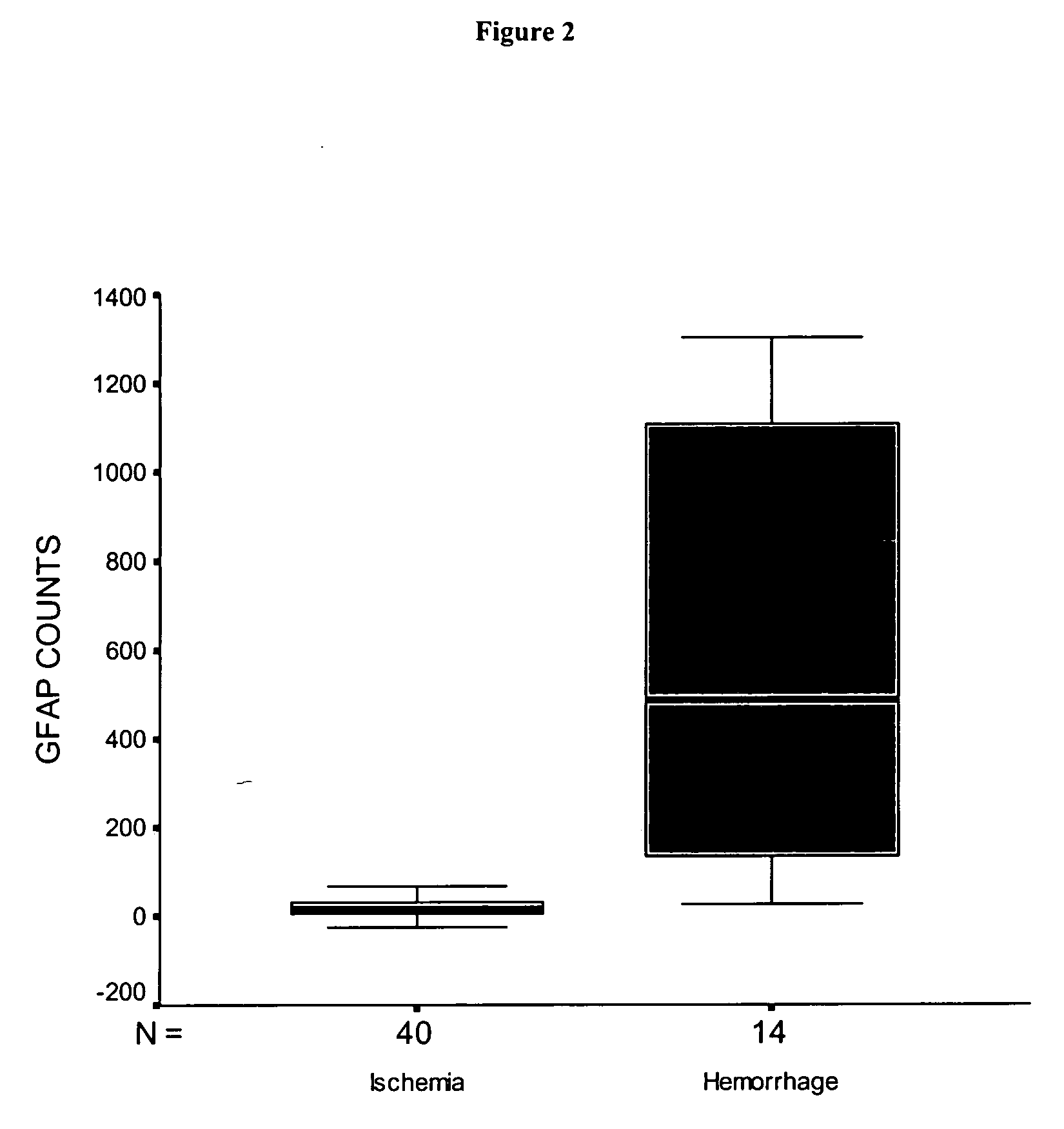
[0105] Analytical and Clinical Data as Measured in the ELECSYS Assay:

TABLE 121-fold determination of a negative and a positive serum (blocked / normal format)SpecificsampletestVal 1Val 2Val 3Val 4Val 5Val 6Val 7UnitNumberAverageSTDEVCVcountspos. serumnormal2783271127062730262027492720Counts212719451.66%108427312730274527242772276027152719275327112671259127282730blocked1645166716301593163116811645Counts211635251.56%16111621161516531625159216591615162116131646168216501648neg. serumnormal1643164316411633162916381618Counts211630140.85%2716281642163916521611161316221593163116481622163116281628blocked1616161016251567162215831605Counts211603221.40%16021620162016211592159015631581164915991617156316111605

[0106]

TABLE 2Results as obtained with the GFAP calibrators usedStandards: CSF-Samplediluted in ELECSYSStd.Specific“Universaldiluent”Test formatVal 1Val 2NumberAverageDev.CVcounts1:50000 UD (169 pg / ml)blocked174317562175090.50%1:50000 UD (169 pg / ml)normal6109621226161731.18%44111:100000 UD (8...


PUM

PropertyMeasurementUnit
concentrationaaaaaaaaaa
timeaaaaaaaaaa
timeaaaaaaaaaa

Abstract

The present invention relates to the use of glial fibrillary acidic protein (GFAP) as a diagnostic marker for intracerebral hemorrhage. The invention especially relates to methods for the early detection of intracerebral hemorrhage. Such early and rapid detection can be performed rapidly e.g. by a test strip format assay. GFAP can be used as a stand-alone marker or in combination with one or more other markers.

Description

RELATED APPLICATIONS [0001] This application is a continuation of PCT / EP2004 / 010711 filed Sep. 24, 2004, and claims priority to European application EP 03021571.9 filed Sep. 24, 2003. FIELD OF THE INVENTION [0002] The present invention relates to the use of serum glial fibrillary acidic protein (GFAP) as a diagnostic marker for intracerebral hemorrhage. The invention especially relates to methods for the very early assessment of intracerebral hemorrhage. Such early and rapid detection can be immediately performed e.g. by a test strip format assay. GFAP can be used as a stand-alone marker or in combination with one or more other markers. BACKGROUND OF THE INVENTION [0003] Stroke is a manifestation of vascular injury to the brain which is commonly secondary to arteriosclerosis or cardiac disease, and is the third leading cause of death (and the second most common cause of neurological disability) in the United States. [0004] Stroke can be categorized into two major types, “ischemic st...

Claims


Application Information

Patent Timeline
Patent Type & Authority Applications(United States)
IPC IPC(8): G01N33/53G01N33/543G01N33/68
CPCG01N2800/2871G01N33/6896G01N33/6893
Inventor CURDT, INGOSITZER, MATTHIASFOERCH, CHRISTIAN
Owner FOERCH CHRISTIAN
Why Eureka
Try Eureka