System and method for spectral-encoded high-rate hemodynamic tomography

a hemodynamic tomography and spectral encoded technology, applied in mammography, medical science, diagnostics, etc., can solve the problems of requiring significant acquisition time to gather sufficient path attenuation and scattering data, and the use of electromagnetic radiation of these wavelengths for imaging

Inactive Publication Date: 2006-11-02
TRUSTEES OF DARTMOUTH COLLEGE THE
View PDF17 Cites 39 Cited by
  • Summary
  • Abstract
  • Description
  • Claims
  • Application Information

AI Technical Summary

Problems solved by technology

The usefulness of electromagnetic radiation of these wavelengths for imaging deep soft tissues has been limited because of the high degree of scattering that occurs, and resultant poor image quality with ordinary optical techniques.
As a result, it can take significant acquisition time to gather sufficient path attenuation and scattering data to develop a high-resolution parameterized tissue model.
Oxygenation of soft tissue is not always uniform.
For example, in atherosclerotic disease, impaired blood flow may result in portions of tissue becoming ischemic, or having less than normal oxygen saturation, especially during activity.
For example, it is known that many malignant tumors require so much oxygen that portions of the tumor may become ischemic and necrotic despite their increased vascularity.
These zones of ischemia tend to be more prone to forming slow or non-healing ulcers than normally oxygenated tissue.

Method used


Image

  • System and method for spectral-encoded high-rate hemodynamic tomography
  • System and method for spectral-encoded high-rate hemodynamic tomography
  • System and method for spectral-encoded high-rate hemodynamic tomography

Examples


Embodiment Construction

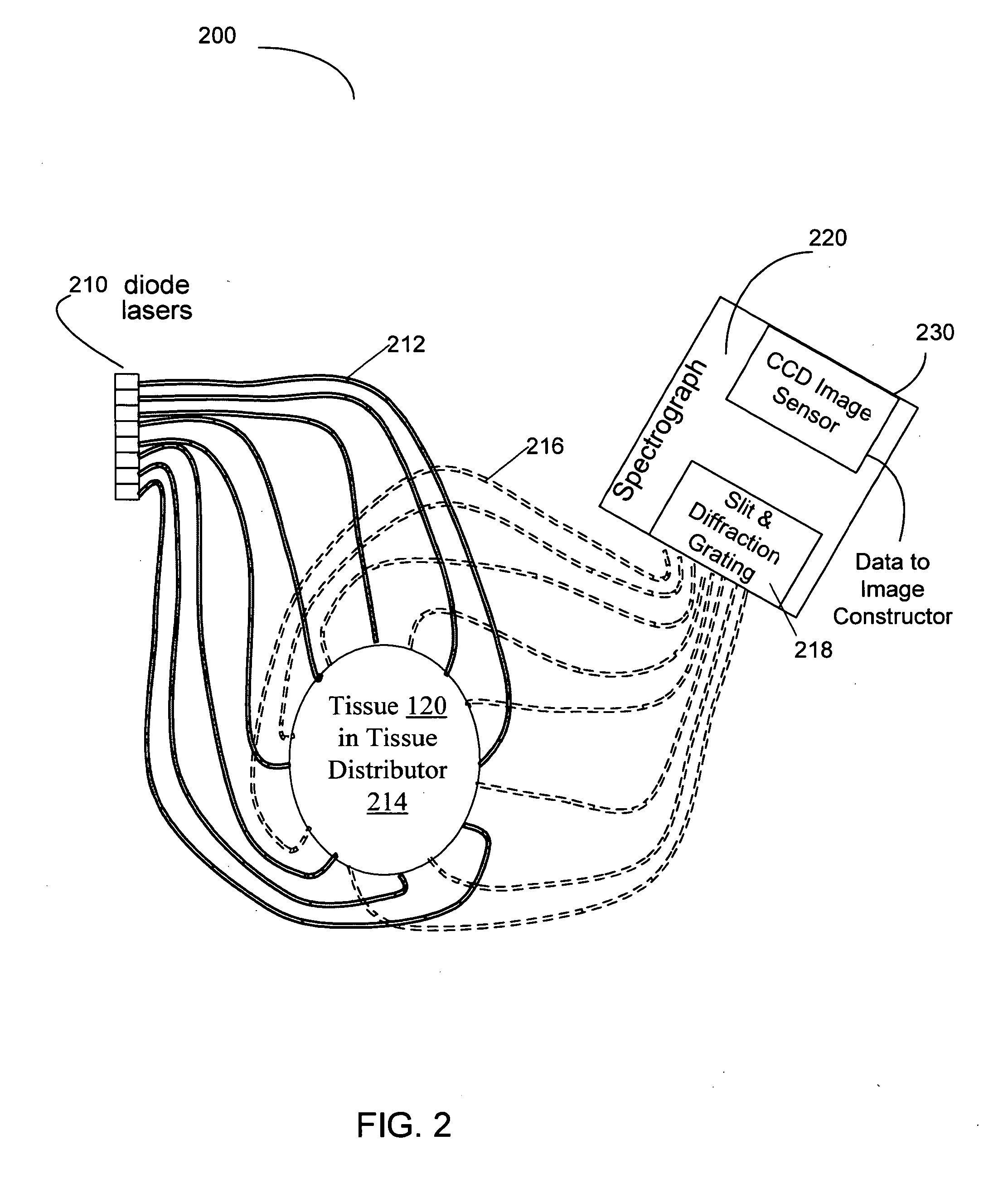
[0032] It has been found that the spectral characteristics of features within mammalian tissue are sufficiently broad that, if visible or near-infrared light from a group of single-mode lasers are used to interrogate tissue with small wavelength separation between lasers, radiation from each laser suffers levels of attenuation and scattering largely similar to radiation from the other lasers of the group.

[0033] When lasers at separate wavelengths are associated with different source positions, a spectral encoding of source origin occurs in a way that can be detected and decoded in parallel at any reception point.

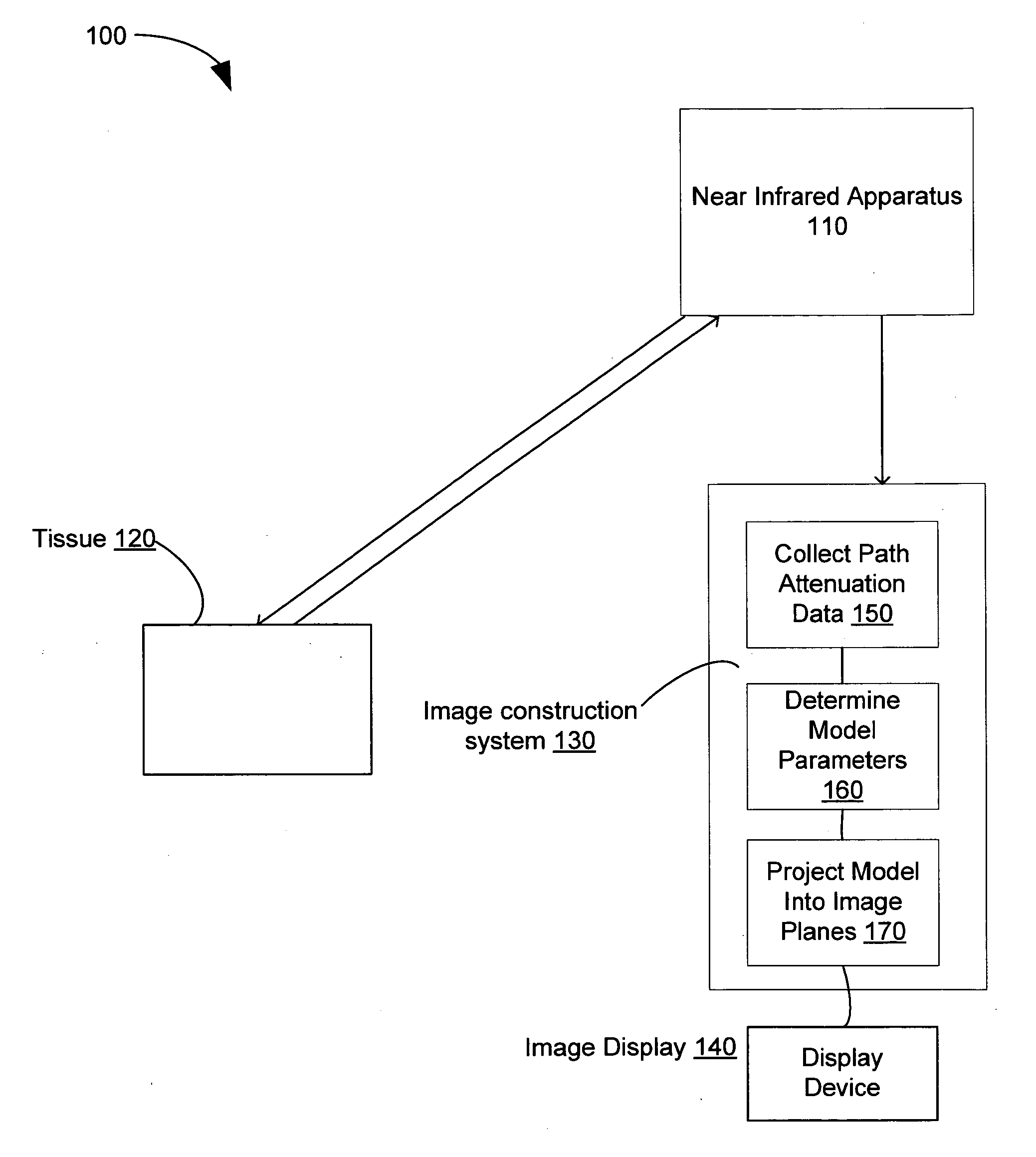
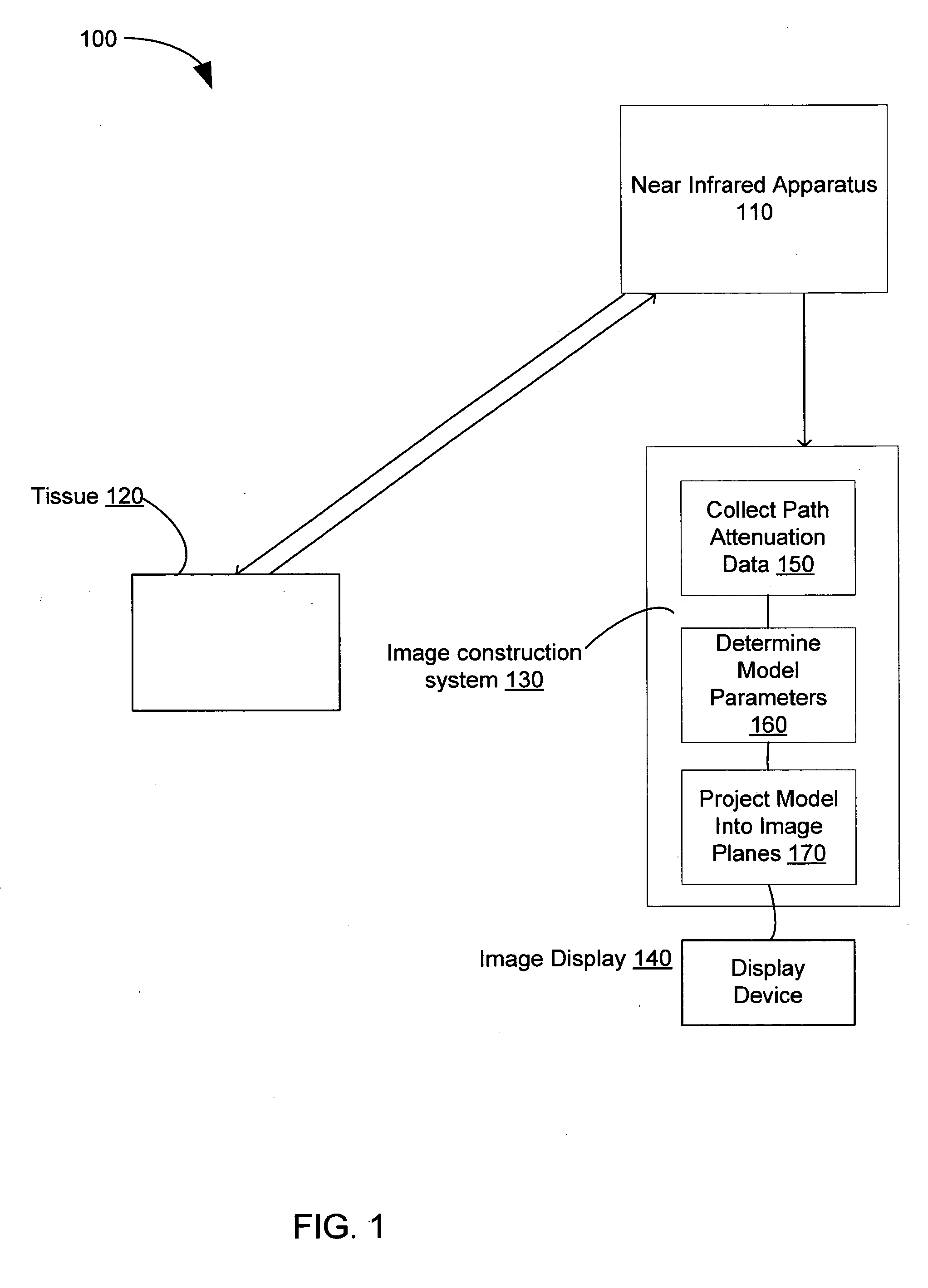
[0034]FIG. 1 shows a System 100 of Spectral-Encoding for High-Rate Hemodynamic Tomography. The System 100 includes a near-infrared apparatus 110, mammalian tissue 120, an image construction system 130, and an image display 140. A tissue model, or phantom, may be used in place of tissue 120 during development and calibration. The near-infrared apparatus 110 generates path-a...


PUM


Abstract

An optical tomography system has a group of lasers for generating light of multiple distinct wavelengths within a first wavelength band. Light from these lasers is directed into mammalian tissue at laser-specific locations on the tissue, and light from the mammalian tissue is collected at a plurality of reception points. Collected light from each reception point is separated according to its wavelength, and received by a photodetector to produce path attenuation signals representing attenuation along paths between the laser-specific locations on the tissue and the reception points. Image construction apparatus generates a tomographic image of heme concentrations in the mammalian tissue from the path attenuation signals. In an alternative embodiment, there is a second group of lasers operating in a second wavelength band, and the image construction apparatus can generate an image of heme oxygenation In the mammalian tissue.

Description

STATEMENT REGARDING FEDERALLY SPONSORED RESEARCH [0001] The United States Government has certain rights in this invention pursuant to contract number 1R21CA100984-01A1 awarded by the National Institutes of Health.BACKGROUND [0002] 1. Field [0003] The present application relates to the field of Near-Infrared (NIR) tomography for imaging mammalian tissue. This method introduces a method for imaging in parallel with multiple sources simultaneously activated. [0004] 2. Description of the Related Art [0005] Visible light and near infrared radiation is absorbed by the Heme group of myoglobin and hemoglobin molecules. Further, the spectral characteristics of absorption by heme groups varies noticeably with their degree of oxygenation. Therefore, imaging mammalian soft tissues with visible and near-infrared wavelengths offers potentially high contrast between portions of the tissue containing high (such as blood and muscle) and low (such as fat) amounts of heme, and between highly oxygenate...

Claims


Application Information

Patent Timeline
Patent Type & Authority Applications(United States)
IPC IPC(8): A61B6/00
CPCA61B5/0073A61B5/0091A61B5/7239A61B5/4312A61B5/14551
Inventor POGUE, BRIAN WILLIAMPIAO, DAQING
Owner TRUSTEES OF DARTMOUTH COLLEGE THE
Why Eureka
Try Eureka