Multiple transducers for intravascular ultrasound imaging

a technology of ultrasound imaging and transducer, which is applied in the field of miniature actuators, can solve the problems of difficult to check the condition inside the vessel after surgery, difficult to find the exact location of the stenosis, and very serious coronary artery diseas

Inactive Publication Date: 2007-01-18
VOLCANO CORP
View PDF2 Cites 120 Cited by
  • Summary
  • Abstract
  • Description
  • Claims
  • Application Information

AI Technical Summary

Benefits of technology

[0012] In some embodiments the actuator means comprises a first anchor, a second anchor, at least one movable element, a first SMA actuator connected to the first anchor and a m

Problems solved by technology

Coronary artery disease is very serious and often requires an emergency operation to save lives.
Unfortunately, with X-ray fluoroscopic images, there is a great deal of uncertainty about the exact extent and orientation of the atherosclerotic lesions responsible for the occlusion, making it difficult to find the exact location of the stenosis.
In addition, though it is known that restenosis can occur at the same place, it is difficult to check the condition inside the vessels after surgery.
Existing intravascular imaging devices are too large and insufficiently flexible to be placed simultaneously with other devices.
These devices have limitations in incorporating other interventional devices into a combination device for therapeutic aspects.
They require a large space inside catheter such that there is not enough room to accom

Method used


Image

  • Multiple transducers for intravascular ultrasound imaging
  • Multiple transducers for intravascular ultrasound imaging
  • Multiple transducers for intravascular ultrasound imaging

Examples


Embodiment Construction

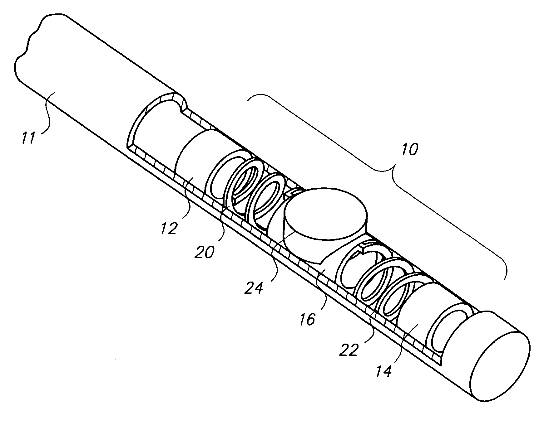
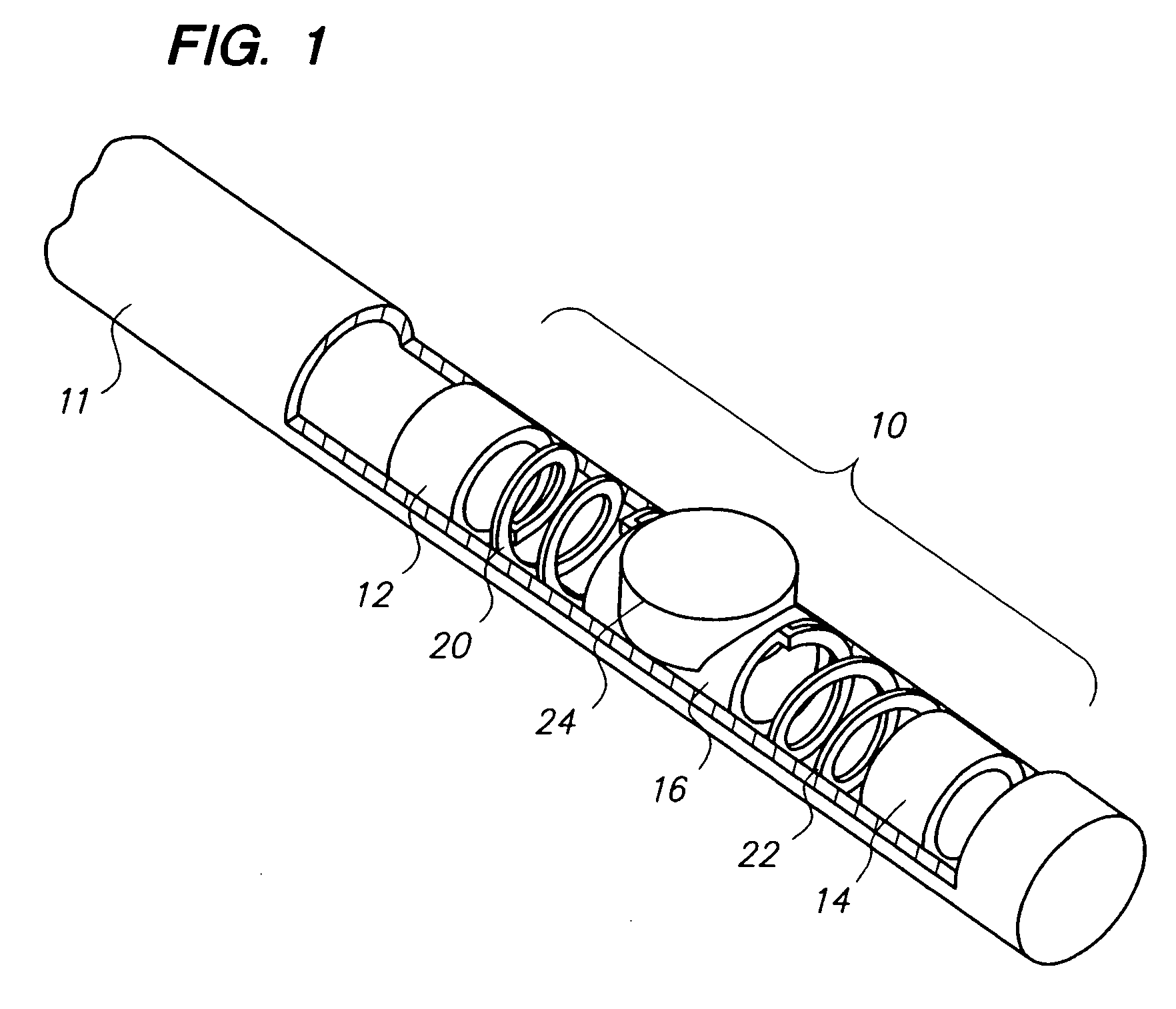
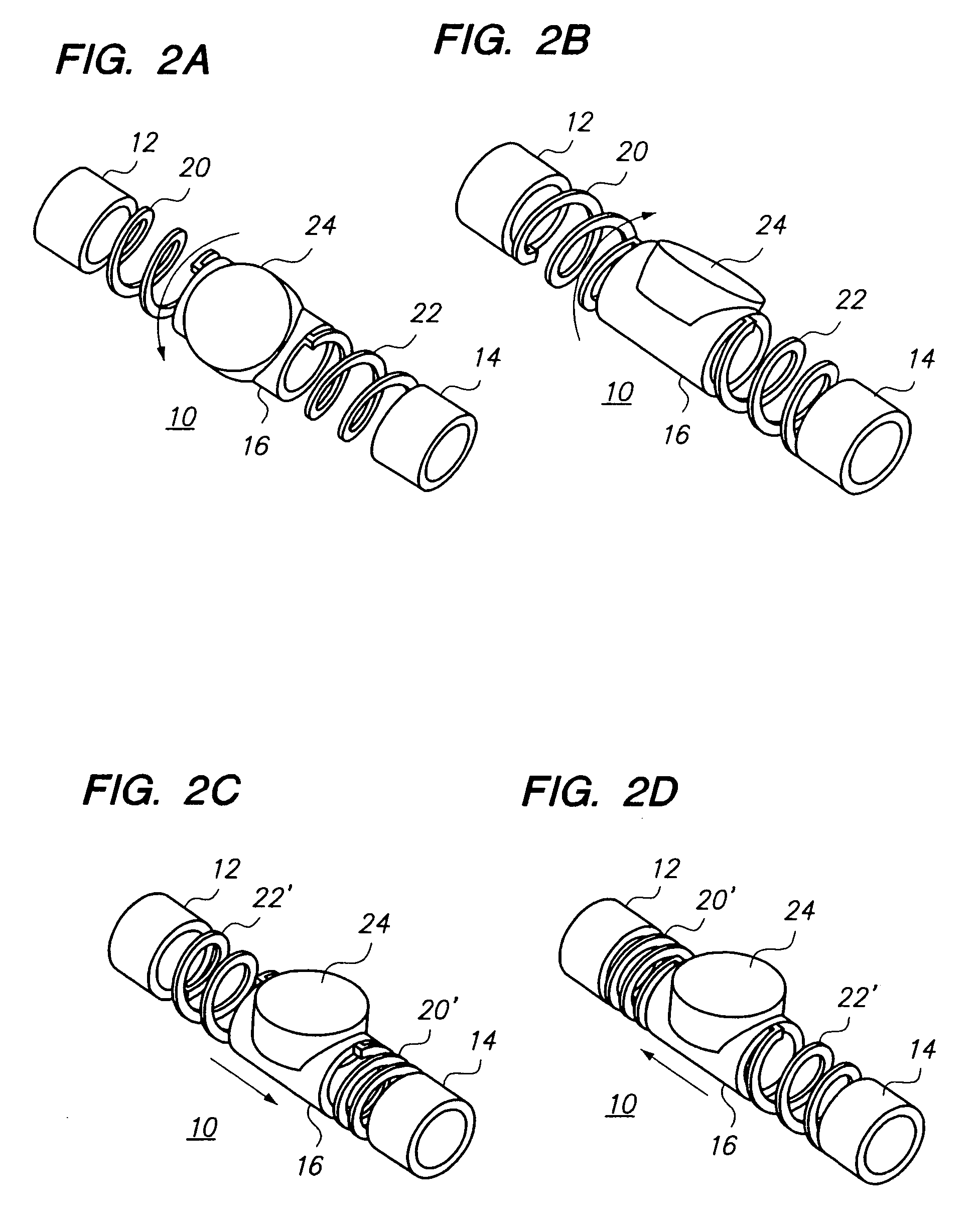
[0029] The present invention relates to imaging devices for intravascular imaging, although the present invention is not limited to this preferred application. Imaging of the intravascular space, particularly the interior walls of the vasculature can be accomplished by a number of different means. Two of the most common are the use of ultrasound energy, commonly known as intravascular ultrasound (IVUS) and optical coherence tomography (OCT). Both of these methods are optimized when the instruments (IVUS or OCT) used for imaging a particular portion of the vasculature are repeatedly swept over the area being imaged.

[0030] To address the limitations in current devices, a new intravascular imaging device is described based on a Shape Memory Alloy (SMA) actuator mechanism embedded inside an elongate member such as a guide wire or catheter. The present invention utilizes a novel SMA mechanism to provide side-looking imaging by providing movement for an ultrasound transducer or OCT eleme...


PUM


Abstract

The present invention relates to a new intravascular ultrasound imaging device with an increased field of view. By using more than one ultrasound transducer crystal that is capable of producing an ultrasonic signal for imaging, and preferably aligning the individual crystals on a shared backing with at least one edge of the crystals in contact, the field of view generated by a given actuator mechanism is increased without substantially increasing the overall dimensions of the device. Also disclosed are methods of using the same.

Description

CROSS-REFERENCE TO RELATED APPLICATIONS [0001] This application claims priority under 35 U.S.C. 119 to U.S. Provisional Application Ser. No. 60 / 678,676, filed May 4, 2005, titled “Multiple transducers for large field of view in intravascular ultrasound imaging,” U.S. Provisional Application Ser. No. 60 / 677,944, filed May 4, 2005, titled “Shape memory alloy (SMA) mechanism for side-looking intravascular imaging,” U.S. Provisional Application Ser. No. 60 / 710,304, filed Aug. 22, 2005, titled “Guide wire enabled with intravascular ultrasound imaging for interventional applications,” and U.S. Provisional Application Ser. No. 60 / 711,653, filed Aug. 25, 2005, titled “Miniature mirror-based intravascular ultrasound imaging device for interventional applications,” and U.S. Provisional Application Ser. No. 60 / 781,786, filed Mar. 13, 2006, titled “Electrically driven miniature intravascular optical coherence tomography imaging device,” the entire contents of each of which are incorporated here...

Claims


Application Information

Patent Timeline
IPC IPC(8): A61B8/14
CPCA61B2019/5217F16C2202/28A61M25/0138A61M25/0158G01S7/52079G01S15/8943G10K11/004A61B5/0066A61B5/02007A61B8/12A61B8/145A61B8/445A61B8/4461A61B8/4483A61B2019/528F03G7/065A61B2090/3614A61B2090/3784
Inventor PARK, BYONG-HORUDY, STEPHEN M.
Owner VOLCANO CORP
Why Eureka
Try Eureka