Reinforced matrices

a matrix and reinforcement technology, applied in the field of reinforcement matrix, can solve the problems of cartilage matrix stiffness reduction, cartilage matrix injury, and matrix rupture,

Inactive Publication Date: 2007-02-01
ASCULAI SAMUEL +1
View PDF12 Cites 2 Cited by
  • Summary
  • Abstract
  • Description
  • Claims
  • Application Information

AI Technical Summary

Benefits of technology

[0013] In one embodiment, the present invention provides a method for the reinforcement of matrices with an internal scaffold. One embodiment of the present invention is directed to a method for making a collagen-based matrix comprising incubating collagen with one or more scaffold-forming proteins to form a collagen-protein suspension, lyophilizing the suspension to form a fleece-like material, and pressing the fleece-like material into sheets to form a matrix. In one

Problems solved by technology

Injuries to the cartilage of the knee or other joints often result from abnormal mechanical loads which deform the cartilage matrix.
The loads applied to the joint can rupture the collagen network in the matrix and decrease the stiffness of the cartilage matrix.
Cartilage injuries are diffi

Method used


Image

  • Reinforced matrices
  • Reinforced matrices
  • Reinforced matrices

Examples


example 1

[0052] Chondrocytes were grown in minimal essential culture medium containing HAM F12, 15 mM Hepes buffer and 5 to 7.5% autologous serum in a CO2 incubator at 37° C. and handled in a Class 100 laboratory at Verigen Transplantation Service ApS, Copenhagen, DK. Other compositions of culture medium may be used for culturing the chondrocytes.

[0053] The cells were trypsinised using trypsin EDTA for 5 to 10 minutes and counted using Trypan Blue viability staining in a Bürker-Türk chamber. The cell count was adjusted to 7.5×10⁵ cells per ml. One NUNCLON™ plate was uncovered in the Class 100 laboratory.

[0054] Six Pieces of a size of 1 cm2 each of commercially available collagen I / III fleece (Chondro-Gide®, Geistlich, CH) were placed under aseptic conditions into the bottom of the well in the NUNCLON™ cell culture tray.

[0055] Approximately 5×10⁶ of the chondrocytes in 5 ml of the culture medium were placed directly on top of the carrier material and dispersed over the surface. The plate w...

example 2

[0059] Chondrocytes were grown in minimal essential culture medium containing HAM F12, 15 mM Hepes buffer and 5 to 7.5% autologous serum in a CO2 incubator at 37° C. and handled in a Class 100 laboratory at Verigen Transplantation Service ApS, Copenhagen, DK. Other compositions of culture medium may be used for culturing the chondrocytes.

[0060] The cells were trypsinised using trypsin EDTA for 5 to 10 minutes and counted using Trypan Blue viability staining in a Bürker-Türk chamber. The cell count was adjusted to 7.5×10⁵ cells per ml. One NUNCLON™ plate was uncovered in the Class 100 laboratory.

[0061] Six Pieces of a size of 1 cm2 each of a collagen I / III matrix (Immedex, France) was cut to a suitable size fitting into the bottom of the well in the NUNCLON™ cell culture tray and placed under aseptic conditions on the bottom of the well.

[0062] Approximately 5×10⁵ of the chondrocytes in 5 ml of culture medium were placed directly on top of the carrier material and dispersed over th...

example 3

[0065] Six pieces, 1 cm2 each in size, of the collagen I / III matrix of Example 2 were incubated for 2 hours at a temperature of 50° C. under gentle stirring with a solution of soluble elastin (EPC Inc., USA) in a suitable buffer such as phosphate buffer (0.02M KH2PO4, pH 7.4) and the pH value was then brought down to 5.0 by adding acetic acid under gentle stirring. The coacervation reaction was allowed to occur for 5 hours.

[0066] The suspension was then lyophilized at a temperature of 25° C. and a pressure of 0.05 mbar.

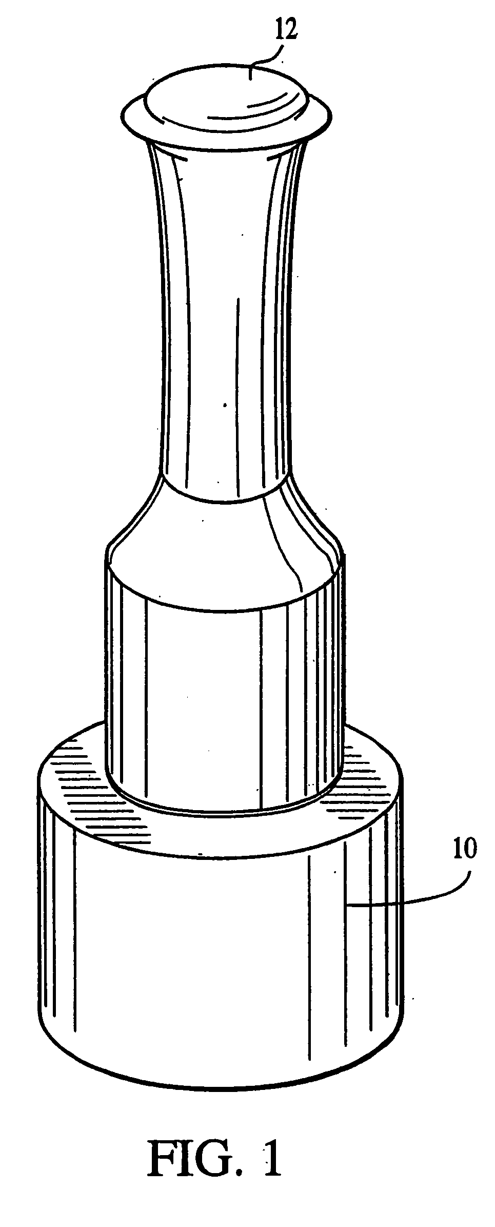
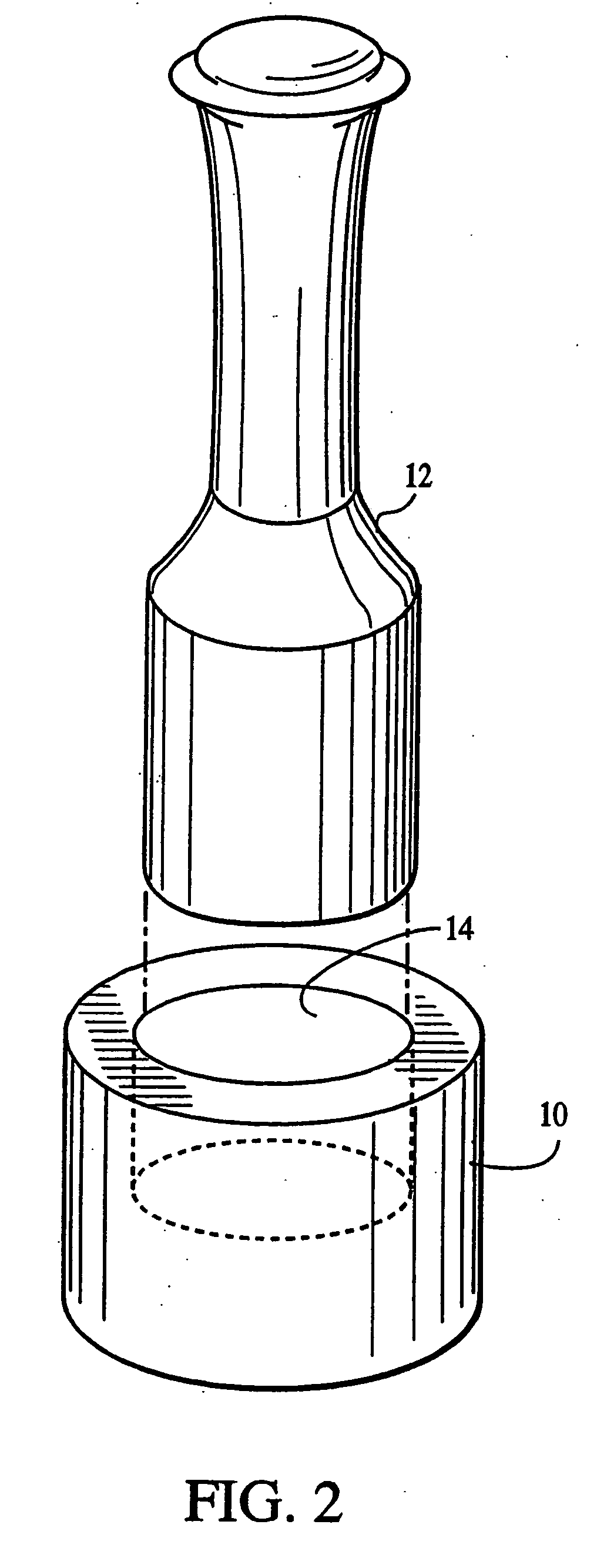
[0067] The lyophilization yielded a fleece-like material which was pressed mechanically using the apparatus in FIGS. 1 and 2 into sheets for use with cells as an implantation article. The material was pressed for about 24 hours until a sheet-like material which resisted tearing upon being handled was obtained.

[0068] Six Pieces, each 1 cm2 in size, of the fleece matrix were cut to a suitable size fitting into the bottom of the well in the NUNCLON™ cell culture tray ...


PUM

PropertyMeasurementUnit
Concentrationaaaaaaaaaa

Abstract

A reinforced matrix membrane containing one or more scaffold-forming proteins suitable for cell growth and for use in chondrocyte cell transplantation, and method of making same. The scaffold is incubated with the collagen matrix in solutions, colloidal dispersions, or suspensions of stabilizing proteins. The reinforced matrix may be used in tissue engineering, cartilage transplantation, bone and cartilage grafting, healing, joint repair and the prevention of arthritic pathologies.

Description

FIELD OF INVENTION [0001] The present invention relates to a reinforced matrix, and a method to stabilize and reinforce matrices. BACKGROUND OF THE INVENTION [0002] Injuries to the cartilage of the knee or other joints often result from abnormal mechanical loads which deform the cartilage matrix. The loads applied to the joint can rupture the collagen network in the matrix and decrease the stiffness of the cartilage matrix. [0003] Cartilage injuries are difficult to treat because human articular cartilage has a limited capacity for regeneration once it has been damaged. Type II collagen is the main structural protein of the extracellular matrix in articular cartilage. Type II collagen, similar to other types of collagen, is comprised of three collagen polypeptides which form a triple helix configuration. The polypeptides are intertwined with each other and possess at each end telopeptide regions that provide the cross-linking between the collagen polypeptides. Collagen matrices in t...

Claims


Application Information

Patent Timeline
IPC IPC(8): A61K38/39A61K35/12A61F2/00A61L27/00A61K35/32A61L27/26
CPCA61K35/32A61K38/012A61L27/26A61L27/3817A61L27/3843A61L27/38A61L27/227C08L89/06A61K2300/00C08L5/08
Inventor ASCULAI, SAMUELGIANNETTI, BRUNO
Owner ASCULAI SAMUEL
Why Eureka
Try Eureka