In situ polypeptide identification

a polypeptide and in situ technology, applied in the field of in situ polypeptide identification, can solve the problems of contaminant problems, increased time to prepare homogenization,

Inactive Publication Date: 2007-03-29
F HOFFMANN LA ROCHE INC
View PDF1 Cites 19 Cited by
  • Summary
  • Abstract
  • Description
  • Claims
  • Application Information

AI Technical Summary

Problems solved by technology

However, the need for tissue homogenization in these methods present problems with regard to incre

Method used


Image

  • In situ polypeptide identification
  • In situ polypeptide identification
  • In situ polypeptide identification

Examples


example 1

Model System for Nephrotoxicity

Example 1a)

Animal Treatment

[0059] Permission for animal studies was obtained from the local regulatory agencies, and all study protocols were in compliance with animal welfare guidelines. Male HanBrl: Wistar rats of approximately 12 weeks of age (300g±20%) were obtained from BRL, Füllinsdorf, Switzerland. The animals were housed individually in Macrolone cages with wood shavings as bedding at 200C and 50% relative humidity in a 12-hr light / dark rhythm with free access to water and Kliba 3433 rodent pellets (Provimi Kliba AG, Kaiseraugst, Switzerland).

[0060] Animals were treated for 7 consecutive days with 100 mg / kg / day Gentamicin (dissolved in saline) or vehicle by subcutaneous (sc) injections and sacrificed 24 hours after the last application by CO2 inhalation. Immediately preceding sacrifice, terminal blood samples for clinical chemistry investigations were collected from the retroorbital sinus under isofluoran anesthesia.

example 1b

)

Histology

[0061] Representative kidney samples were fixed in 10% neutral buffered formalin. All samples were processed using routine procedures and embedded in Paraplast. Tissue sections approximately 2-3 microns were cut and stained with hematoxylin-eosin (HE) or periodic acid-Schiff (PAS).

example 2

MALDI-IMS of Rat Kidney Cortex Treated with Gentamicin

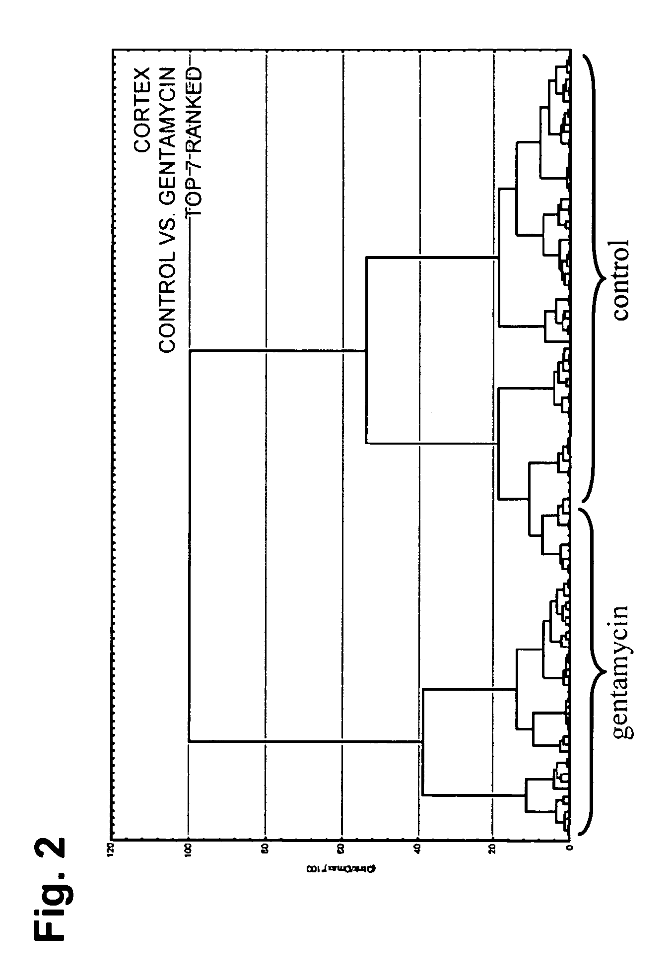
[0062] The differential protein expression was studied within the areas of the kidney (Cortex, Medulla, Papilla) to investigate whether the histopathologically-observed toxicity of gentamicin could be correlated by IMS. Several protein peaks in the cortex area were significantly modulated upon gentamicin administration.

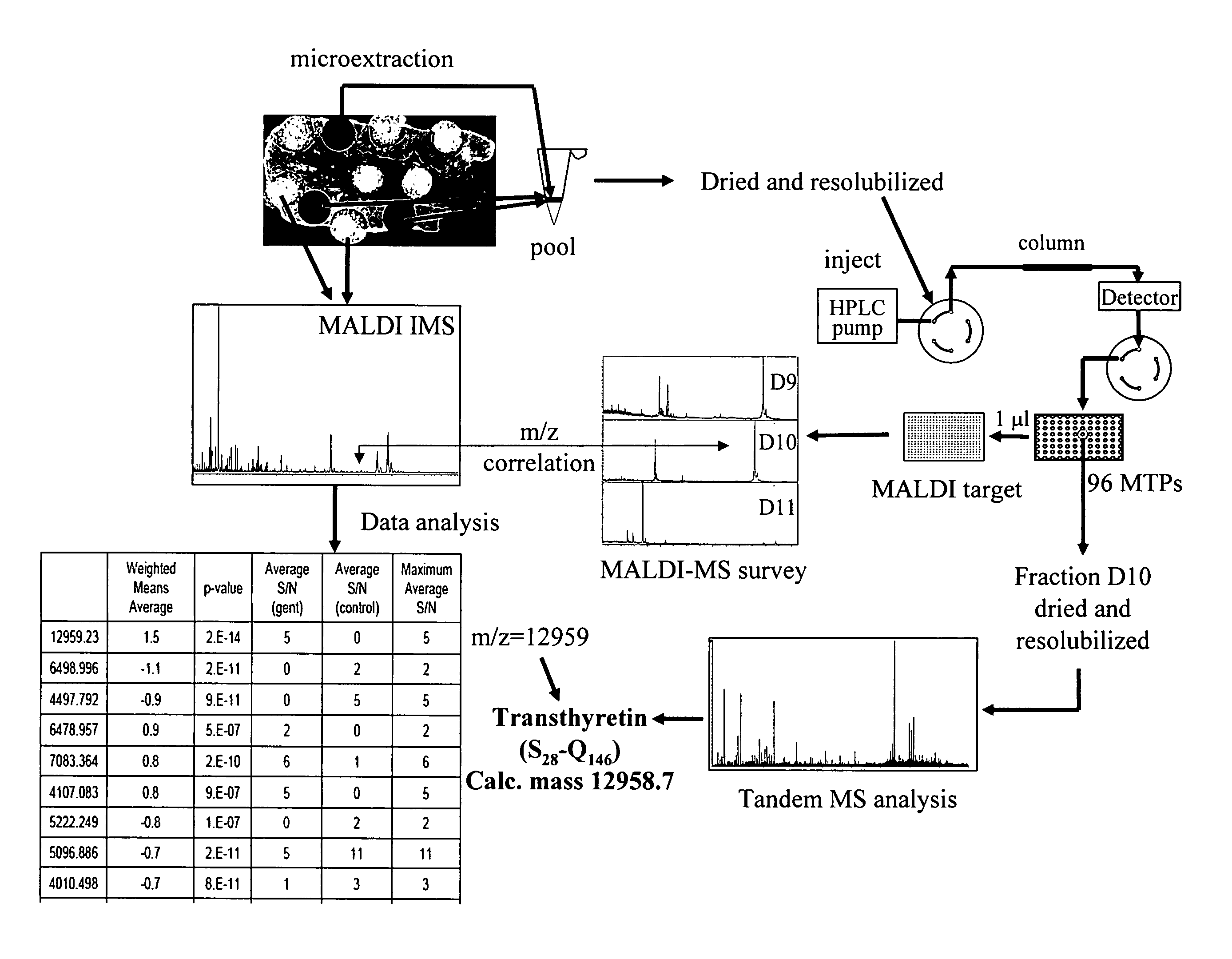
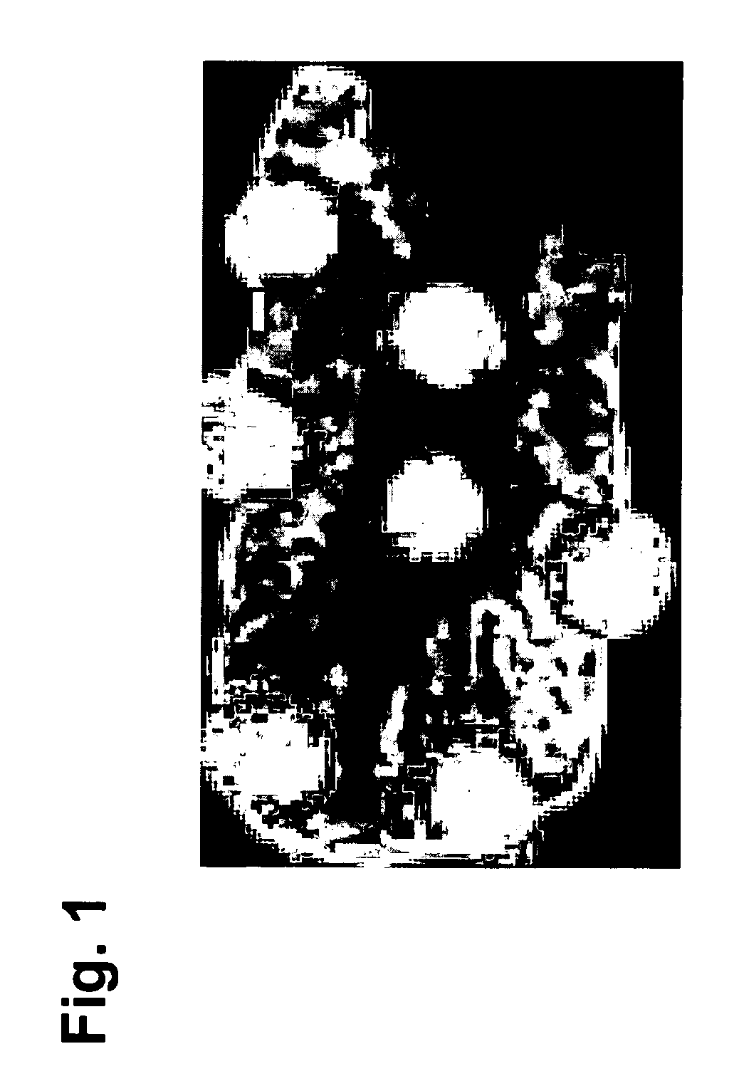
[0063] Marked degeneration / regeneration of proximal tubules (grade 4) was clearly observed in rats treated with 100 mg / kg / day gentamicin for 7 days. This finding was correlated to the analysis of rat kidney thin sections by MALDI IMS. 9 control and 9 gentamicin-treated rat kidneys thin sections (3 animals, 3 sections from each animal; the thin section was spotted with matrix as shown in FIG. 1) were subjected to mass spectrometric analysis as described below.


PUM


Abstract

The invention relates to a method for identifying protein biomarkers directly in a specific region of interest of a tissue which comprises the identification of a mass of interest by analysis of MALDI imaging mass spectrometry results, followed by the identification of the protein represented by said mass of interest by direct elution from the specific region of interest of a tissue, fractionation and tandem mass spectrometry.

Description

PRIORITY TO RELATED APPLICATIONS [0001] This application claims the benefit of European Application Nos. 05104829.6, filed Jun. 3, 2005 and 06111461.7, filed Mar. 21, 2006, both of which are hereby incorporated by reference in their entirety. This application also claims the benefit of co-filed US Application No. 23145, entitled “Transthyretin as a Marker for Nephrotoxicity”, which is likewise incorporated by reference in its entirety. FIELD OF THE INVENTION [0002] The present invention relates to methods for de novo in situ protein biomarker identification, and more particularly, methods for de novo in situ protein identification (protein biomarker) wherein proteins are directly eluted from the tissue region of interest (without any need for tissue homogenization). BACKGROUND OF THE INVENTION [0003] Proteomics techniques have the potential to correlate differential protein expression with environmental disease or toxicant exposure. High resolution two-dimensional gel electrophoresi...

Claims


Application Information

Patent Timeline
IPC IPC(8): H01J49/00
CPCG01N27/622H01J49/164H01J49/004
Inventor AUGUSTIN, ANGELIQUEDUCRET, AXELLANGEN, HANNOMEISTERMANN, HELENE
Owner F HOFFMANN LA ROCHE INC
Why Eureka
Try Eureka