Diaphragm entry for posterior surgical access

Active Publication Date: 2007-04-12
ATRICURE
View PDF60 Cites 115 Cited by
  • Summary
  • Abstract
  • Description
  • Claims
  • Application Information

AI Technical Summary

Benefits of technology

[0016] The devices described herein allow for creating a temporary cavity between organs in a body. Generally, the devices include an elongate member having at least one working channel extending therethrough, the elongate member having a distal portion adapted for insertion into the body and a proximal portion, the elongate member having sufficient column strength to allow insertion of the distal portion between organs, at least one opening at a distal end of the elongate member, where the working channel(s) exits the elongate member at the opening(s), and a f

Problems solved by technology

With a median sternotomy, although the heart can be lifted and manipulated by hand, the posterior surface of the heart or other organs is still not readily visible unless the heart is significantly rotated or lifted.
However, significant rotation or lifting of the heart may cause undesirable hemodynamic issues during beating heart procedures.
Any coughing or dramatic movement is extremely painful to the patient because the chest moves.
Clearly, rehabilitation after the medial sternotomy requires a significant amount of time.
Much of the pain during the rehabilitation process is due to the cutting of the muscles.
A thoracotomy provides limited access and visualization to the heart unless endoscopes are used.
Yet, even the use of endoscopes provides limited access to the posterior regions of the heart and lungs because these organs cannot be lifted or rotated easily.
However, this technique does not provide ready access or visualization of posterior regions of the heart or other organs.
Accordingly, subxyphoid techniques are often limited to procedures that target the anterior or apical ventricular regions.
The conventional surgical techniques discussed do not provide the medical practitioner with sufficient visibility of anatomic structures within the thoracic cavity.
For example, these procedures do not provide sufficient visibility for anatomic structures located along or adjacent to the posterior surface of the heart or lungs.
Accordingly, surgical practitioners may be hesitant to treat tissues located along or adjacent to the posterior heart or lungs during less invasive procedures, given the inability to visually observe the target area.
As such, minimally invasive cardiothoracic surgery has been limited to those anatomic structures located along the anterior surface of the heart.
A

Method used


Image

  • Diaphragm entry for posterior surgical access
  • Diaphragm entry for posterior surgical access
  • Diaphragm entry for posterior surgical access

Examples


Embodiment Construction

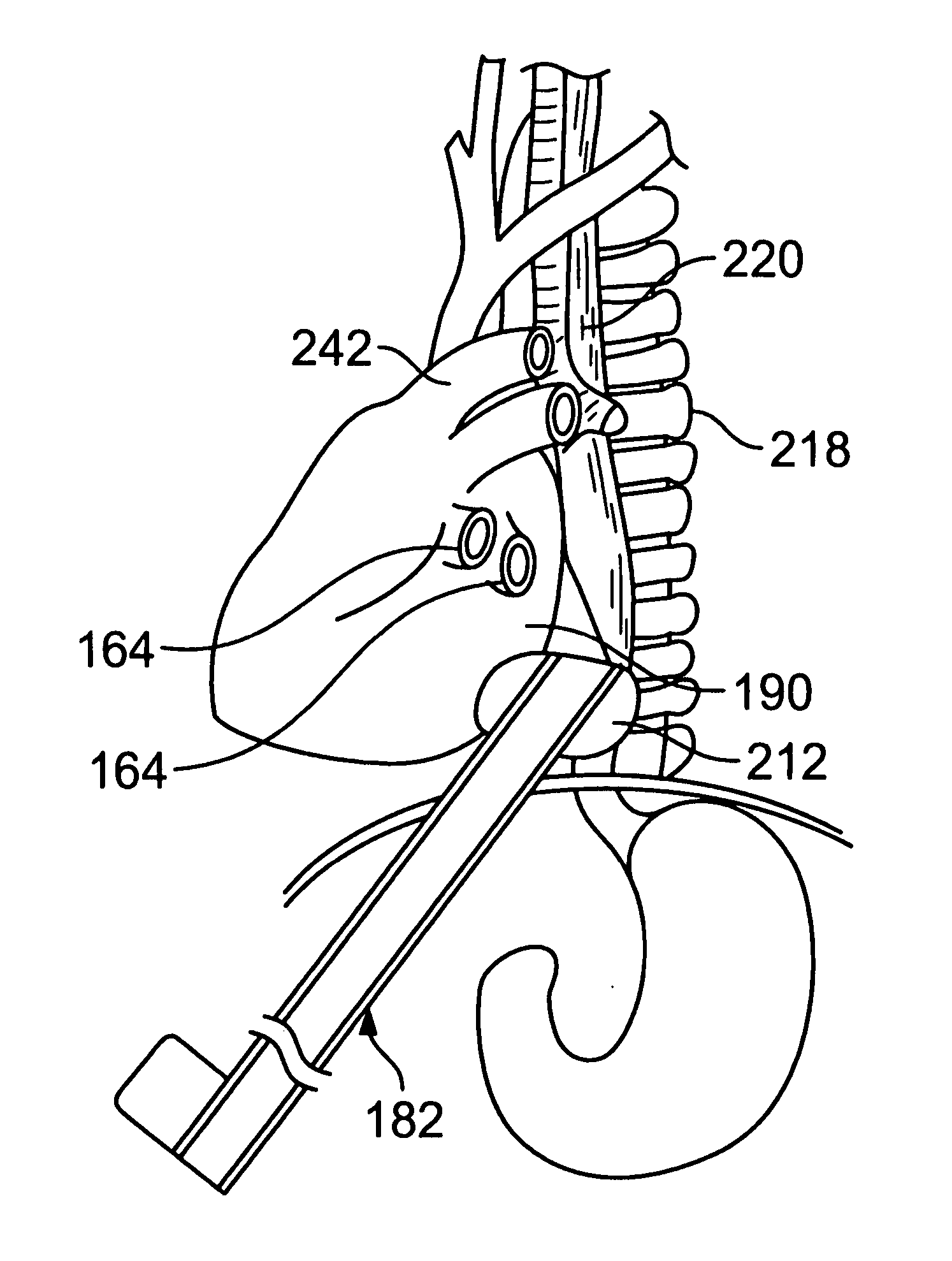
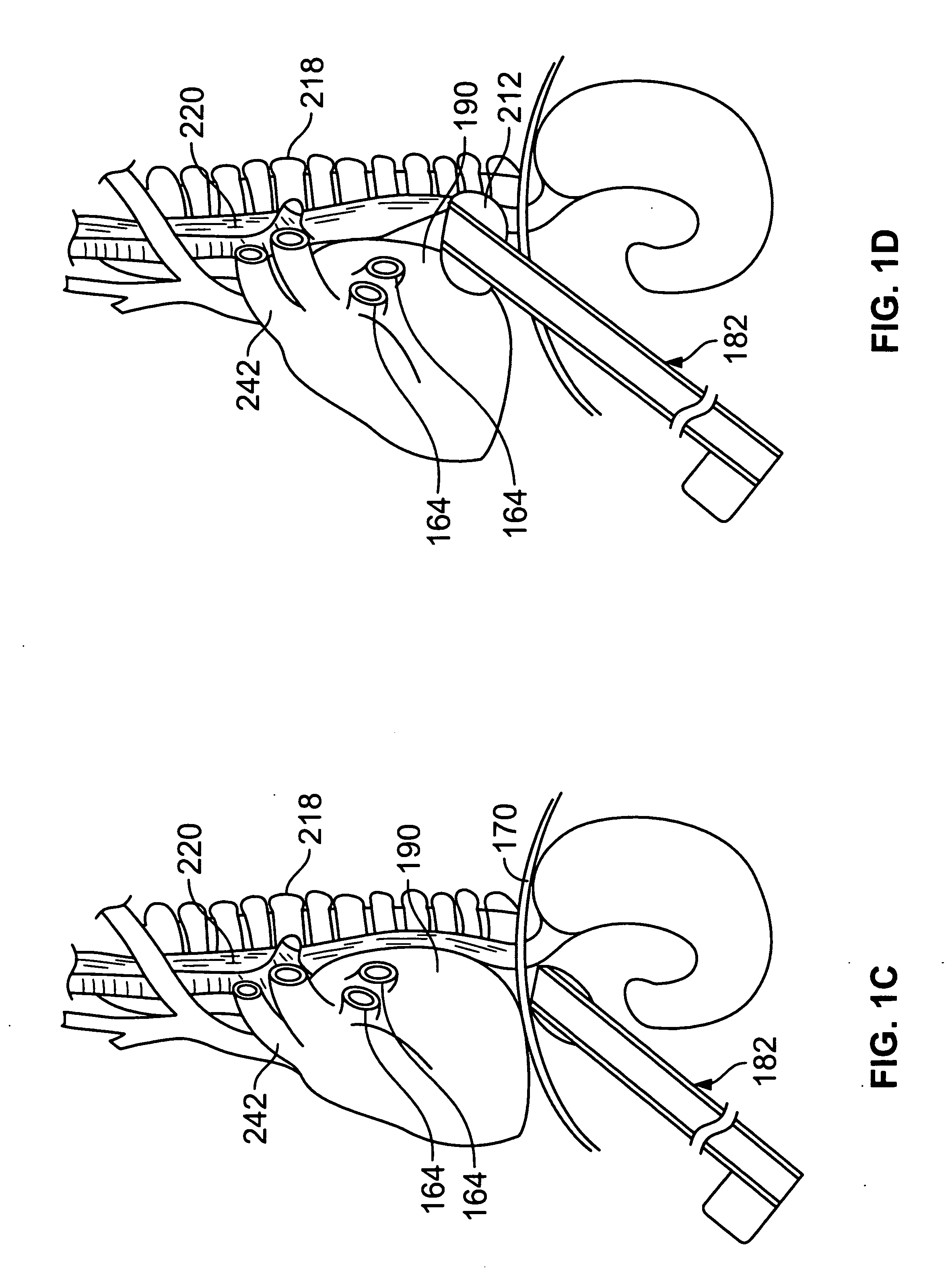
[0063] Methods and devices described herein provide for improved manipulation of organs and / or instruments in the thoracic cavity. The methods and devices may allow for direct visualization along the posterior region of the heart and other anatomic structures not attainable with conventional thoracic approaches. Furthermore, the methods and devices described herein may be used in conjunction with, or as an alternative to the conventional approaches described herein. In general, the surgical approaches and procedures described herein rely on entry through the diaphragm of a patient to access a posterior region of that patient (the procedure hereafter referred to as “Diaphragm Entry for Posterior Access” or simply “DEPA”). The DEPA procedure may also be referred as VAPS (Video-Assisted Pericardiac Surgery) or TAPS (Trans-Abdominal Pericardiac Surgery).

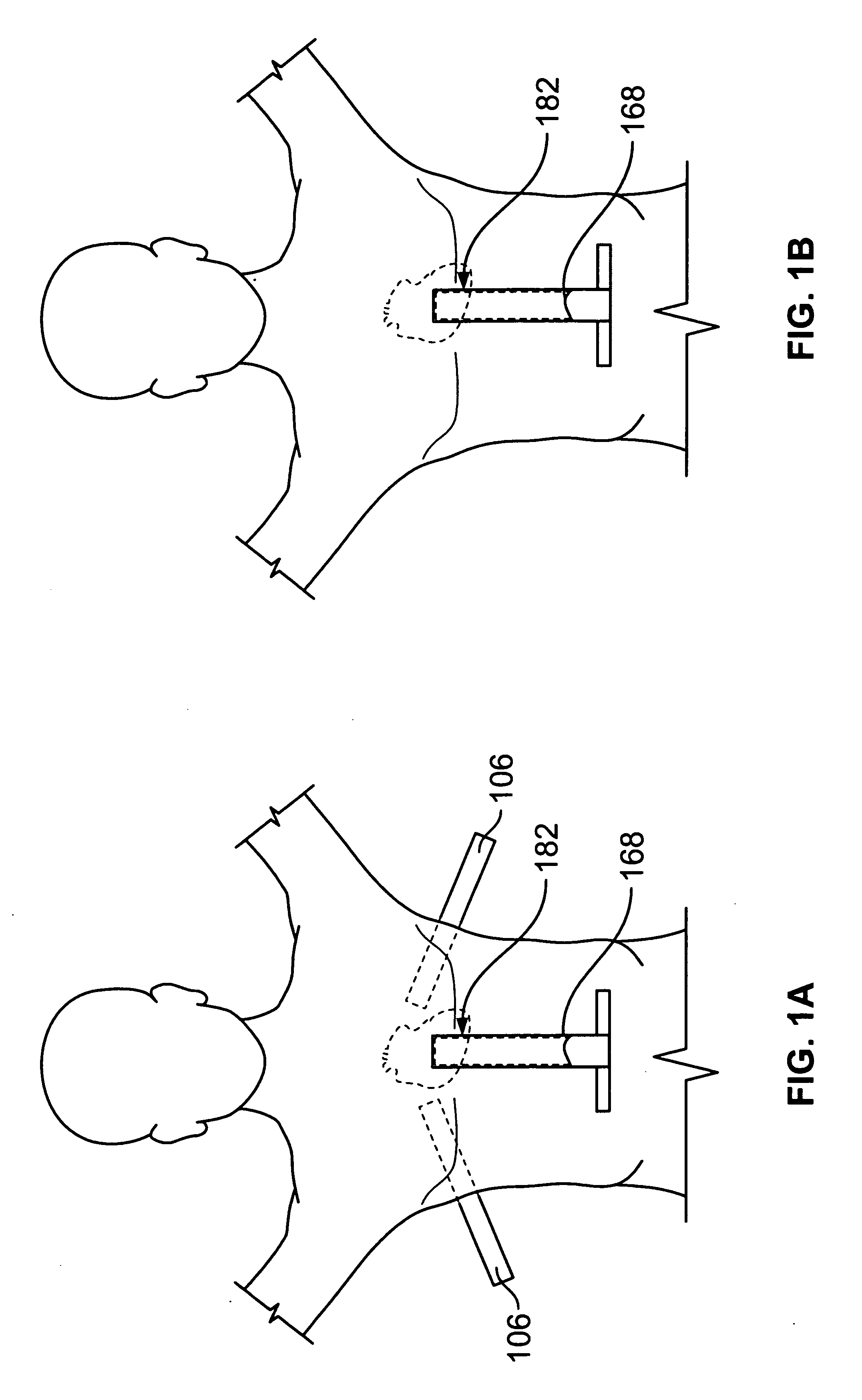
[0064]FIGS. 1A to 1B show examples of placement of access devices 182 (also referred to as a separator or an elevator herein) under th...


PUM


Abstract

Methods and devices described herein facilitate diaphragm entry for posterior access of body organs.

Description

CROSS-REFERENCE TO RELATED APPLICATIONS [0001] This application claims the benefit under 35 U.S.C. §119(e) of U.S. Provisional Application No. 60 / 726,342 filed Oct. 12, 2005, the contents of which are incorporated herein by reference. [0002] The subject matter of this application may be incorporated with the subject matter in the following applications: U.S. patent application Ser. No. 11 / 208,465 entitled “Vacuum Coagulation & Dissection Probes”; U.S. patent application Ser. No. 10 / 425,251 entitled “Vacuum Coagulation Probes”; U.S. Provisional application No. 60 / 726,342 entitled Diaphragm Entry for Posterior Access Surgical Procedures; and U.S. Pat. No. 6,893,442 the entirety of each of which is hereby incorporated by reference in their entireties.BACKGROUND OF THE INVENTION [0003] 1. Field of the Inventions [0004] Embodiments of the invention relate to methods for minimally invasive surgery and devices useful in such methods. More particularly, methods and devices described herein ...

Claims


Application Information

Patent Timeline
IPC IPC(8): A61M29/00
CPCA61B1/04A61B17/068A61B17/29A61B17/3421A61B17/3468A61B18/14A61B19/5225A61B2017/00243A61B2017/00278A61B2017/0237A61B2017/291A61B2017/2927A61B2017/308A61B2017/320044A61B2017/320048A61B2017/3419A61B2017/3445A61B2017/3484A61B2017/3486A61B2017/3488A61B2018/00291A61M25/1002A61M25/1011A61M29/02A61N7/02A61B2017/2906A61B90/37
Inventor KISER, ANDY C.WHAYNE, JAMES G.FLEISCHMAN, SIDNEY D.ROGERS, EARL W.NEUBERT, REBECCA
Owner ATRICURE
Why Eureka
Try Eureka