Three-dimentional tissue structure

a three-dimensional structure and tissue technology, applied in the field of three-dimensional structure applicable to the heart, can solve the problems of not having the effect of clinically used pharmaceutical agents or treatments on the replacement of myocardial scars with functional contraction tissue, the effect of reducing the size of the cardiac muscle, etc., to achieve excellent strength and large size

Inactive Publication Date: 2007-04-26
CELLSEED
View PDF3 Cites 42 Cited by
  • Summary
  • Abstract
  • Description
  • Claims
  • Application Information

AI Technical Summary

Benefits of technology

[0044] Hereinafter, the present invention will be described by way of preferred embodiments. It will be understood by those skilled in the art that the embodiments of the present invention can be appropriately made or carried out based on the description of the present specification and commonly used techniques well known in the art. The function and effect of the present invention can be easily recognized by those skilled in the art.
[0045] The present invention provides an implantable prosthetic tissue. This tissue has a larg

Problems solved by technology

No clinically used pharmaceutical agent or treatment has an efficacy on the replacement of myocardial scars with functional contraction tissue.
However, the graft hardly attaches to the myocardium, resulting in the least possible benefit for the improvement of the cardiac function (Leor J., Etzion S. A., Dar A. et al., Circulation, 2000; 102 [suppl.
However, it is unknown whether or not cardiomyocyte sheets retain their functions after in vivo implantation.
Such grafts are often made from non-biological mater

Method used


Image

  • Three-dimentional tissue structure
  • Three-dimentional tissue structure
  • Three-dimentional tissue structure

Examples


example 1

Production and Utilization of Prosthetic Tissue and Three-dimensional Structure Made of Cardiomyocyte Sheet—Tissue Engineered Contractile Cardiomyocyte Sheet Regenerates Impaired Myocardium

[0336] (Bioengineered Contractile Cardiomyocyte Sheet Regenerates Infarcted Myocardium)

[0337] In Example 1, the present inventors investigated (1) whether or not a cardiomyocyte sheet (prosthetic tissue) survives after implantation and shows histological electrical connection with impaired myocardium; (2) whether or not the implanted cardiomyocyte sheet (prosthetic tissue) can induce an improvement in a cardiac function. As a result, it was demonstrated that the present invention provides the electrical connection and the improvement of cardiac function.

[0338] The present inventors introduced the concept of bioengineered tissue implantation into the treatment of impaired myocardium. It was demonstrated that a tissue engineered contractile cardiomyocyte sheet without a scaffold provides histolog...

example 2

Self-derived Myoblast Sheet Regenerates Impaired Myocardium: A Way to Clinical Application—Demonstrating Examples Using Rats

[0396] Recent progress in tissue engineering is likely to provide implantable functional tissue comprising various cells other than myocardial cells, and extracellular matrices. The present inventors designed autologous myoblast sheets. The present inventors considered that these sheets are beneficial for clinical applications. In this example, therefore, myoblasts were used as material to construct a prosthetic tissue or three-dimensional structure and an effect of the prosthetic tissue or three-dimensional structure on clinical applications was demonstrated.

[0397] (Methods)

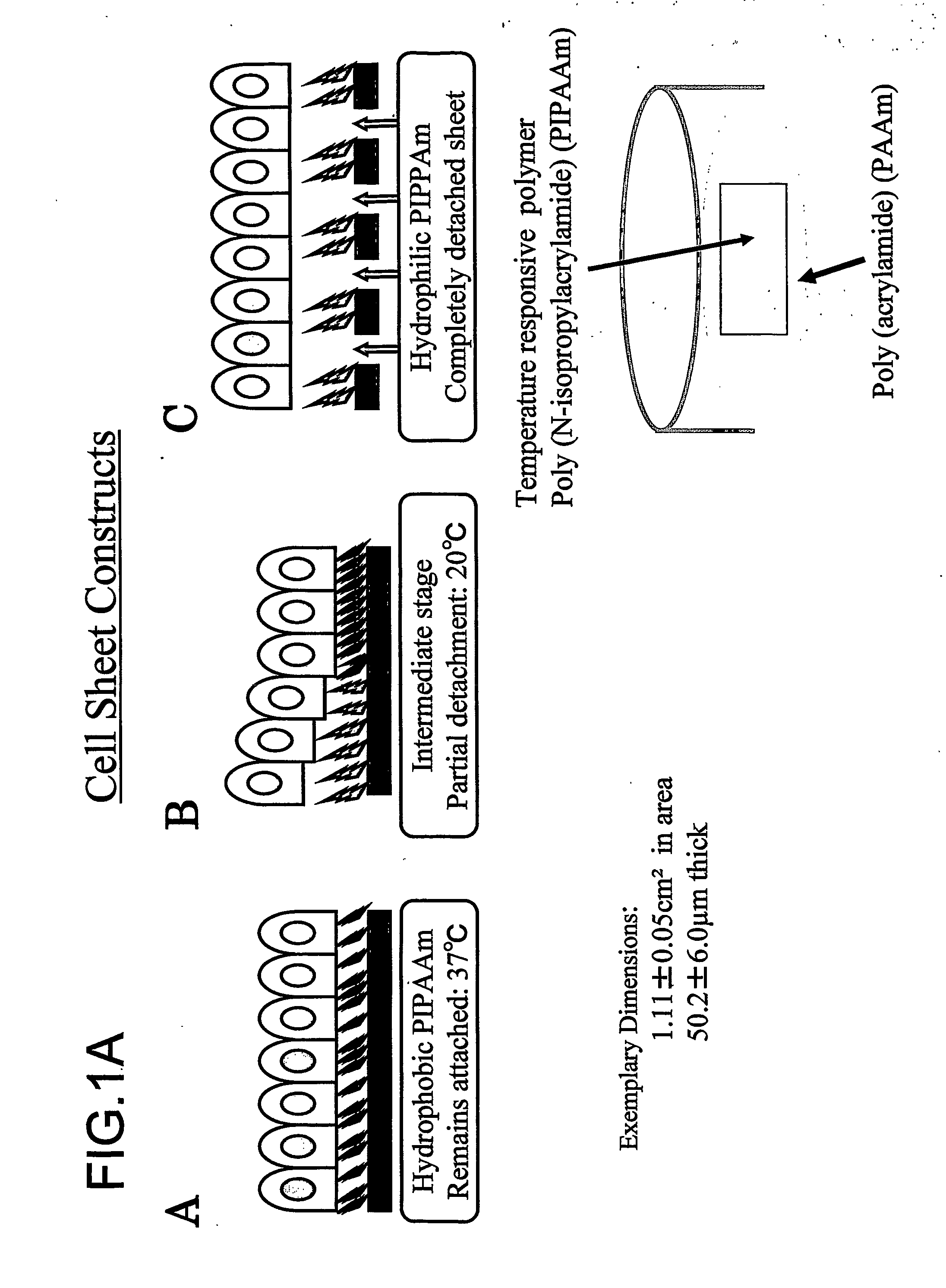
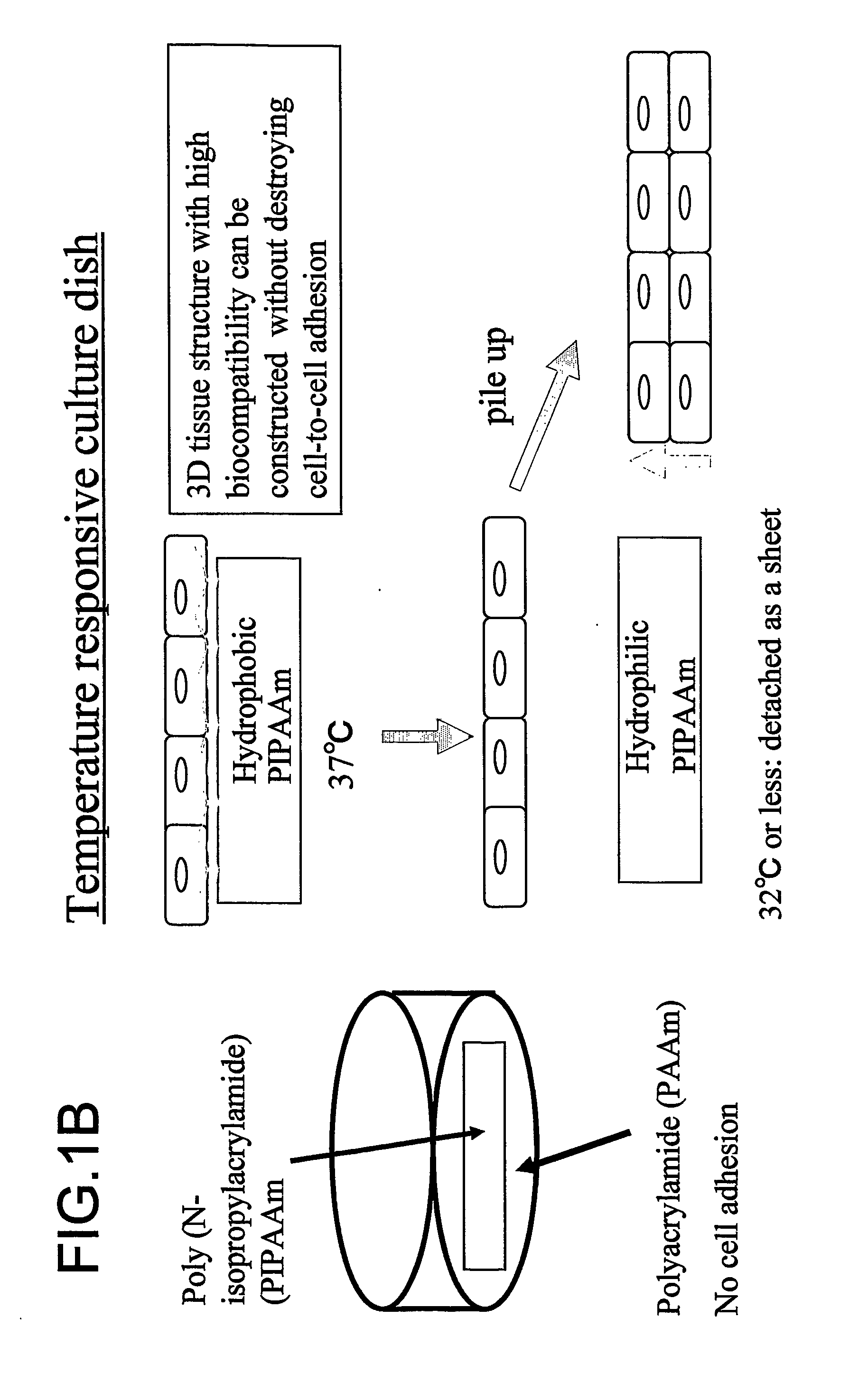
[0398] An impaired heart was created in 28 rats by ligating the left anterior descending (LAD) for 2 weeks. A temperature responsive domain made of a polymer (N-isopropylacrylamide) was coated on culture dishes. Skeletal myoblasts (SMs) isolated from leg muscle were cultured and detached...

example 3

Skeletal Myoblast—Implantation of Tissue-engineered Myoblast Sheet Improves Cardiac Function with Attenuation of Cardiac Remodeling in Cardiomyopathic Hamsters

[0518] Next, it was examined whether or not a prosthetic tissue or three-dimensional structure produced using skeletal myoblasts ameliorates cardiomyopathy.

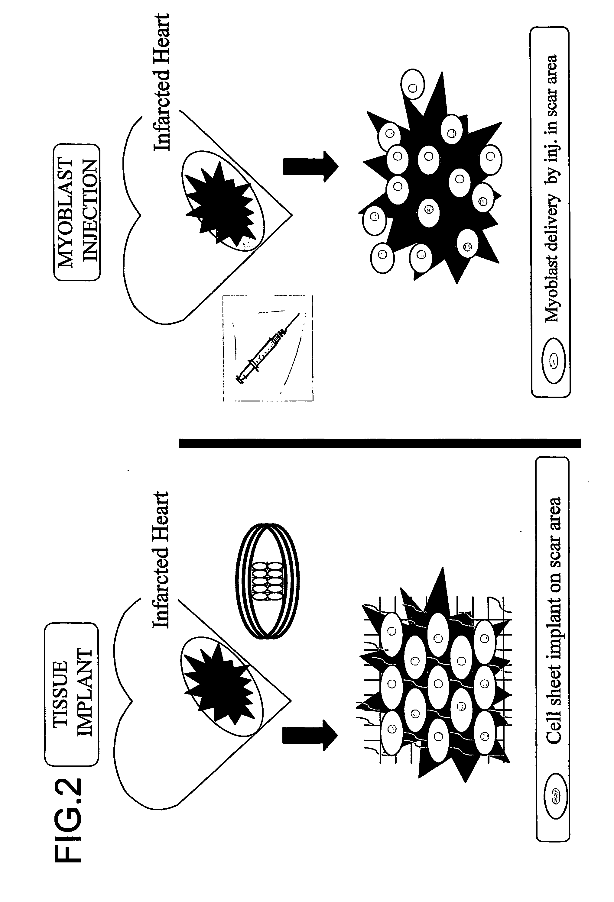
[0519] Cell therapy is a promising strategy for ischemic cardiomyopathy. However, direct injection methods seem to have limitations for generalized cell delivery in dilated cardiomyopathy (DCM). Given this body of evidence, the present inventors considered that a tissue-engineered myoblast sheet implantation might be a superior and promising method to ameliorate the cardiac function in DCM. Therefore, the present inventors carried out this example.

[0520] (Method)

[0521] Male 27-week old BIO TO-2 (dilated cardiomyopathy (DCM)) hamsters which showed moderate cardiac remodeling were used as recipients. Myoblasts isolated from BIO FIB hamsters (FIB) were cultured on dishes g...


PUM


Abstract

The present invention provides a prosthetic tissue or sheet capable of withstanding implantation operations, which can be used in actual operation and can be produced by culture. The present invention also provides a novel therapy which can substitute for cell therapy. Particularly, the present invention provides a method for producing a prosthetic tissue comprising a cell derived from a part other than myocardium and capable of withstanding implantation operation. The above-described objects of the present invention were partially achieved by finding that by culturing cells under specific culture conditions, the cells are unexpectedly organized into a tissue, and the resultant prosthetic tissue is capable of being detached from culture dishes. The present invention also provides a three-dimensional structure applicable to heart, comprising a cell derived from a part other than the myocardium of an adult.

Description

TECHNICAL FIELD [0001] The present invention relates to a three-dimensional structure applicable to the heart, and more particularly, to a three-dimensional structure applicable to the heart, which comprises cells derived from parts of an adult other than the heart. The present invention also relates to a method for producing such a three-dimensional structure. BACKGROUND ART [0002] Myocardial infarct is an irreversible injury (Ho K. K., Anderson K. M., Kannel W. B., Grossman W., Levy D., Circulation, 1993; 88: 107-115). Ischemic heart diseases are the cause of death responsible for 50% of all cardiovascular system-related deaths and the major cause of congestive heart failure. The 1-year mortality observed in patients who are diagnosed as having congestive heart failure and eventually die from chronic heart disease is 20% (American Heart Association, Dallas, Tex.: American Heart Association; 2001). Most therapies currently available to clinicians can significantly improve the progn...

Claims


Application Information

Patent Timeline
IPC IPC(8): A61K35/34C12N5/06C12N5/08A61F2/00A61L27/38C12N5/077
CPCA61K35/12A61L27/3804A61L27/3834A61L27/3839A61L27/3895C12N2539/10C12N5/0657C12N5/0658C12N2506/02C12N2506/1392C12N2533/40A61L2430/20A61P9/04A61P9/10A61K35/34C12N5/0068C12N2513/00C12N2523/00
Inventor MATSUDA, HIKARUSAWA, YOSHIKITAKETANI, SATOSHIMIYAGAWA, SHIGERU
Owner CELLSEED
Why Eureka
Try Eureka