Method of forming multicellular spheroids from the cultured cells

Inactive Publication Date: 2007-06-28
YANG MEI JU +4
View PDF2 Cites 14 Cited by
  • Summary
  • Abstract
  • Description
  • Claims
  • Application Information

AI Technical Summary

Benefits of technology

[0013] The present inventors have determined that cells cultured by the conventional hanging drop technique or by use of low attachment dishes (Petri dish) failed to generate spheroids in an efficient manner. As such, in one aspect, the present invention provides a method of forming spheroids or embryoid bodies,

Problems solved by technology

However, in some circumstances, such as forming embryoid bodies from embryonic stem cells, attachment is a problem as it may cause unwanted differentiation of the cells.
However, current cell therapy suffers a major drawback of unable to produce enough number of uniform phenotypic cells for wide applications such as embryo research, drug screening and cell transplant.
Although several approaches as described above have been proposed, yet each of them suffers at least one of the following drawbacks, such as time consuming, high manpower, complicating system, unable to scale up, cost inefficient, low spheroids formation rate, low differentiation rate and etc.

Method used


Image

  • Method of forming multicellular spheroids from the cultured cells
  • Method of forming multicellular spheroids from the cultured cells
  • Method of forming multicellular spheroids from the cultured cells

Examples


example 1

Formation and Phenotypic Characterization of Embryoid bodies (EBs) of HES cells

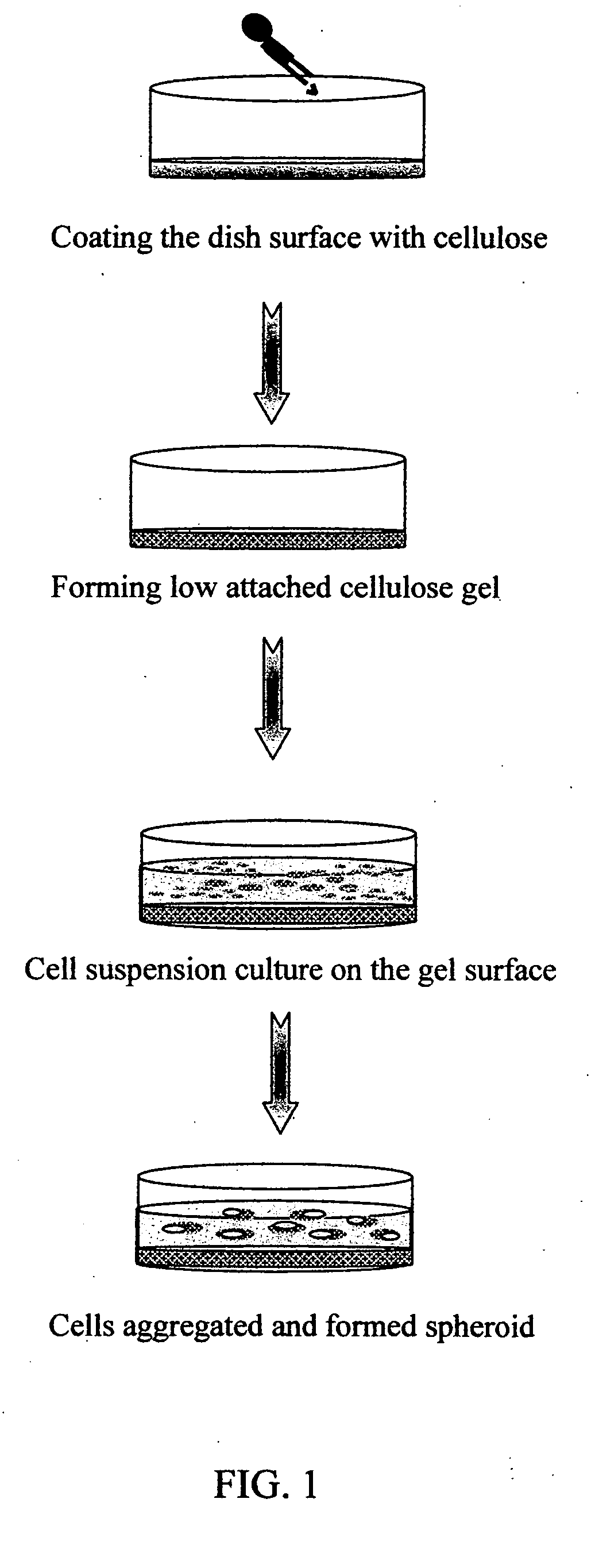
1.1 Culturing of HES Cells

[0052] 1.1.1 Preparation of Conditioned Medium

[0053] Conditioned medium for maintaining the culture of HES cell was prepared according to the following procedure. Briefly, primary mouse embryonic fibroblasts (PMEF) were plated in Dulbecco's Modified Eagle Medium (DMEM, obtained from Gibco Invitrogene) supplemented with 10% fetal bovine serum (FBS). Cell cultures were maintained at 37° C. and 5% CO2 and in a water-saturated atmosphere until they reached confluence, then 10 μg / ml mitomycin C was added to inactivate the fibroblasts. The inactivated fibroblasts were then re-grown in DMEM medium supplemented with 20% FBS, 1 mM β-mercaptoethanl (obtained from Gibco Invitrogene), 1% non-essential amino acids (obtained from Gibco Invitrogene), 1% glutamine (obtained from Gibco Invitrogene), and 1% insulin- transferrin-selenium G supplement (ITS G supplement, obtained from Gibco Invit...

example 2

Formation of Hepatic Spheroids

[0072] Human hepatoma cell line, i.e., C3A cells, were plated in DMEM medium supplemented with 10% FBS. Cells were maintained at 37° C. and 5% CO2 and in a water-saturated atmosphere until they reached 90% confluence, then the cells were trypsinized, counted and re-plated onto the cell culture dishs, conventional low attachment Petri dishes (Falcon) or the MC-gel coated culture dishes of Example 1.2.2. FIG. 6A illustrated the morphology of C3A cells attached culture on cell culture dish, FIG. 6B illustrated the partial formation of spheroids for cells maintained in a low attachment Petri dishes for 4 days, most cells showed attached growth on the photograph. In stark contrast, most of the cells maintained in the MC-gel coated culture dish of this invention for 4 days formed spheroids (FIG. 6C), and their expression of hepatic-specific genes such as α-FP, albumin, G6P and CYP 3A4 were illustrated in FIG. 7. (Lane 2)

[0073] Hepatic Enzyme Activity Measur...


PUM


Abstract

Provided herein are methods for producing multicellular spheroids or embryoid bodies suitable for providing cells in large scale for various medical applications. In one embodiment, a method of forming embryoid bodies is provided, which comprises culturing undifferentiated HES cells in a culture vessel pre-coated with cellulose and / or its derivatives. In another embodiment, a method of forming hepatic spheroids is provided, which comprises culturing hepatocytes in a culture vessel pre-coated with cellulose and / or its derivatives.

Description

FIELD OF THE INVENTION [0001] The present invention relates to methods of producing multicellular spheroids or embryoid bodies that are suitable for providing cells for various medical applications such as cell therapy, drug screening and etc. More particularly, the present invention provides methods for the formation of multicellular spheroids or embryoid bodies by culturing cells in a culture vessel pre-coated with cellulose and / or its derivatives. BACKGROUND OF THE INVENTION [0002] Three-dimensional (3-D) cell culture approach offers researchers a means to study cell growth, proliferation, and differentiation under conditions that emulate an in vivo environment and, to varying degrees, allow cell-cell and cell-extracellular matrix (ECM) interactions that might otherwise be severely constrained or precluded entirely in 2-dimensional (2-D) culturing condition (Edlman and Keefer, Experimental Neurology 2005, 192: 1-6). [0003] Prior uses of the three-dimensional spheroid culture syst...

Claims


Application Information

Patent Timeline
IPC IPC(8): C12N5/08C12N5/06C12N5/071C12N5/0735
CPCC12N5/0068C12N5/0606C12N5/067C12N2533/78
Inventor YANG, MEI-JUCHEN, WANN-HSINLIN, PEI-JUCHEN, CHUN-HUNGSUNG, HSING-WEN
Owner YANG MEI JU
Why Eureka
Try Eureka