Micronized device for the delivery of biologically active molecules and methods of use thereof

a micro-device and biological active technology, applied in the field of encapsulated cell therapy, can solve the problems of loss of transplant function, eventual necrosis of transplanted tissue or cells, and limited application, and achieve the effect of long-term functional transplantation and limited application

Active Publication Date: 2007-07-05
NEUROTECH USA
View PDF3 Cites 39 Cited by
  • Summary
  • Abstract
  • Description
  • Claims
  • Application Information

AI Technical Summary

Benefits of technology

[0018] Also provided are methods of treating ophthalmic disorders in patients suffering therefrom by implanting one or more of the micronized devices of the invention into an eye of the patient. For example, the ophthalmic disorder to be treated may be a retinal degeneration disease such as retinopathy of prematurity, glaucoma, cataract formation, retinoblastoma, retinal ischemia, uveitis, retinitis pigmentosa, forms of wet and dry age-related macular degeneration, diabetic retinopathy, and / or choroideremia.
[0019] Unless otherwise defined, all technical and scientific terms used herein have the same meaning as commonly understood by one of ordinary skill in the art to which this invention belongs. Although methods and materials similar or equivalent to those described herein can be used in the practice or testing of the present invention, suitable methods and materials are described below. All publications, patent applications, patents, and other references mentioned herein are incorporated by reference in their entirety. In the case of conflict, the present specification, including definitions, will control. In addition, the materials, methods, and examples are illustrative only and are not intended to be limiting.

Problems solved by technology

However, while transplantation can provide dramatic benefits, it is limited in its application by the relatively small number of organs that are suitable and available for grafting.
In general, transplantation patients must be immunosuppressed in order to avert immunological rejection of the transplant, which results in loss of transplant function and eventual necrosis of the transplanted tissue or cells.
Likewise, in many cases, the transplant must remain functional for a long period of time, even for the remainder of the patient's lifetime.
It is both undesirable and expensive to maintain a patient in an immunosuppressed state for a substantial period of time.
One major problem in treatment of such diseases is the inability to deliver therapeutic agents into the eye and to maintain them there at therapeutically effective concentrations.
However, none of these approaches have been satisfactory for providing long-term transplant function.

Method used


Image

  • Micronized device for the delivery of biologically active molecules and methods of use thereof
  • Micronized device for the delivery of biologically active molecules and methods of use thereof
  • Micronized device for the delivery of biologically active molecules and methods of use thereof

Examples


example 1

Comparison of “First Generation” and Micronized ECT Devices

Materials and Methods

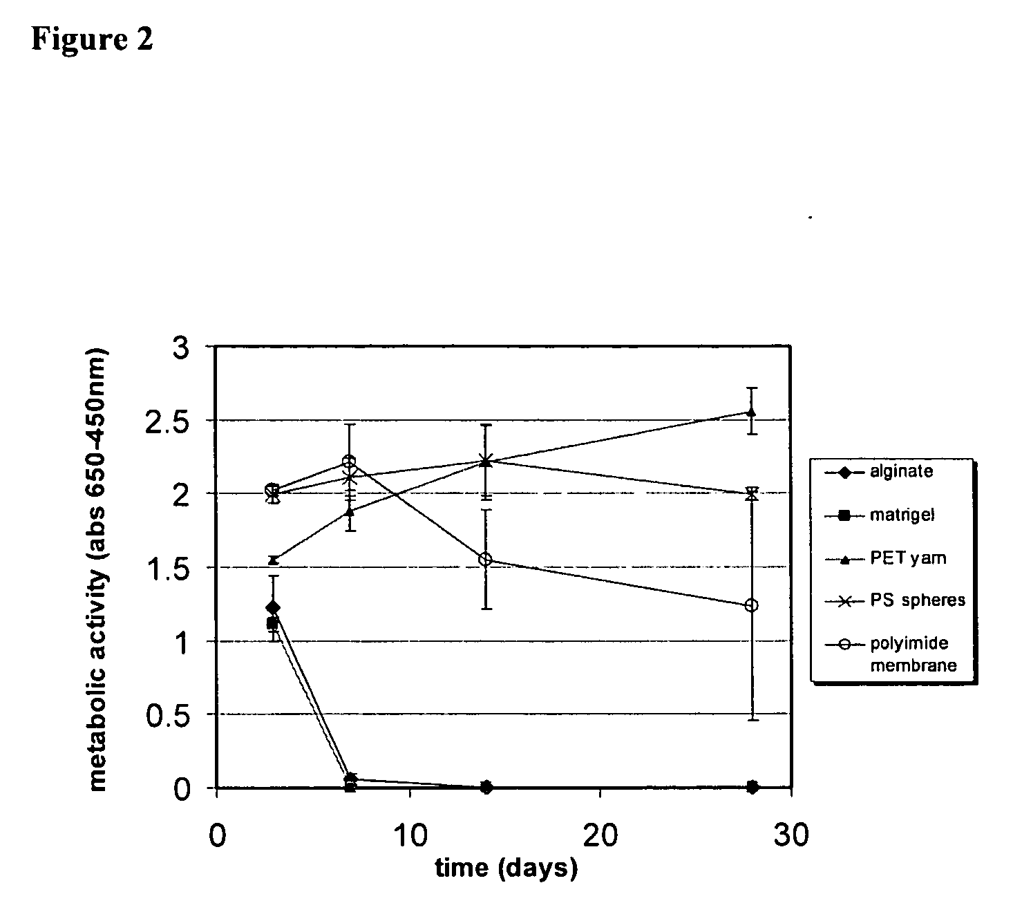
[0150] ECT devices were fabricated to allow intravitreal implantation into the eye. The first generation devices totaled 600 μl in displaced volume (1.1 mm diameter, 6 mm length). Micronized ECT devices were fabricated with a total displacement volume of 0.05 μl (200 mm diameter, 1 mm length). A comparison of the size differences between the first generation and micronized ECT devices is provided in FIG. 7. Encapsulated cell lines used in these studies were genetically modified to secrete either ciliary neurotrophic factor (CNTF) or interleukin-10 (IL-10). Devices were designed to produce a high and a low dose delivery for both CNTF and IL-10. Implant periods ranged from 2 weeks to 18 months in the rabbit model and were 2 weeks in the rat model. Protein delivery levels were quantified over the course of the studies and clinical exams were conducted to assess ocular irritation and surgical wound healin...

example 2

Delivery of Encapsulated Cell Technology (ECT) Micronized Device Implants Using a Small Gauge Needle

[0160] Implantation of first generation ECT devices, which are currently in Phase II human clinical trials for retinitis pigmentosa and age-related macular degeneration, requires a 2.0 mm sclerotomy and three sutures to close the incision site. Thus, development of an ECT micronized device capable of producing comparable protein levels that could be implanted through a small gauge needle would improve the surgical procedure and minimize surgical risk.

Methods

[0161] Micronized devices were prepared that contained encapsulated cells transfected to produce either ciliary neurotrophic factor (CTNF), interleukin-10 (IL-10) or pigment epithelial derived factor (PEDF). Implantation of such micronized devices delivering CNTF was tested using a 23-gauge needle having a modified syringe. CNTF-producing devices were surgically implanted into New Zealand white rabbits and evaluated at 2 weeks ...

example 3

Safety and Pharmacokinetics of an Injectable Micronized Encapsulated Cell Technology Device

[0166] Three concurrent studies of a first generation encapsulated cell technology (ECT) device delivering ciliary neurotrophic factor (CNTF) are in human clinical for early and late-stage retinitis pigmentosa and age-related macular degeneration. ECT devices used in these trials are implanted in patients eyes using conjunctival excision, sceral incision, and inter-ocular placement followed by securing the device followed by suture closure of the sclera and conjunctiva. The current study reports on the research efforts to develop the long-term pharmacokinetics and safety profile of a smaller profile, micronized ECT device capable of delivering efficacious therapeutics, implanted using sutureless, 23-gauge injection procedures.

Methods

[0167] Human retinal pigment epithelial cells genetically modified to continuously secrete CNTF were encapsulated in 300 micron diameter hollow fiber semi-perm...


PUM

PropertyMeasurementUnit
lengthaaaaaaaaaa
lengthaaaaaaaaaa
lengthaaaaaaaaaa

Abstract

The invention provides micronized encapsulated cell therapy devices that are capable of delivering a biologically active molecule to the eye. Also provided are methods of using the same to deliver biologically active molecules to the eye and to treat ophthalmic disorders in patients suffering there from.

Description

RELATED APPLICATIONS [0001] This application claims priority to U.S. Ser. No. 60 / 755,478, filed Dec. 30, 2005, which is herein incorporated by reference in its entirety.FIELD OF THE INVENTION [0002] The present invention relates generally to the field of encapsulated cell therapy. BACKGROUND OF THE INVENTION [0003] Many clinical conditions, deficiencies, and disease states can be remedied or alleviated by supplying to the patient one or more biologically active molecules produced by living cells or by removing from the patient deleterious factors which are metabolized by living cells. In many cases, these molecules can restore or compensate for the impairment or loss of organ or tissue function. Accordingly, many investigators have attempted to reconstitute organ or tissue function by transplanting whole organs, organ tissue, and / or cells, which provide secreted products or affect metabolic functions. However, while transplantation can provide dramatic benefits, it is limited in its...

Claims


Application Information

Patent Timeline
Patent Type & Authority Applications(United States)
IPC IPC(8): A61F2/02A61K35/30A61K35/12
CPCA61K9/0048A61K9/0051A61K38/185A61K38/2066A61K38/57A61K2035/128A61K2300/00A61P27/02A61P27/06A61P27/12Y02A50/30
Inventor KAUPER, KONRADSTABILA, PAULTAO, WENG
Owner NEUROTECH USA
Why Eureka
Try Eureka