
Novel g-csf conjugates

a technology of conjugates and g-csf, applied in the field of new g-csf conjugates, can solve the problem of limit the short half-life in the body, and achieve the effect of simplifying the mixture of products and selective conjugation

Inactive Publication Date: 2007-07-19
NEKTAR THERAPEUTICS INC
View PDF30 Cites 18 Cited by
  • Summary
  • Abstract
  • Description
  • Claims
  • Application Information

AI Technical Summary

Benefits of technology

[0088] In another aspect of this invention, bi-functionally activated PEG derivatives (OPSS-PEG-OPSS; VS-PEG-VS; VS-PEG-OPSS, MAL-PEG-MAL, MAL-PEG-OPSS, MAL-PEG-VS) may be used to generate dimeric G-CSF (i.e. G-CSF-PEG-G-CSF) in which two G-CSF monomers are linked together at the thiol group of Cys 17 via PEG. The in vitro activity of such obtained dimers are comparable to those of the corresponding pegylated form of G-CSF. The main advantage of these dimers is the reduction of the in vivo elimination rate (if compared with the corresponding pegylated form of G-CSF) due to the increasing of their molecular mass.

Problems solved by technology

Well known to the expert of the art in the application of proteins for therapy, and of G-CSF among these, is the limitation of short half-life in body, due to the high clearance rate, the proteolytic degradation, and furthermore immunogenic reactions.

Method used


Image

  • Novel g-csf conjugates
  • Novel g-csf conjugates
  • Novel g-csf conjugates

Examples


example 2

Reversible Denaturation of rhG-CSF

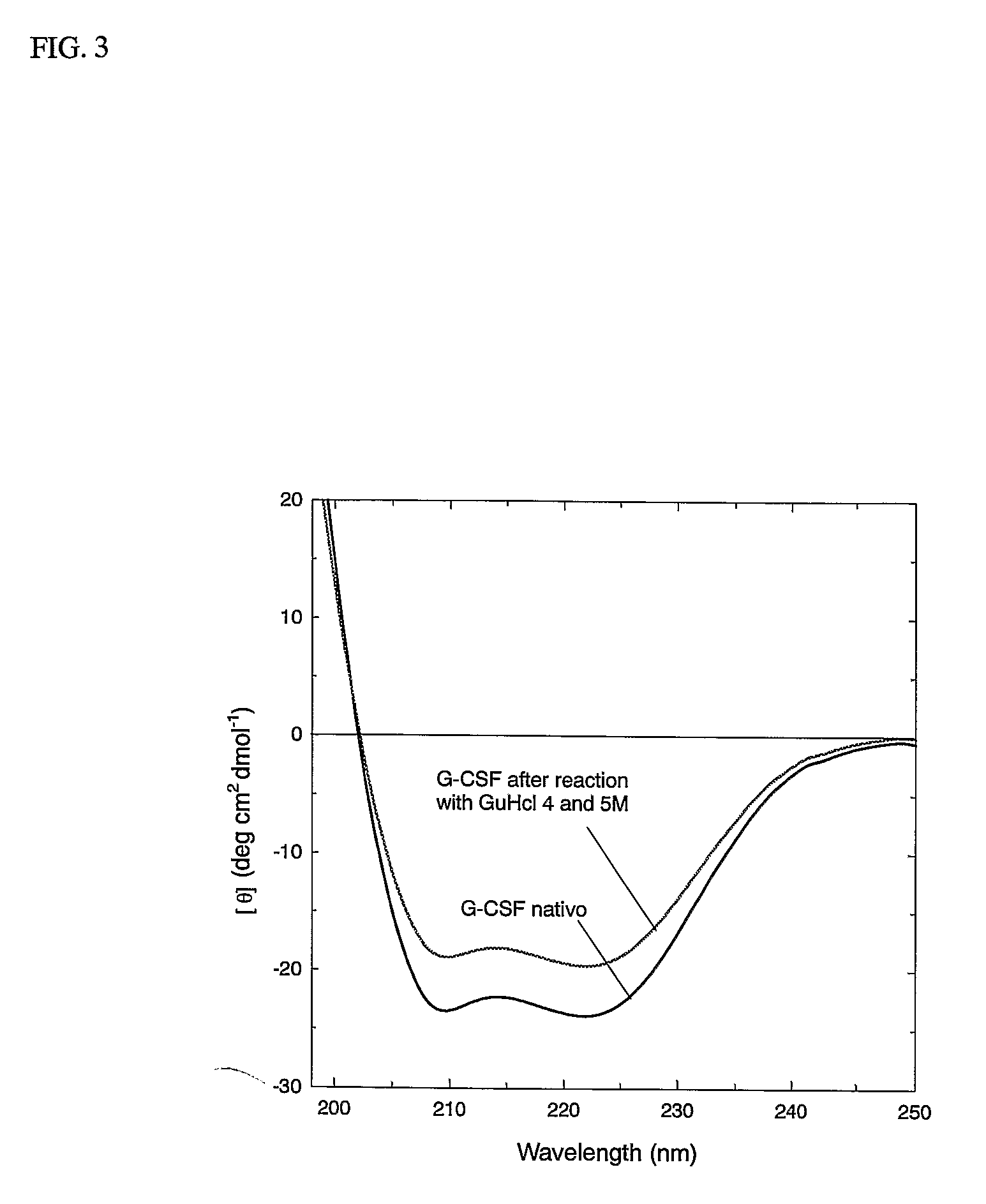
[0110] Guanidine HCl 8M in phosphate buffer 0.1 M, pH 7.27 were added at different amount to a rh-GCSF buffer solution in order to reach a final concentration of 2, 4 e 5 Molar. After 4 hours incubation at room temperature the mixture was dialyzed against an aqueous acidic solution, to remove the denaturant and to reach the G-CSF stable condition of pH 3.5.

[0111]FIG. 3 reports the CD spectrum of the starting proteins and after the denaturation-renaturation process. The regain of the secondary structure is evident from the 220-208 mm ellypticity ratio of before and after treatment. The following experiment demonstrates that G-CSF may be subject to denaturation and renaturated to the original structure.

example 3

Preparation of 5 kDa PEG rhG-CSF Conjugate by Thiol Reagents in Denaturant Conditions

Conjugation of 5 kDa PEG-OPSS and PEG-VS to rhG-CSF.

[0112] 3a: To a G-CSF buffer solution Tris 0.5 M, 6 M Urea, pH 7.2 was added in order to reach a final concentration of 3 M urea. To this solution MPEG-OPSS or mPEG-VS were added at a molar ratio of 10 moles of reagent per mole of protein. The reaction was allowed to proceed at room temperature for 6 hours and stopped by 0.1 N HCl addition. [0113] 3b: G-CSF was added to a solution of 8 M Guanidine HCl in phosphate buffer 0.1 M, pH 7.27, to have a final denaturant concentration of 2, 4, 6M. The denaturated G-CSF was added to PEG-OPSS or PEG-VS solution in order to reach molar ration of 10 moles of reagents per mole of protein. After 4 hours at room temperature the reaction was stopped with HCl 0.1 N.

example 4

Characterisation of 5 kDa PEG Conjugated to rh-G-CSF

[0114] 1. RP-HPLC Chromatography.

[0115] The reaction mixture was analysed by reverse phase HPLC using a C4 Vydac column according to the following elution condition. Eluent A, 0.05% TFA in water, and B, 0.05% TFA in acetonitrile. The following linear gradient was used: 40% B for 4 minutes, up to 70% B in 18 minutes, 95% B in 2 minutes, stay at 95% B for 6 minutes and than 40% B in 2 minutes. The flow was 0.8 ml / min and UV lamp at 226 nm.

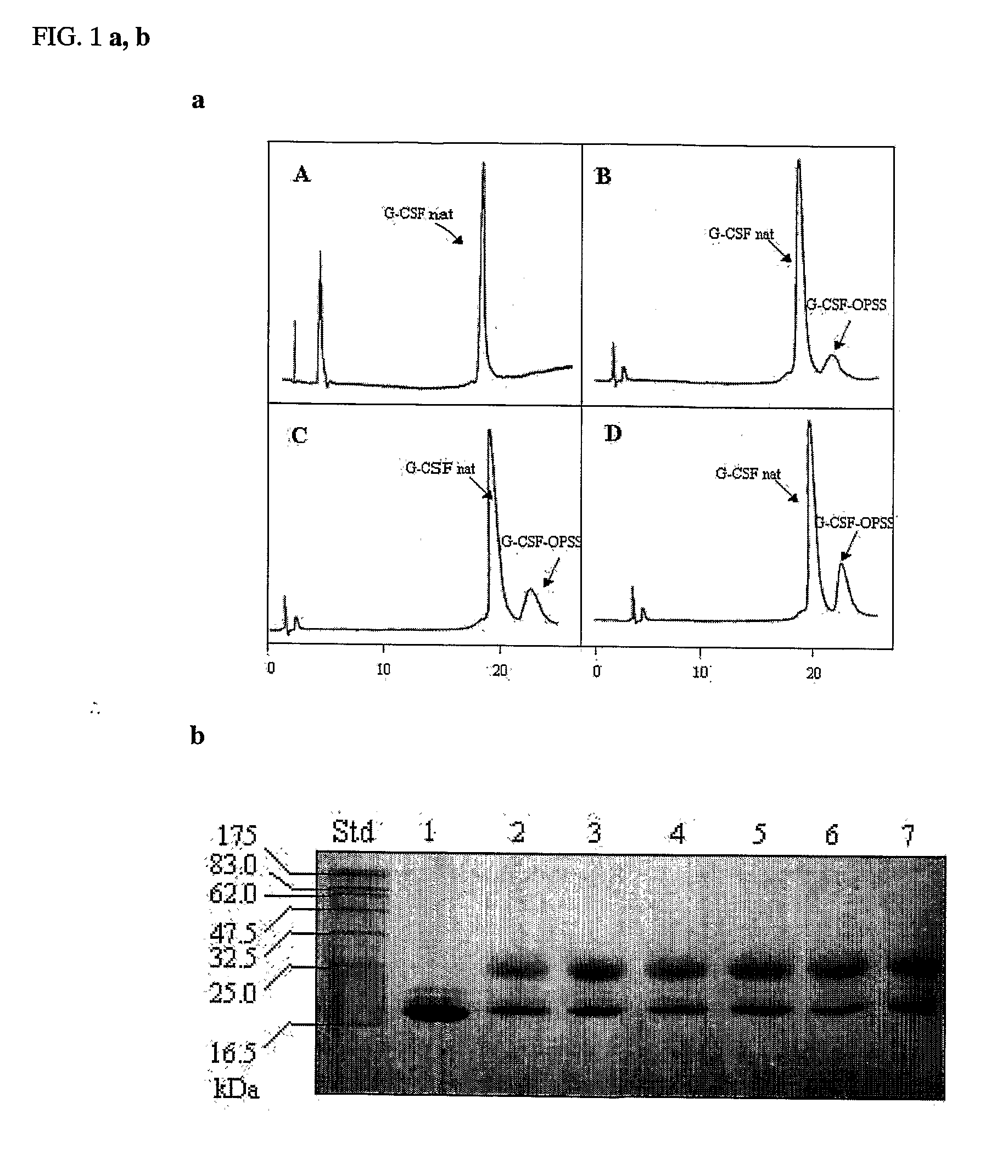
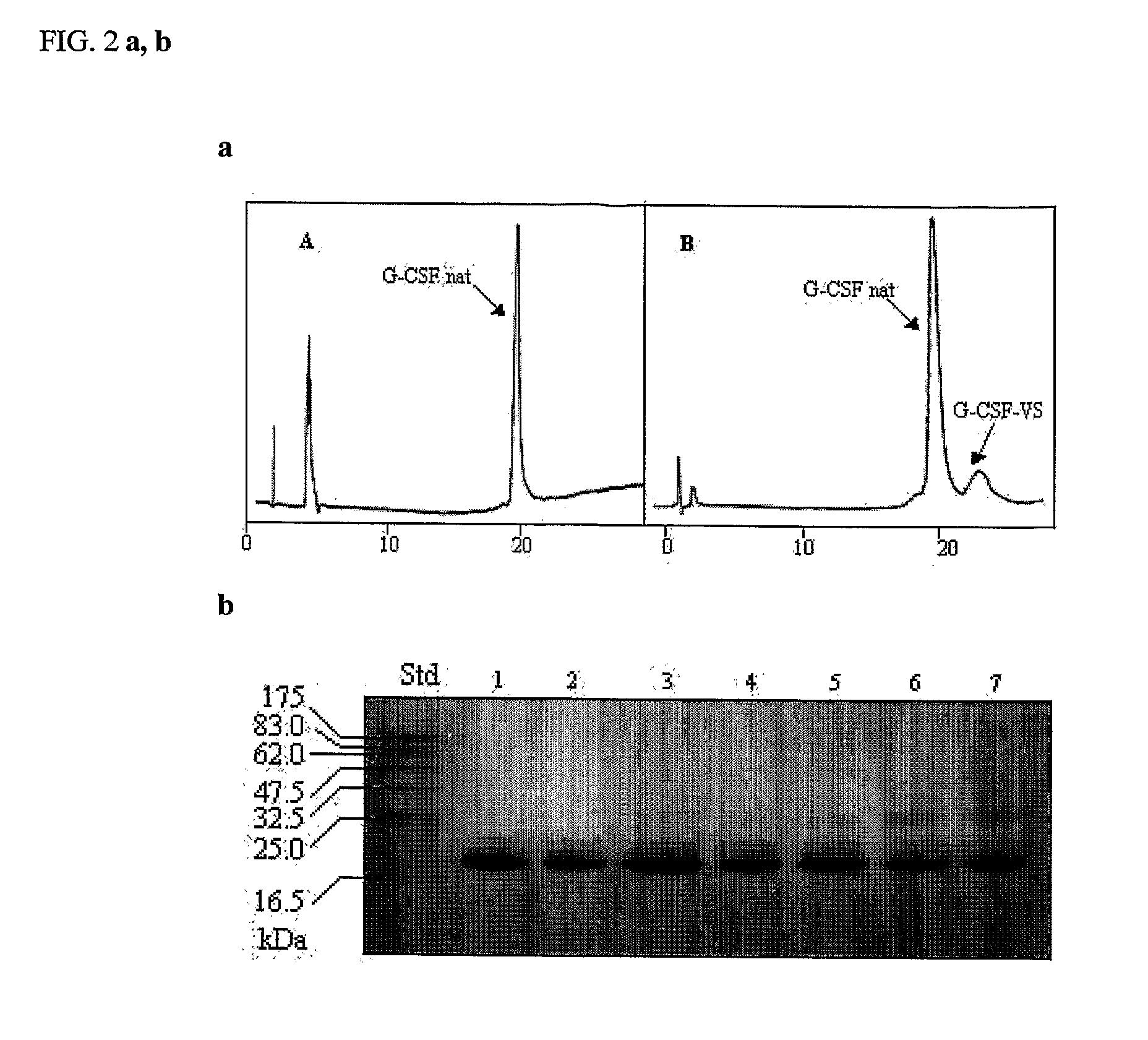
[0116]FIG. 4A shows the chromatogram of the native G-CSF. FIG. 4B shows the formation of the conjugate (PEG-OPSS) using urea as denaturant. FIGS. 4C, D, E show the formation of the conjugate (OPSS) in different reaction condition, namely in FIG. 4C the amount of guanidine HCl was 2 M, in 4D, 4M, and in 4E, 6M. As it possible to see in FIG. 4C-E the percentage of conjugate is increased following the concentration of guanidine. FIGS. 5B, C, D show the formation of the conjugate (PEG-VS) respectivel...


PUM

PropertyMeasurementUnit
MWaaaaaaaaaa
MWaaaaaaaaaa
pHaaaaaaaaaa

Abstract

The present application relates to novel PEG-G-CSF conjugates in which a PEG molecule is linked to the cyteine residue in position 17 of native G-CSF primary sequence or to the cysteine residue of the corresponding position of a G-CSF analogue. The present application also describes a process for the manufacture of such conjugates, such process comprising the following steps: (i) subjecting the G-CSF protein to conditions inducing reversible denaturation of the protein, (ii) conjugation of the denatured protein obtained is step i with a thiol-reactivc PEG under denaturing conditions, (iii) subjecting the conjugates obtained in step ii to conditions promoting renaturation of the conjugate yielding biologically active G-CSF-PEG conjugate.

Description

[0001] The present invention relates to a site-specific chemical modification of granulocyte colony-stimulating factor (G-CSF). BACKGROUND OF THE INVENTION [0002] Granulocyte-colony stimulating factor (G-CSF), the major regulator of granulopoiesis in vivo, represents now a pharmaceutically active protein that stimulates proliferation, differentiation and survival of cells of the granulocyte lineage. Human G-CSF is a glycoprotein of about 20 kDa in size produced by macrophages and stromal cells in bone marrow (Fibbe et al, 1989 Interleukin 1 and poly(rI).poly(rC) induce production of granulocyte CSF, macrophage CSF, and granulocyte-macrophage CSF by human endothelial cells, Exp Hematol., Mar 17(3), 229-34), that was first purified from the conditioned medium of a human bladder carcinoma cell line denominate 5637 (Welte et al., 1985, Purification and biochemical characterization of human pluripotent hematopoietic colony-stimulating factor, Proc. Natl. Acad. Sci. USA 82, 1526-1530). Th...

Claims


Application Information

Patent Timeline
Patent Type & Authority Applications(United States)
IPC IPC(8): A61K38/19C07K14/53A61K47/48
CPCA61K47/48215A61K47/60A61P35/00A61P43/00A61P7/00A61K47/50
Inventor VERONESE, FRANCESCO M.BERNA, MANUELA
Owner NEKTAR THERAPEUTICS INC
Learn More