

Use of focused ultrasound for vascular sealing
a vascular catheter and focused ultrasound technology, applied in the field of vascular catheter entry wound sealing, can solve the problems of ischemia and/or thrombosis, time-consuming procedure, and discomfort for patients, and achieve the effect of safe sealing of vascular catheter entry wounds and little operator training
- Summary
- Abstract
- Description
- Claims
- Application Information
AI Technical Summary
Benefits of technology
Problems solved by technology
Method used
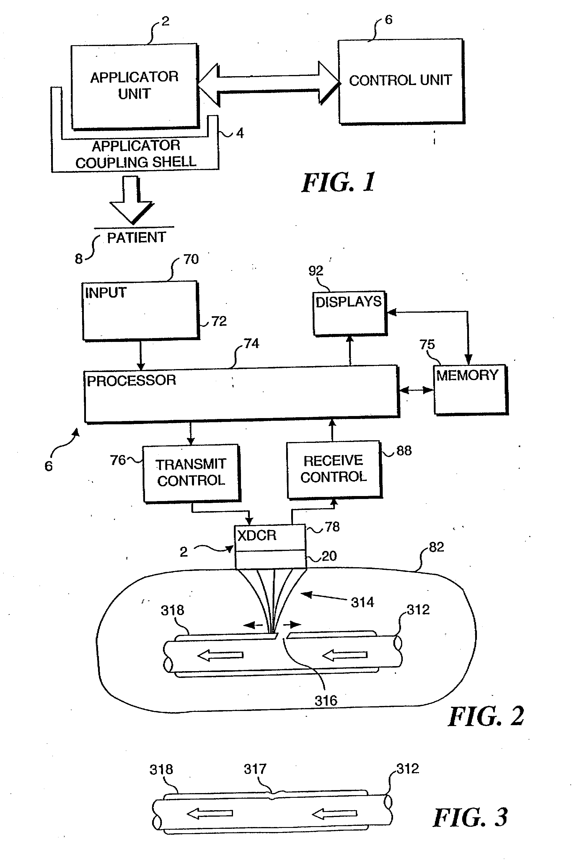
Image

Examples
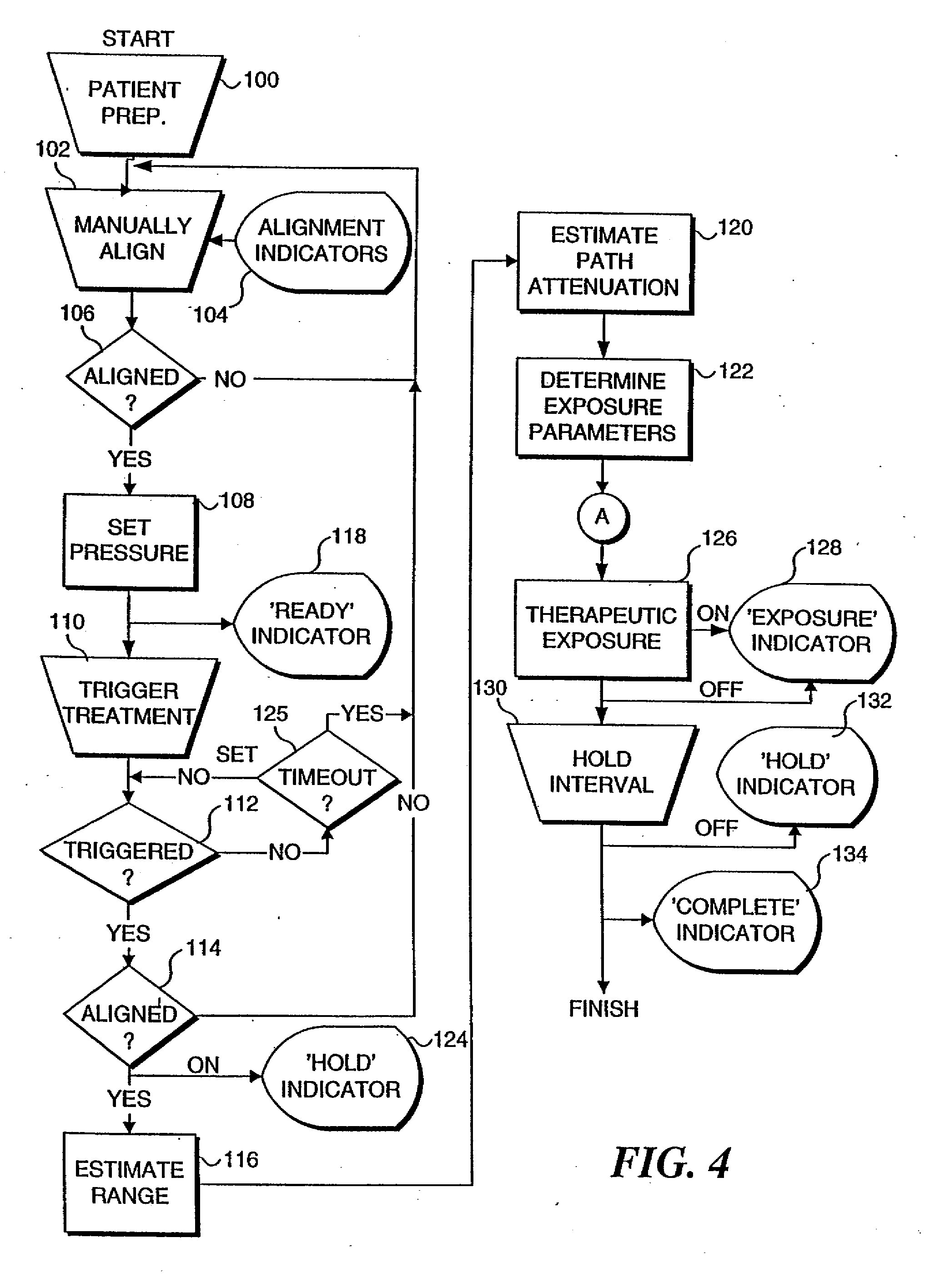
Embodiment Construction
Use of Ultrasound for Sealing Punctures
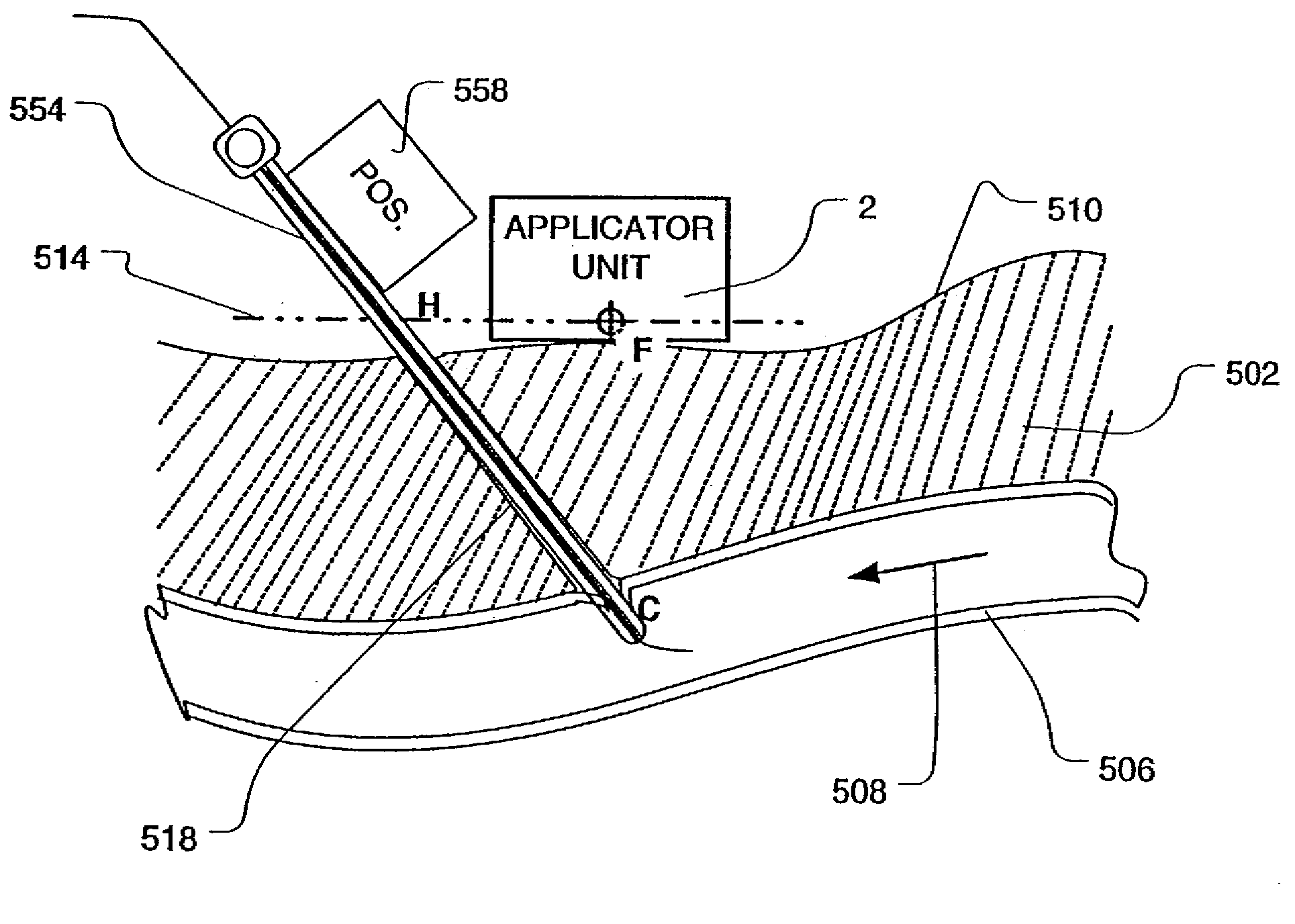
[0044] Because of its unique properties in soft tissue, ultrasound can be brought to a tight focus at a distance from its source. If sufficient energy is radiated within the ultrasound beam, cells located in the focal volume can be rapidly heated, while intervening and surrounding tissues are spared. Surrounding tissues are unaffected in the unfocused portion of the ultrasound beam because the energy is spread over a correspondingly larger area and associated heating is minimized.
[0045] Whereas ultrasound intensities on the order of 0.1 Watts / cm2 are employed in diagnostic imaging applications, intensities in excess of 1,000 Watts / cm2 are typical in high-intensity focused ultrasound (HIFU) applications. At the focal point, these high intensities result in large, controlled temperature rises within a matter of seconds.
[0046] It has been demonstrated in numerous in vivo animal studies that HIFU can rapidly seal blood vessel punctures and lace...
PUM
 Abstract
Description
Claims
Application Information