Apparatus and Methods for Creating an Opening in a Tissue Membrane

a tissue membrane and orifice technology, applied in medical science, surgery, vaccination/ovulation diagnostics, etc., can solve the problems of clogging the catheter, affecting the operation, and affecting the operation, so as to increase the procedure time and the expense and complexity of the etv procedure. , the problem of faulty pressure valves or one-way valves

Inactive Publication Date: 2007-08-30
MEDTRONIC VASCULAR INC
View PDF21 Cites 9 Cited by
  • Summary
  • Abstract
  • Description
  • Claims
  • Application Information

AI Technical Summary

Benefits of technology

[0017] Embodiments of the invention can include a vacuum or suction lumen in the delivery device and a vacuum or suction source for stabilizing the tissue membrane relative to the apparatus and/or for removing sections of portions that are cut from the membrane (throughout this document, the term “vacuum” should be taken to mean “vacuum or suct

Problems solved by technology

However, the VP shunts have an extremely high failure rate, e.g., in the range of 30 to 40 percent.
Failure includes clogging of the catheter, infection, and faulty pressure valves or one-way valves.
Thi

Method used


Image

  • Apparatus and Methods for Creating an Opening in a Tissue Membrane
  • Apparatus and Methods for Creating an Opening in a Tissue Membrane
  • Apparatus and Methods for Creating an Opening in a Tissue Membrane

Examples


Embodiment Construction

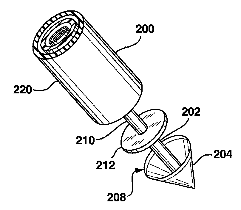
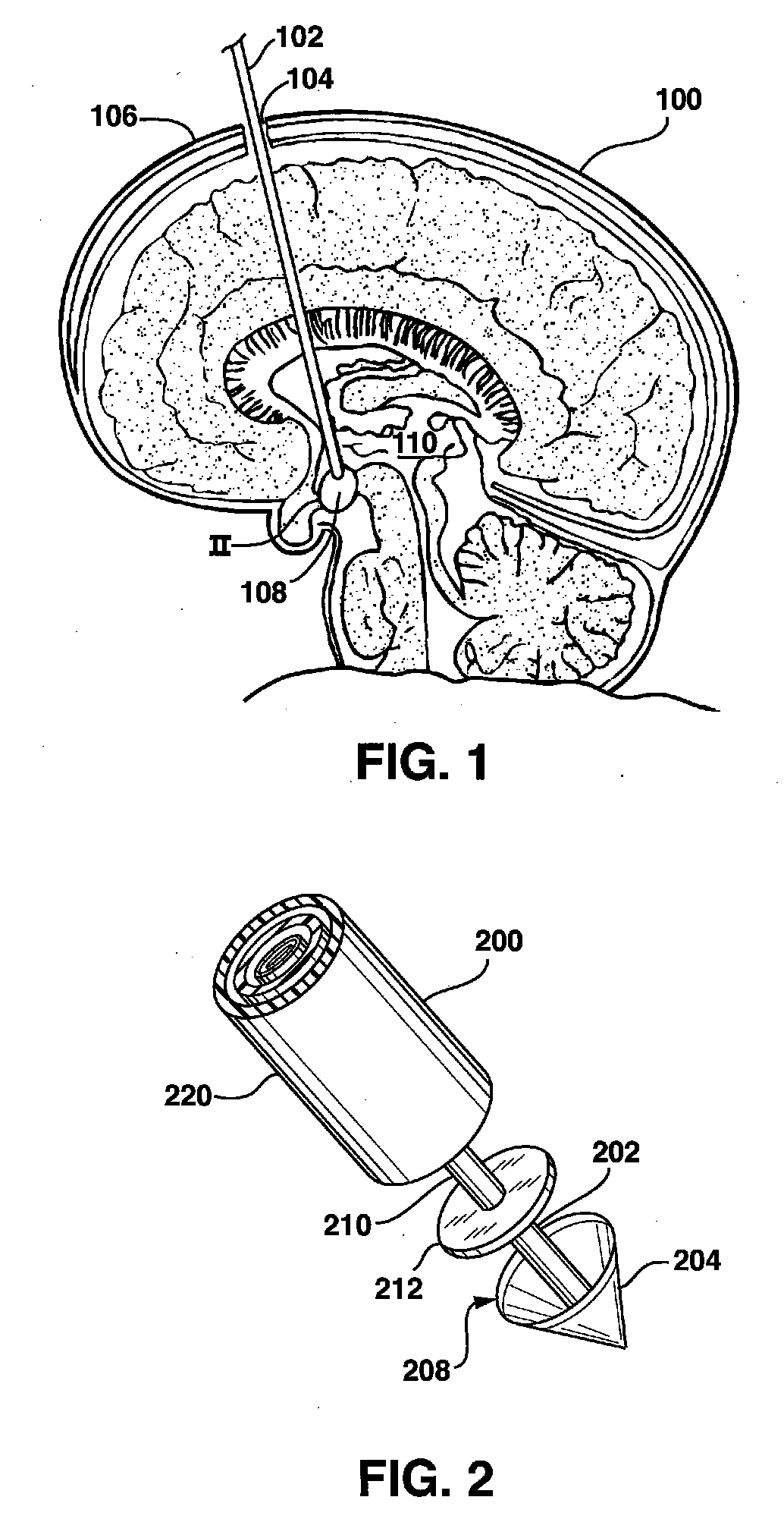
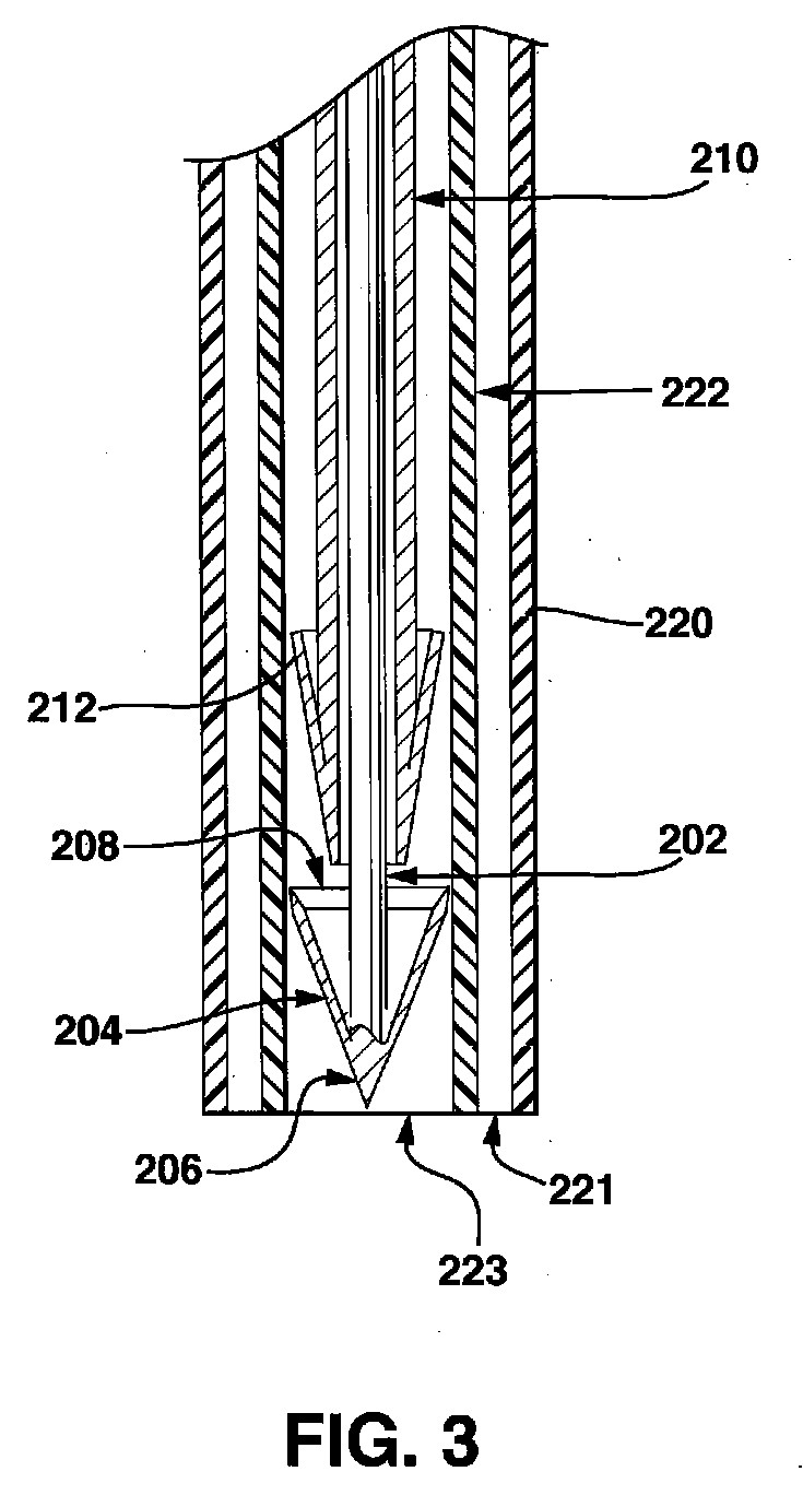
[0026] The invention will now be described in detail below by reference to the drawings, wherein like numbers refer to like structures. Referring to FIG. 1, one embodiment of an ETV procedure, using the devices and methods disclosed herein, includes forming a burr hole 104 (FIG. 1) in a skull 106, passing an endoscopic third ventriculostomy (ETV) device 102 through burr hole 104, and cutting an opening in the floor 108 of the third ventricle 110 with a cutting device (FIG. 2) to form an opening in floor 108. The ETV procedure can further include deploying a membrane eyelet into the opening.

[0027] The procedure can also include measuring the flow of CSF through the opening with a flow sensor, or using a pressure sensor to measure the pressure gradient across the opening. While not depicted in the drawings, the devices described herein can also include a lumen for injecting a contrast medium into the third ventricle such that a clinician can use fluoroscopy or other imaging modalitie...


PUM


Abstract

The current invention discloses an apparatus for creating openings in tissue membranes and methods for using the apparatus. The apparatus comprises a cutting device that can include a cutting element and a resisting element located on the ends of elongate members. The cutting device can be manipulated so that the cutting element and resisting element are on opposite sides of the target tissue membrane. The cutting device is delivered to a tissue membrane using an elongate delivery device that can be routed through a working lumen in an endoscopic probe. The delivery device can also access the tissue membrane via means other than an endoscopic probe. Embodiments of the invention can include a vacuum lumen in the delivery device and a vacuum source for stabilizing the tissue membrane relative to the apparatus or for securing sections of tissue that are removed from the membrane.

Description

TECHNICAL FIELD [0001] The present invention relates to a medical device and method. More particularly, the present invention relates to an apparatus and method for creating an opening or orifice in a septum (or membrane). Specifically, the invention discloses an apparatus and methods for using the apparatus to make an opening in the membrane floor of a ventricle in the brain during an endoscopic third ventriculostomy (ETV) procedure. BACKGROUND OF THE INVENTION [0002] Non-communicating hydrocephalus is a condition that results in the enlargement of the ventricles caused by abnormal accumulation of cerebrospinal fluid (CSF) within the cerebral ventricular system. [0003] In non-communicating hydrocephalus there is an obstruction at some point in the ventricular system. The cause of non-communicating hydrocephalus usually is a congenital abnormality, such as stenosis of the aqueduct of Sylvius, congenital atresia of the foramina of the fourth ventricle, or spina bifida cystica. There ...

Claims


Application Information

Patent Timeline
IPC IPC(8): A61B17/34
CPCA61B10/0283A61B10/04A61B2017/00867A61B17/32075A61B17/32053
Inventor NAYAK, ASHA
Owner MEDTRONIC VASCULAR INC
Why Eureka
Try Eureka