Functional Immunohistochemical Cell Cycle Analysis as a Prognostic Indicator for Cancer

a cell cycle analysis and immunohistochemical technology, applied in the field of immunohistochemical cell cycle analysis as a prognostic indicator of cancer, can solve the problems of invariably fatal multi-myeloma, one of the most common lymphoid cancers in adults, and is difficult to predict the outcome of pre-malignant lesions, and the mechanism that underlies the loss of cell cycle control in each cancer type is not understood, so as to achieve the effect of monitoring and predicting treatment

Active Publication Date: 2007-09-13
CORNELL RES FOUNDATION INC
View PDF2 Cites 50 Cited by
  • Summary
  • Abstract
  • Description
  • Claims
  • Application Information

AI Technical Summary

Benefits of technology

[0022] The present invention provides oncology professionals with the tools for early diagnosis and personalized prognostication of precancerous or cancerous conditions, and the ability to monitor and predict treatment efficacy based on an individual patient's response to therapy at the cellular level. The present invention provides for an improvement over the current standard of cancer care. For example, periodic testing of the patient's pS-Rb status according to the present invention can provide evidence that the patient's cells have begun re-entry into the proliferative stage following a quiescent period. The presence of pS-Rb in greater than 10% of the cells signifies an eminent relapse and triggers a treatment decision. Further characterization of the patient's disease status can be obtained from immunostaining for other cell-cycle proteins (as described hereinafter).

Problems solved by technology

Similarly, among blood cancers, lymphoblastic leukemia, the most common malignancy in childhood, can be cured by multi-agent chemotherapy, but multiple myeloma, one of the most common lymphoid cancers in adults, is invariably fatal.
It is particularly difficult to predict the outcome of pre-malignant lesions.
However, the mechanism that underlies the loss of cell cycle control in each cancer type is not understood.
However, there is no direct evidence that loss of a single INK inhibitor is sufficient to cause cancer.
Chromosomal changes in large cohorts correlate statistically with prognosis, but the associations are inconsistent and not explained by biologic data.
However, the method is costly, labor intensive, and requires specialized equipment not available in most hospitals.
Also, because it requires live PCs, it can only be performed immediately after an aspirate is drawn.

Method used


Image

  • Functional Immunohistochemical Cell Cycle Analysis as a Prognostic Indicator for Cancer
  • Functional Immunohistochemical Cell Cycle Analysis as a Prognostic Indicator for Cancer
  • Functional Immunohistochemical Cell Cycle Analysis as a Prognostic Indicator for Cancer

Examples


example 1

ow Specimen Procurement

[0055] Bone marrow specimens were obtained from volunteers without hematological disease at the Hospital for Special Surgery and from MM patients at the New York-Presbyterian Hospital under informed consent as part of an Institutional Review Board approved study. The posterior and superior iliac spines were identified before beginning the procedure. The patient was placed in a ventral or lateral decubitus position. The field was cleaned with Betadine and the area of interest was anesthetized with 1% Lidocaine. A bone marrow aspirate and biopsy was performed unilaterally using a modified Jamshidi bone marrow aspiration / biopsy needle, 11 gauge×4 inch. Several 5-7 cc aspirates were obtained by relocating the needle in each aspirate to obtain a total of approximately 30 cc of marrow. The core biopsy was then obtained. The bone marrow core biopsies were fixed in 10% neutral-buffered formalin and paraffin-embedded. CD138+ normal and malignant plasma cells (greater t...

example 2

tochemistry

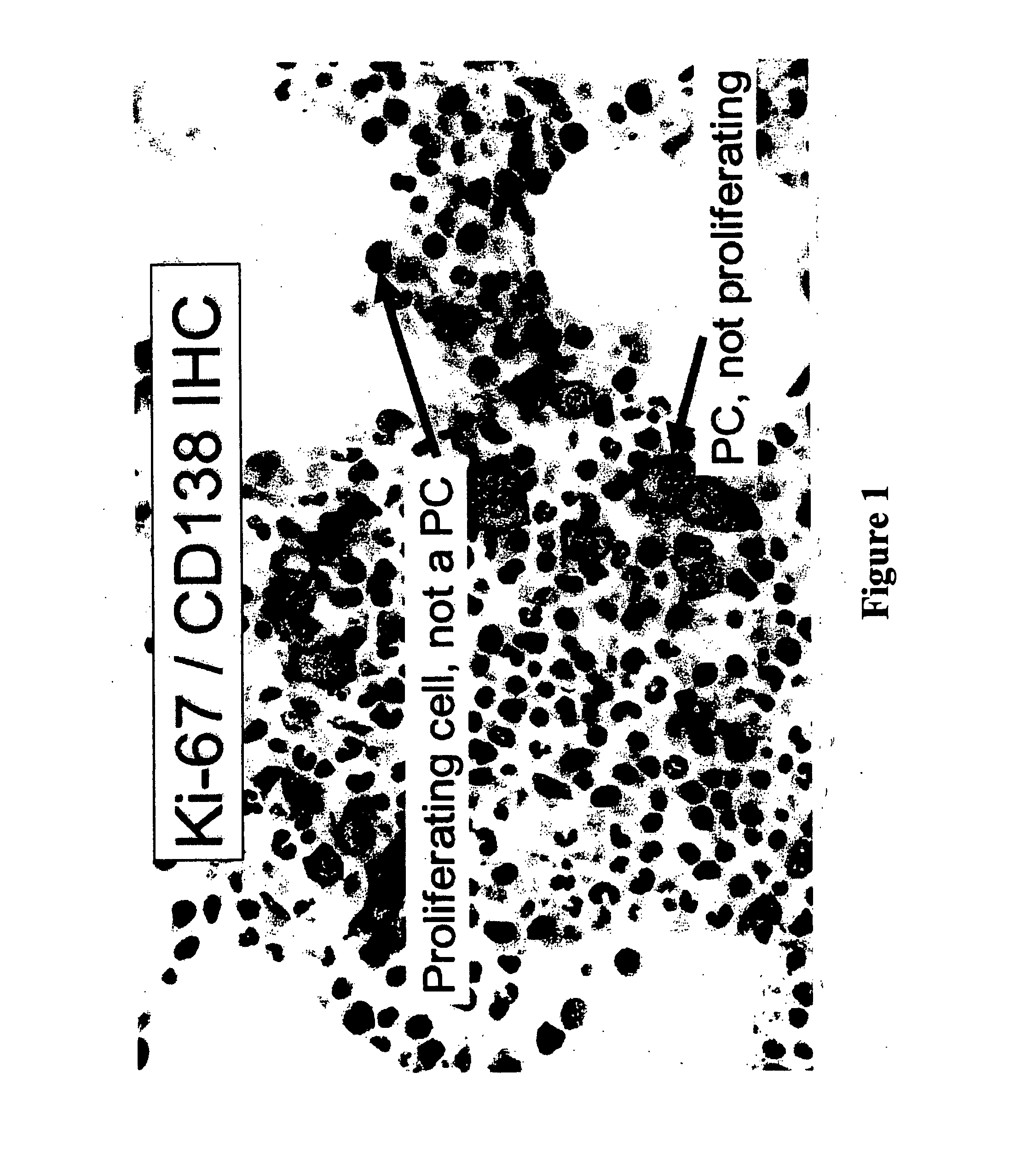
[0056] Plasma cells were identified with an anti-CD138 antibody and a detection system yielding a red membranous signal. To ensure that hematopoietic background cells did not express CD138 and that all plasma cells in the samples did express CD138, and to ensure that any background non-neoplastic plasma cells were not included in the analysis, the CD138 stained slides in all biopsies were compared to serial sections immunostained for cytoplasmic immunoglobulin expression (IgM, IgD, IgG, IgA, IgK or IgX); these slides were also compared to routine hematoxylin and eosin stained serial sections. The concordance between routine histologic examination, monotypic patient-myeloma isotype specific cytoplasmic immunoglobulin expression, and CD138 expression was very high; rare myeloma specimens showed weak CD138 expression in minor subpopulations of cells but none of the myeloma specimens was negative for CD138. CD138 negative plasma cells were very rare. Non-neoplastic background...

example 3

of BrdU-Uptake

[0064] BrdU uptake in primary bone marrow CD138+ plasma cells ex vivo was detected by immunofluorescence staining (Morse et al., “Induction of Cell Cycle Arrest and B Cell Terminal Differentiation by CDK Inhibitor p18(INK4c) and IL-6,”Immunity 6:47-56 (1997), which is hereby incorporated by reference in its entirety), using a Mab FITC-anti-BrdU (Roche Diagnostics, Indianapolis, Ind.) and Texas red-conjugated rabbit anti-human Ig κ and λ antibodies (Southern Biotechnology). The frequency of BrdU+ cells was determined by counting 250 cells in three areas.


PUM

PropertyMeasurementUnit
half lifeaaaaaaaaaa
pHaaaaaaaaaa
timeaaaaaaaaaa

Abstract

The present invention relates to a method of determining the presence of a retinoblastoma protein (Rb)-mediated cancerous or pre-cancerous condition in a sample of fixed cells that involves exposing the sample to an antibody that recognizes phosphorylation of serine on the retinoblastoma protein (pS-Rb), and identifying cells in the sample that are recognized by the anti-O body to pS-Rb, where the recognition of pS-Rb in the sample determines the presence of an Rb-mediated cancerous or pre-cancerous condition. The method also involves identifying the presence of specific cell cycle markers and positive and negative cell cycle regulators to assess the stage of disease progression in a cancer patient having an (Rb)-mediated cancer. The present invention also relates to methods of assessing the efficacy of cancer therapy for an Rb-mediated cancer, determining the mechanism of cell cycle progression in an Rb-mediated cancer, and identifying cells predisposed to developing an Rb-mediated cancerous condition.

Description

[0001] This application claims the benefit of U.S. Provisional Patent Application Ser. No. 60 / 560,687, filed Apr. 8, 2004, which is hereby incorporated by reference in its entirety.FIELD OF THE INVENTION [0002] The present invention relates to immunohistochemical methods for functional assessment of cell cycle progression of cells in situ as a prognostic indicator of disease progression in hematopoietic cancers and as a measure of cancer therapy efficacy in cancer patients. BACKGROUND OF THE INVENTION [0003] Cancers are, in general, classified by the cell type and site of origin, such as lymph node, lung, bone marrow, etc. In addition, they are sub-classified based on histologic characteristics and the proteins expressed by the cancer cells, as determined by immunophenotyping. These sub-types define biologically distinct diseases, each of which has its own distinct clinical behavior and, hence, its own specific therapy. For example, the most common testicular cancer, seminoma, is tr...

Claims


Application Information

Patent Timeline
Patent Type & Authority Applications(United States)
IPC IPC(8): G01N33/574
CPCG01N33/57484
Inventor CHEN-KIANG, SELINAELY, SCOTTDI LIBERTO, MAURIZIO
Owner CORNELL RES FOUNDATION INC
Why Eureka
Try Eureka