
Glucose sensor and uses thereof

Inactive Publication Date: 2007-09-20
MCGILL UNIV
View PDF0 Cites 82 Cited by
  • Summary
  • Abstract
  • Description
  • Claims
  • Application Information

AI Technical Summary

Benefits of technology

[0019] An object of the present invention is to provide a glucose sensor comprising a glucokinase protein, wherein the catalytic enzymatic activity has been disabled.

Problems solved by technology

While the use of such glucose monitors has improved insulin strategy, actual insulin delivery remains inflexible, i.e. a fixed dose via a systematic route.
While glucose sensors have been developed using physical chemical approaches, such sensors tend to lack both specificity and sensitivity.
For example, an infrared device has been developed which measures blood glucose, however, this device is reliant on complex computer analysis of the emission spectra to enhance the relatively weak glucose signal and distinguish it from background noise [Robinson, M. R. et al., Clin. Chem. 38:1618-1622 (1992)].
A major drawback inherent in these systems is the fact that enzyme-catalysed reactions are greatly affected by the concentration, and therefore the availability, of their reactants.
Thus, if access of either glucose or oxygen to the device containing the glucose oxidase is compromised in any way, the results obtained from measuring the catalytic activity of the enzyme will be inaccurate.
In the blood, for example, the glucose concentration is typically much higher than the concentration of available oxygen, therefore, the rate of the enzyme-catalysed oxidation of glucose will be controlled by the oxygen concentration and will not accurately reflect the concentration of glucose.
This biosensor has been limited to investigational use only by U.S. law.
Of these, the most significant is that both GGBP and GBP, like glucose oxidase, are bacterially derived and are not, therefore, necessarily optimized for detection of physiological concentrations of glucose in a human subject.
Both biosensors require incorporation of detectable labels or reporter systems into the protein and the resultant requirement for an appropriate light source for the reporter systems limits the ability of these sensors in an implantable device.
Notable drawbacks to the use of this protein include the fact that no known human counterpart exists and thus its use may have unfavourable antigenic consequences.

Method used


Image

  • Glucose sensor and uses thereof
  • Glucose sensor and uses thereof
  • Glucose sensor and uses thereof

Examples


example 1

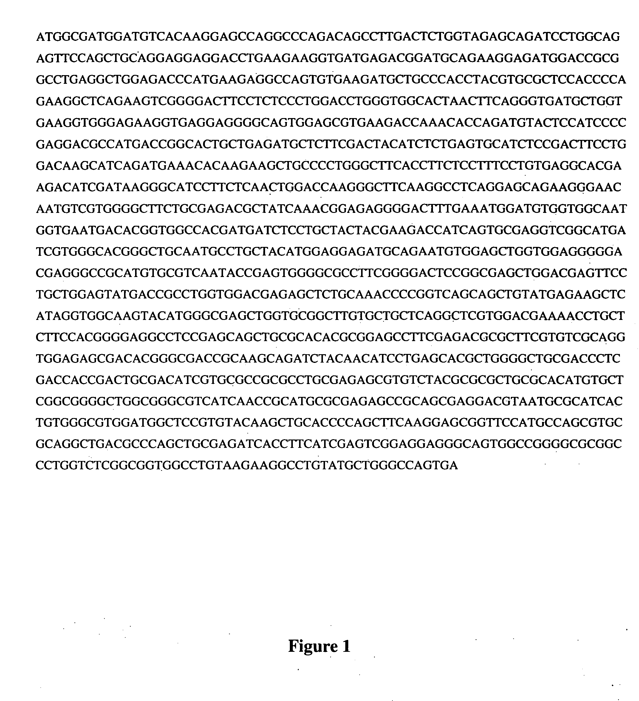
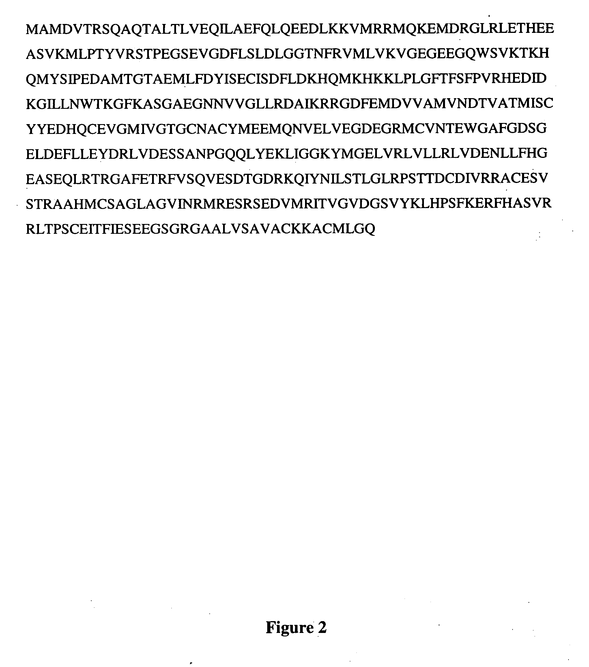
Cloning Human Glucokinase

[0130] The human liver glucokinase was cloned from the Hep 3B liver cell line. Following isolation of total mRNA from the cell line using standard techniques, RT-PCR was employed to generate sufficient glucokinase cDNA. Expand reverse transcriptase, a genetically engineered version of MoMuLV-RT that has negative RNase H activity, and an Oligo (dT)15 primer were used for the reverse transcription step. Pwo DNA Polymerase was used for the PCR step. PCR was performed in three separate reactions. The first reaction amplified a 5′ portion of the glucokinase cDNA, the second reaction amplified a 3′ portion of the glucokinase cDNA and the third reaction amplified the complete glucokinase sequence from the combined products of the first and second reactions. Primers were used that incorporated convenient restriction enzyme sites to facilitate cloning into appropriate vectors. Primers used to amplify the glucokinase for cloning into plasmid pcDNA3 (digested with Bam...

example 2

Site-Directed Mutagenesis of the Cloned Human Glucokinase

[0134] In vitro site-directed mutagenesis of the glucokinase was achieved by PCR-based techniques to create mutations at position 336 (Ser->Val; Ser->Leu and Ser->Ile) and at position 205 (Asp->Ala). The PCR reactions employed complementary primers containing mutagenic sequences, and a set of upstream and downstream primers. The sequences of the mutagenic primers were as follows (nucleotides that are different from those that occur in the wild type sequence are underlined):

Ser336ValPrimer A5′-TCGTGGTCCAGGTGGAGAGCG-3′[SEQ ID NO:9]Primer B5′-CGCTCTCCACCTGGACCACGA-3′[SEQ ID NO:10]Ser336LeuPrimer A5′-TCGTGCTGCAGGTGGAGAGCG-3′[SEQ ID NO:11]Primer B5′-CGCTCTCCACCTGCAGCACGA-3′[SEQ ID NO:12]Ser336IlePrimer A5′-TCGTGATTCAGGTGGAGAGCG-3′[SEQ ID NO:13]Primer B5′-CGCTCTCCACCTGAATCACGA-3′[SEQ ID NO:14]Asp205AlaPrimer A5′-GGTGAATGCAACGGTGGCCACG-3′[SEQ ID NO:15]Primer B5′-CGTGGCCACCGTTGCATTCACCC-3′[SEQ ID NO:16]Primer GLK-35′-CTGAATTCACTGGC...

example 3

Generation of Wild-Type and Mutant Glucokinase Vectors

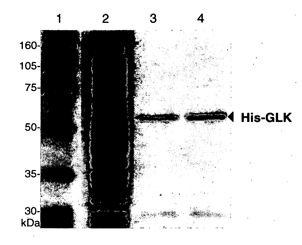
[0140] The Ser336 and Asp205 mutant glucokinases produced by the above PCR reactions were first cloned into pcDNA3 (as indicated above). Wild-type glucokinase and the mutant glucokinases were each subsequently subcloned into the pGEX-KG and pET-15b expression vectors using Xhol and BamHI restriction enzymes (blunt end) for the wild-type and Sac II and BsrGI for the mutants. The pGEX-KG and pEt-15b vectors were used in order to express wild-type and mutant glucokinases containing GST and polyhistidine (His) affinity tags, respectively.

[0141] The following plasmids were generated in this manner. All plasmids have been sequenced to confirm the presence of the appropriate mutant sequence and the absence of any abnormalities.

TABLE 1List of PlasmidsPlasmidClone #GlucokinasepGEX-KG20Wild-type23Ser336Val32Ser336Leu43Ser336Ile53Asp205AlapCDNA320Wild-type7Ser336Val15Ser336Leu25Ser336Ile33Asp205AlapET-15b14Wild-type1Ser336Val9Ser336Leu1...


PUM

PropertyMeasurementUnit
Catalytic activityaaaaaaaaaa
Affinityaaaaaaaaaa

Abstract

The present invention provides a glucokinase protein in which the catalytic activity has been disabled in order to enable its use as a glucose sensor. The catalytically disabled glucokinase protein can be used as the glucose sensor in hand-held glucose monitors and in implantable glucose monitoring devices. The glucose sensor can also be incorporated into biomedical devices for the continuous monitoring of glucose and administration of insulin.

Description

RELATED US APPLICATION DATA [0001] This application is a Continuation-in-Part of application Ser. No. 10 / 421,360 filed on Apr. 22, 2003.FIELD OF THE INVENTION [0002] The present invention pertains to the field of glucose sensors, in particular, to a glucokinase protein, wherein the catalytic enzymatic activity has been disabled, yet the protein retains a high specific affinity for and ability to bind glucose. BACKGROUND [0003] Glucose control in diabetics is of paramount importance. While poor glucose control leads to morbidity and associated mortality, good glucose control has been shown to reduce cardiovascular, retinal, and kidney diseases by almost 50%, in addition to considerably reducing other complications [The Diabetes Control and Complications Trial Research Group. N. Engl. J. Med. 329:977-986 (1993)]. [0004] The push for better management of glucose control in the past led to the development of conventional hand-held glucose monitors. While the use of such glucose monitors...

Claims


Application Information

Patent Timeline
IPC IPC(8): C07K14/605C12N9/12
CPCC12Y207/01001C12N9/1205
Inventor TRIFIRO, MARK A.
Owner MCGILL UNIV
Learn More